肩袖是冈上肌、冈下肌、小圆肌和肩胛下肌四块肌肉及其肌腱组成的复合体.它们环绕肱骨头上端,共同协调肩关节运动.在年龄、创伤、劳损等外在因素以及肌腱退变、血运不足、肩峰下撞击等内在因素作用下,肩袖易发生损伤,尤其是肌腱撕裂,进而引发肩部疼痛、力量减弱、活动范围减少.肩袖撕裂通常需手术修复,术后大部分疼痛减轻,肩关节功能改善,但发生再撕裂的比率却很高.这其中腱骨仿生愈合是一个重要的临床问题.为此,出现了很多新技术、新方法,这其中,生物疗法因其较好的应用前景一直是医生和患者关注的热点.目前的文献也反映多种生物制品,如富血小板血浆(platelet-rich plasma,PRP)、凝胶蛋白、生物补片或支架、干细胞、细胞因子和生物信号分子可以在肩袖撕裂中腱骨愈合方面起到作用.本综述主要讨论:(1)骨髓刺激技术;(2)PRP;(3)干细胞;(4)生物补片或支架;(5)细胞因子生物疗法的基础和临床研究,旨在总结生物疗法在肩部病变中的应用现状及发展,为医生和患者提供可靠的临床经验和技术应用参考.
患者男,21 岁,1 年前无明显诱因出现右腕部肿胀、疼痛,活动受限.疼痛肿胀加重2 个月,遂来解放军总医院就诊.专科检查:右腕部及手掌掌侧肿胀明显,可触及8 cm × 3 cm大小的肿物,局部皮温不高,压痛,有波动感,可触及异物滑动.Tinel征阴性,右腕部屈伸活动受限伴疼痛,右手各手指感觉、末梢血运正常.右腕部 X 线片检查未见骨质异常.右腕关节MRI检查示右腕指深浅屈肌腱鞘内不同程度囊性异常信号影,腱鞘内见多发粟粒样结节影,考虑为良性病变腱鞘囊肿伴(米粒样)滑膜炎可能性较大;相应腕管内正中神经受压,形态欠规则,信号似尚可,不能排除腕管综合征;初步诊断为右腕关节米粒体滑囊炎(图1 A).
[目的]探讨前交叉韧带(anterior cruciate ligament,ACL)重建失败后翻修进行翻修手术的主要原因.[方法]回顾性2009年3月-2020年3月解放军总医院骨科收治的因重建失败而行翻修术的36例患者的临床资料,其中男31例,女5例,年龄(29.39±6.63)岁;31例于外院行初次ACL重建术,5例于本院初次重建.结合患者临床病例及影像学资料研究分析ACL重建失败的原因.[结果]36例翻修患者中,33例初次重建术移植物使用自体肌腱,2例使用同种异体肌腱,1例使用LARS人工韧带.在患者初次ACL重建失败的因素中,19例为创伤导致的翻修;10例为单纯的技术原因;3例因生物学原因而翻修;7例为组合因素,其中6例同时存在创伤因素及技术因素,1例为生物学因素合并技术因素;另有3例为其他原因.翻修手术中,23例使用自体肌腱,10例使用LARS人工韧带,2例使用同种异体肌腱,1例患者因自体肌腱过细(≤6mm)而使用自体肌腱加同种异体肌腱进行翻修.[结论]导致前交叉韧带重建手术失败的因素包括骨隧道定位错误、移植物选择及合并损伤等,但主要与手术技术有关.
Objective To compare clinical outcomes of arthroscopic therapy for tibial eminence avulsion fracture with loop ligature suture and absorbable suture anchor. Methods From June 2012 to February 2018 62 patients with avulsion fractures of the anterior cruciate ligament injured within three weeks in First Medical Center of PLA General Hospital were selected in the study. All the affected knees have normal function before injury. The patients with open fracture, multiple fracture, tibial plateau fracture, meniscus and ligament injury were excluded in the study. All the patients were divided into two groups according to different operation methods: 30 patients were treated with arthroscopic reduction and fixation using loop ligature suture, as the suture group; 32 patients were treated with arthroscopic reduction and fixation using absorbable suture anchor, as the anchor group. The operation time and intraoperative blood loss of the two groups were compared. Knee joint stability of two groups was compared by front drawer test, Lachman test and axial shift test. Knee joint function of two groups was compared by Lysholm score and International Knee Documentation Committee (IKDC) 2000 subjective score. The independent sample t test was used to compare the quantitative data between two groups. The paired t test was used to compare the quantitative data within groups, and the chi-square test was used for compare the qualitative data between the two groups. Results All the incisions healed primarily. Radiographic evaluation showed bone union within three months postoperatively in both groups. The mean follow-up period after the operation was (22±8)months in suture group and (19±7)months in anchor group. The operation time in anchor group was lower than that in suture group, and the difference was statistically significant (t=2.491, P 0.05). The Lysholm score were both improved in two groups after operation for six months. The difference was statistically significant(suture group: t=26.265, P 0.05). Conclusion In the arthroscopic therapy for tibial eminence avulsion fracture, the loop ligature suture fixation and absorbable suture anchor fixation have their own characteristics but equivalent clinical effects, which could both provide satisfying reduction, rigid fixation, well healing of avulsed fragment and effective functional rehabilitation. Key words: Arthroscopy; Anterior cruciate ligament; Fractures, avulsion; Sutures
镁金属材料是一种新型的可降解医用材料,因其可降解、生物相容性好、力学性能佳、可诱导骨生长等优点,具有良好的应用前景.但是由于耐腐性差其临床应用目前仍受到限制.在镁金属材料表面进行表面处理是目前研究中常用的提高耐腐性的方法 ,钙磷复合物涂层是其中最常见的表面处理方式之一.本文从涂层制备方法 、成骨活性、机械性能、生物学性能等几方面对近年来镁金属材料表面钙磷复合物涂层处理的研究进行了总结,发现钙磷复合物涂层是一种很有潜力的能够将镁金属材料推向临床应用的表面处理方法 ,但在应用于临床前仍需进行深入、系统的研究.
Objective To construct the orthopedics arthroscopic computer image collection and teaching system so as to improve the quality and efficiency of clinical and education work .Methods By integrating the latest server technology , computer digital multimedia technology , local and remote network technology , we developed this comprehensive application system that integrates data acquisition , process-ing, storage, editing, research and teaching of orthopedics arthroscopic computer images .Results This system , applicable to orthopedic arthroscopic surgery and related clinical education , is convenient , stable and powerful .Conclusion This system is practical and valuable in clinical work and teaching .
腱鞘囊肿发生在肩关节比较少见,是引起肩关节疼痛不常见的病因。然而,肩关节腱鞘囊肿临床上通常表现为疼痛和力弱症状,容易和肩袖损伤相混淆,使得诊断困难。为准确诊断和正确的治疗,必须知道这种肩关节疼痛的罕见原因[1]。大多数学者认为,肩关节腱鞘囊肿的发生机制是关节囊盂唇复合体损伤,关节液不断渗入损伤的软组织,形成单向活瓣效应,最终形成囊肿[2-4]。因此,通常伴有上盂唇前后部(superior labrum anterior and posterior,SLAP )损伤。MRI 检查是诊断肩关节腱鞘囊肿的主要手段,MRI 检查可以显示肩关节腱鞘囊肿的位置和大小,同时显示是否合并有肩袖损伤,这对于术前准备十分重要[5-7]。如果发现囊肿位于肩胛骨切迹,则需要进行肌电图检查,进一步排除是否有肩胛上神经损伤表现。肩关节腱鞘囊肿的治疗方法分为保守治疗和手术治疗,手术治疗包括切开手术、关节镜下手术、关节镜联合切开手术,文献均有报道。现分析一例肩关节腱鞘囊肿患者进行全关节镜下手术,治疗过程及术后随访效果并结合文献复习,探讨关节镜诊断和治疗肩关节腱鞘囊肿的疗效。
目的:构建带Myc标签的CCAAT增强子结合蛋白β(C/EBPβ)的真核表达载体,并检测其对骨肉瘤细胞增殖的影响.方法:应用PCR技术从人乳腺文库中扩增C/EBPβ全长编码区基因,并克隆到pXJ-40载体中;将重组质粒转染人胚胎肾293T细胞,采用SDS-PAGE和Western印迹鉴定蛋白表达;另将重组C/EBPβ质粒转染人骨肉瘤细胞系U2OS,生长实验检测其对肿瘤细胞增殖的影响.结果:基因测序和双酶切鉴定表明,Myc-C/EBPβ真核表达质粒构建成功;SDS-PAGE和Western印迹结果显示,Myc-C/EBPβ转染人胚胎肾293T细胞后获得表达;生长实验证明C/EBPβ具有抑制骨肉瘤细胞增殖的功能.结论:构建了Myc-C/EBPβ的真核表达载体,为全面了解C/EBPβ的生物学功能及调控机理奠定了基础.
Objective To construct the eukaryotic expression vector of CCAAT / enhancer binding protein-α ( C/EBPα ) labeled with Myc tag ( pXJ-40 ), and to examine the inhibitory effects of C/EBPα on osteosarcoma cell growth.Methods Human C/EBPα coding gene region was amplified from mammary gland cDNA library by polymerase chain reaction ( PCR ), which was inserted into the pXJ-40-myc vector. Myc-C/EBPα was constructed and transfected into 293T cells. Western blotting analysis was carried out to detect their expressions. In addition, U2OS cells transfected with Myc-C/EBPα were divided into the experimental group, and U2OS cells only transfected with Myc were divided into the control group. The growth of U2OS cells in both groups was identiifed using growth curve experiment.Results The coding region of C/EBPα was successfully ampliifed by PCR and cloned into pXJ-40-myc vector. Myc-C/EBPα was successfully expressed in human 293T cells. Growth curve experiment showed that the growth of U2OS cell lines transfected with Myc-C/EBPα was apparently inhibited. With the extension of the culture time, the inhibitory effects were gradually enhanced. At the 5th day, the inhibition ratio of U2OS cell growth was 58%in the experimental group. The growth rate of U2OS cell lines transfected with Myc-C/EBPα [ optical density ( OD )=0.495 ] was apparently lower than that of the control group ( OD=1.179 ), and the differences between them were statistically signiifcant (P<0.01 ).Conclusions The eukaryotic expression vector Myc-C/EBPα can effectively inhibit osteosarcoma cell growth.
Objective: To construct the eukaryotic expression vector of human casein kinase 1(CK1) labeled with FLAG tag, obtain the expressed product, and measure its cellular localization in osteosarcoma U2OS, breast cancer ZR-75-1, hepatocellular carcinoma HepG2 cells. Methods: Human CK1 coding gene region, amplified from mammary cDNA library by PCR, was inserted into the pCMV-Tag2B vector. The resulting recombinant plas-mid pCMV-Tag2B-CK1 was transfected into U2OS, ZR-75-1 and HepG2 cells and cell lysates were examzined by SDS-PAGE and Western blotting. In addition, immunofluorescence was applied to determine the cellular local-ization. Results: The CK1 was successfully amplified by PCR and cloned into pCMV-Tag2B, as evidenced by dou-ble enzyme digestion and gene sequencing. U2OS, ZR-75-1 and HepG2 cell lines transfected by the recombinant plasmid pCMV-Tag2B-CK1 successfully expressed the protein, according to SDS-PAGE and Western blotting. Im-munofluorescence indicated that CK1 protein was expressed in all three cell lines and localized in both the nucle-us and cytoplasm, predominantly in the nucleus. Conclusion: The CK1 eukaryotic expression vector was successful-ly obtained, and the CK1 protein could be expressed in both the nucleus and cytoplasm in different tumor cell lines, which lays foundation for further research on cell regulation by CK1.
临床上肩关节疼痛是骨骼肌肉疾病中仅次于背部疼痛和膝关节疼痛的第三大常见症状,发病率约26%[1]。如果不及时治疗,可能会导致明显的肩关节功能障碍,影响患者生活质量,甚至无法工作[2-3]。由于导致肩关节疼痛、无力和功能障碍的原因很多,所以要获得准确的诊断并不容易。因此,临床医生必须进行全面体格检查,以判断出肩关节功能障碍的不同病因[4-5]。本文将对肩关节疾病的相关有效体格检查方法作一综述。
Objective To construct the eukaryotic expression vector of cyclin-dependent kinase ( CDK) 7 labeled with Myc tag, obtain the expressed product , and identify its interaction with FLAG-P53 at the protein level .Methods Human CDK7 coding gene region amplified from the mammary cDNA library by PCR was inserted into the pXJ -40 vector.The recombinant plasmid Myc-CDK7 transfected into human 293T cell lines was investigated and examined by SDS-PAGE and Western blotting.In addition,assay was applied to determine the interaction between Myc-CDK7 and FLAG-p53.Results The coding region of CDK7 was successfully amplified by PCR and cloned into pXJ-40 vector, which was identified by double enzyme digestion and gene sequencing .Myc-CDK7 was successfully expressed in human 293T cell lines according to SDS-PAGE and Western blotting assay indicated that Myc-CDK7 could interact with P53 protein, which verified its known function .Conclusion The eukaryotic expression vector Myc-CDK7 is successfully obtained , which will contribute to further research on CDK 7-mediated cell cycle regulation .
OBJECTIVETo study the efficacy of self-made, antibiotic-loaded cement articulating spacer in the treatment of infected total knee arthroplasty.METHODSThe self-made molds were used to form the spacer during the operation. From March 2002 to March 2007, 22 patients with infected knee arthroplasty (10 males with 10 knees, 12 females with 12 knees) were treated with this kind of spacer in our center. The mean age of the patients was 59.6 years old (33 to 75 years old). The interval time between primary arthroplasty and first onset of infective syndrome was 6.7 months (1 to 14 months). The diagnosis was established by the clinical presentation,serum laboratory inflammatory markers (white blood cell count,erythrocyte sedimentation rate and C-reactive protein) and knee aspiration. The serum laboratory inflammatory markers were used to measure the systemic response to infection. Clinical and radiographic follow-up was regularly performed by HSS score system and X-ray.RESULTSAll the patients were followed, the average interval between debridement and reimplantation was 4.7 months (3 to 9 months) and the infection control rate was 100% after the implantation of spacer. The average follow-up duration after reimplantation was 29.8 months (10 to 64 months) and there was no recurrence of infection at the latest follow-up. The HSS score increased from 40.5+/-5.9 to 65.8+/-7.5 after the implantation of spacer, furthermore, the score reached 88.7+/-5.1 in average at the latest follow-up. The patient satisfaction rate was 95.3%.CONCLUSIONThis self-made molds and spacers is a reliable approach for the management of infected knee arthroplasty with some virtues, such as providing a mobile and functional joint through the treatment course, decreasing the difficulty of reimplantation, avoiding of a long-term post-operative infusion and high effective for eradicating infection.
Objective To provide basis for the improvement of arthroscopic reconstruction of the anterior cruciate ligament(ACL) through comparison of coverage rate and tunnel positions between single and double-bundle on tibial plateau by dual-source computed tomography(DSCT) three-dimensional imaging.Methods DSCT scans were performed on 14 pairs of double-bundle and 20 pairs of single-bundle reconstruction knees after arthroscopic reconstruction of ACL.ACL tibial footprints and tunnels were reconstructed on tibial plateau by 64-slice spiral CT workstation(GE,Volume Share2-AW 4.4 version).The area of ACL tibial footprint and tibial tunnel were marked and measured.The coverage rate of single and double-bundle on tibial plateau and the tunnels position were compared and calculated.Results(1) The coverage rate after surgery in single bundle was(50.50±13.58) % while(61.07±11.53) % in double bundle.There was significant difference(t=2.370,P=0.024) between the two groups.(2) The tunnel center in ACL single-bundle reconstruction was located at a mean of 43.80% in sagittal plane and 55.15% in frontal plane,whereas the natural footprint center located at 44.90% in sagittal plane and 51.85% in frontal plane.There was significant difference in frontal plane(t=5.592,P=0.001) but no significant difference in sagittal plane(t=0.631,P=0.536).(3) In ACL double-bundle reconstruction,the center of AMB(anteromedial bundle) tibial tunnel was located at a mean of 37.00% of anterior-to-posterior plane and 53.00% of lateral-to-medial plane.The center of natural footprint was located at 37.43% in sagittal plane and 51.14% in frontal plane.No significant difference was found in sagittal plane(t=0.120,P=0.908) while significant difference was observed in frontal plane(t=4.192,P=0.001).The center of PLB(posterolateral bundle) tibial tunnel was located at a mean of 55.00% in sagittal plane and 56.00% in frontal plane while the center of natural footprint was located at 40.79% in sagittal plane and 51.64% in frontal plane.There was significant difference in sagittal plane(t=9.121,P=0.001) and frontal plane(t=7.280,P=0.001).Conclusions The coverage rate of ACL on tibial tunnel was larger in double bundle reconstruction than in single bundle.In order to anatomically reconstruct ACL,individualized reconstruction technique should be adopted.Moreover,the dual-source three-dimensional computed tomography(DSCT) can help us to evaluate the relative location of tibial tunnel and ACL footprint which can provide meaningful guidance for arthroscopic reconstruction of the ACL.
Objective To reconstruct and measure the tibial footprint of the anterior cruciate ligament(ACL) by dual-source 3D CT(DSCT),so that to provide basis and parameters for anatomic double-bundle anterior cruciate ligament reconstruction.Methods A total of 110 knees from the both knees of 55 volunteers,including 32 male and 23 female,aged from 20 to 50 years old,were scanned and processed with 64-slice spiral CT workstation(GE,Volume Share2-AW 4.4 version).Tibial plateau and the ACL tibial footprint were reconstructed three-dimensionally.Footprint areas,the footprint length and width,and the average geometric midpoint insertion for the ACL located on the tibial plateau were measured.A P value < 0.05 was considered significant.ResultsMale ACL showed significantly larger area and longer length of the minor axis of the tibial footprint [(200.99±30.42) mm2 vs.(170.30±26.94)mm2,t=5.471,P=0.000;(9.25±1.56) mm vs.(8.30±1.46) mm,t=3.235,P=0.002].No significant difference was observed in the length of long axis of the tibial footprint nor the average geometric midpoint insertion for the ACL located on the tibial plateau in sagittal and frontal reconstructions between male and female [(14.00±1.76) mm vs.(13.63±1.65) mm,t=1.116,P=0.267;(48.35±6.13)% vs.(47.53±4.34)%,t=0.778,P=0.439;(49.70±2.37)% vs.(49.00±2.68)%,t=1.446,P=0.151].Conclusions The native ACL tibial footprints can be reconstructed and measured by DSCT.Because of the highly different configuration and position,individualized reconstruction technique should be used to restore the insertion anatomy at the tibial footprint of the ACL.
前交叉韧带(anterior cruciate ligament,ACL)是膝关节重要的静力性稳定结构,对维持膝关节稳定具有重要作用.ACL断裂后自愈能力差,若不及时修复,可导致关节不稳并继发半月板、软骨损伤、关节退行性改变等,影响膝关节功能.
Objective To investigate the effect of percutaneous transhepatic cholangio drainage(PTCD) in hilar biliary obstruction patients using biliary drainage tube with added side holes.Methods A total of 36 patients with malignant hilar biliary obstruction(32 with cholangiocarcinoma and 4 with liver metastasis tumor) underwent PTCD.The biliary drainage tube with added side holes was managed to pass through the obstructed biliary ducts percutaneously.The pigtail shaped end of the tube was put in the common bile duct distal to the obstructed tumor and the side holes were situated in the hepatic duct proximal to the obstruction to achieve internal and external biliary drainage without back flow of the intestinal juice in the whole liver(Bismuth-Corlette typeⅠandⅡ) or half liver(type Ⅲ and Ⅳ).In multiple intrahepatic bile duct obstruction,drainage tube side hole was placed in the intrahepatic bile duct connecting the puncture site,and the end of the tube was put into other obstructed intrahepatic bile duct in order to increase drainage volume.Results The obstruction drainage tube placement was successful in 31 cases,of which 27 were internal and external drainage,and 4 were single drainage in multiple intrahepatic bile duct obstruction.The success rate in technology was 86%.PTCD of hepatic lobe or segment was done for 5 patients because the guide wire failed to pass the biliary duct occlusion.Average total bilirubin(TBIL) 1 week post PTCD decreased to 135±34μmol/L,compared to the average TBIL of 189±53μmol/L before PTCD.Clinical symptoms improved in various degrees.Conclusion Application of biliary drainage tube with added side holes helps PTCD to be effective in patients with hilar biliary obstruction.
Objective To evaluate clinical outcomes of the elderly patients with type C distal radius fractures caused by osteoporosis who were treated by open reduction and internal fixation with 2.4 mm anatomical locking compression plate (LCP). Methods From October 2006 to November 2008, 21 elderly patients with comminuted unstable distal radius fractures caused by osteoporosis were treated by open reduction and internal fixation with 2. 4 mm LCP. They were 7 males and 14 females, with an average age of 76 years (range, 71 to 85 years). According to AO classification, there were 6 cases of type CI, 13 cases of type C2 and 2 cases of type C3. All of them were closed fractures of distal radius. All the fractures were fixed with 2.4 mm LCP by volar approach and dorsal soft tissues were not dissected during the operation. Artificial bones were implanted when the defects were large enough. Results The patients were followed up for 8 to 27 months (average, 10. 6 months) . The X-ray pictures showed primary union in all patients in a mean healing time of 8 weeks. Artificial bones were implanted in 2 cases. No infection, non-union, loosening of nails or carpal tunnel syndrome was found. Functional recovery was achieved in 4 to 36 weeks (average, 11 weeks) . Passive wrist motion, active finger motion and forearm rotation were encouraged immediately after surgery. Active wrist motion was suggested by 7 days postoperatively. The clinical outcomes were evaluated according to the modified Mcbide grading system. There were 14 excellent, 5 good and 2 fair cases, with a good-to-excellent rate of 90. 5%. Conclusion In treatment of unstable distal radius fractures caused by osteoporosis in elderly patients, the 2.4 mm LCP can result in a rigid fixation, early exercise, and an excellent clinical outcome.
Objective: To evaluate the effect of postoperative rehabilitation guidance after Ribbed total hip replacement.Methods: 104 patients were given postoperative rehabilitation guidance after Ribbed total hip replacement and functional rehabilitation of the patients was followed up respectively in the first 3 months,6 months and one year after surgery according to imaging examination and Harris scoring.Results: All patients completed one-month rehabilitation training at least.91 patients were followed up for one year,the average Harris scores were 34.7 before surgery and 92.8 after surgery.The excellent cases were 81.3%,good 11.0%,fair 4.4 % and poor 3.3%.Acetabulum loosenness occurred in one patient.Conclusion: Systematic postoperative rehabilitation guidance is conducive to an early resumption of the function of hip joints and improvement of patient's quality of life after Ribbed total hip replacement.
界面磨损和骨溶解一直是影响全髋关节置换长期效果的主要问题。如何减少全髋关节置换术后界面磨损和骨溶解已成为近年关节外科的主要挑战,尤其是对那些年轻、活动量大的患者。生物氧化铝陶瓷界面材料已在全髋关节置换中应用30多年,是一种为人认同的方法,在很大程度上解决了这些问题。该文就人工关节界面的现状、氧化铝陶瓷的发展及氧化铝陶瓷在美国应用的临床结果作一综述。
 Wei Chai (柴伟)合作论文数Chinese PLA General Hospital5
 Yonggang Zhou (周勇刚)合作论文数Chinese PLA General Hospital5
 Quanbo Ji (冀全博)合作论文数Chinese PLA General Hospital4
 Yan Wang (王岩)合作论文数Nankai University;Chinese People's Liberation Army General Hospital4Yangmu Fu (傅仰木)合作论文数4XiuLi Wang (王秀丽)合作论文数3Hu Xu (徐虎)合作论文数3Ling Li (李玲)合作论文数3Chunli Zhang合作论文数3Xiaojie Xu (徐小洁)合作论文数Academy of Military Medical Sciences3
 Min Wei (魏民)合作论文数Chinese PLA General Hospital3
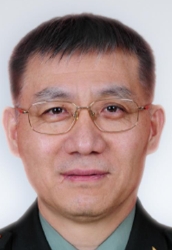 Jiying Chen (陈继营)合作论文数Chinese PLA General Hospital3Yujie Liu (刘玉杰)合作论文数Hainan Hospital of the General Hospital of the People's Liberation Army of China;School of Medicine, Nankai University3
 Guoqiang Zhang (张国强)合作论文数Chinese PLA General Hospital3
 Zhongli Li (李众利)合作论文数Chinese PLA General Hospital3ZHANG Baoxiang (张宝祥)合作论文数2Yan Shen (沈研)合作论文数2JingDong Li (李静东)合作论文数2YingChun Liang (梁迎春)合作论文数2LiYing Zhou (周丽英)合作论文数2Wei Wang (王蔚)合作论文数2Rong Huang (黄蓉)合作论文数2
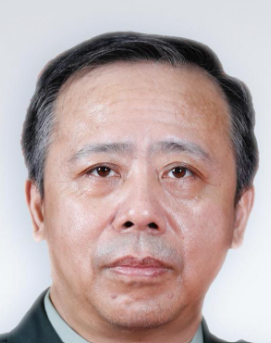 Libo Hao (郝立波)合作论文数Chinese PLA General Hospital2
 Jiyuan Dong (董纪元)合作论文数Chinese PLA General Hospital2
 Feng Lin (林峰)合作论文数Chinese People's Liberation Army General Hospital2
 Qinong Ye (叶棋浓)合作论文数Academy of Military Medical Sciences;Medical College, Guizhou University2
 Zhigang Wang (王志刚)合作论文数Chinese People's Liberation Army General Hospital2
 Chunbao Li (李春保)合作论文数College of Food Science and Technology, Nanjing Agricultural University2Zhigang Jin (金志刚)合作论文数2Zhiqiang Qi (祁志强)合作论文数1Yan, Wang合作论文数1Xing Yun合作论文数1Yameng Xu合作论文数Department of Traditional Chinese Medicine, Xinhua Hospital Affiliated to Shanghai Jiao Tong University School of Medicine, Shanghai, 200025, China.1Jianli Di (邸建丽)合作论文数1
 Yu Wei合作论文数1Ming Liu合作论文数1Yang LIU (刘洋)合作论文数1Yi Yin合作论文数1Yueqiang Wang (王跃强)合作论文数1Ping-an SHI (史平安)合作论文数1Xiaodong Li (李晓东)合作论文数1Li ZHANG (张立)合作论文数1Cui Ma (马毳)合作论文数1Hai Dai (戴海)合作论文数1ZongGui Huang (黄宗贵)合作论文数1




