In the field of digital signal processing (DSP), the execution of vector operations depends on the optimization of Single Instruction Multiple Data (SIMD) technology. However, manual SIMD vectorization method is complex to develop, poorly portable, and costly to maintain. Therefore, we propose a method of SIMD instruction sequence generation based on LLVM. This method builds a hierarchical instruction generation framework, combines the characteristics of the target architecture, and uses LLVM automatic vectorization tool to gradually convert the vectorized intermediate representation into the target architecture instruction sequence containing SIMD instructions. Experiments on FT-M7002 hardware platform show that, compared with the vectorization method of manually calling SIMD built-in functions, the average execution performance of the instruction sequence generated by this method can be improved by up to 70%.
Recurrence of adamantinomatous craniopharyngioma (ACP) is relatively common in clinical practice, yet the mechanisms underlying its recurrence remain poorly understood. Here, we performed spatial transcriptome sequencing analysis on paired formalin-fixed paraffin-embedded (FFPE) samples of primary and recurrent ACP from the same patient to construct spatial cellular maps of ACP in primary and recurrent states. Additionally, we utilized single-cell sequencing technology to conduct transcriptome analysis on fresh primary and recurrent samples to delineate their cellular and tumour microenvironment landscapes. Integrative analysis of these datasets enabled us to group common cell populations into clusters and perform Gene Ontology (GO) and Kyoto Encyclopedia of Genes and Genomes (KEGG) pathway analysis on differentially expressed genes in each cell population between primary and recurrent samples. We observed a significantly increased proportion of regulatory T (Treg) cells in recurrent samples. In macrophages, IL-4R expression was markedly elevated in recurrent samples, indicating enhanced M2 polarisation. Further analysis of immune cells revealed significantly elevated SRGN expression in recurrent samples and activation of the SPP1-CD44 cell communication pathway. These findings suggest that Treg cells, IL-4/IL-4R signalling, SRGN and SPP1-CD44 pathways are interconnected and collectively promote ACP recurrence. Our study delineates the spatial cellular maps of primary and recurrent ACP samples, uncovering potential mechanisms of recurrence. Moreover, we propose that SRGN may serve as a biomarker for predicting recurrence, and identify SPP1-CD44 as a candidate clinical therapeutic target for ACP.
Adamantinomatous craniopharyngioma (ACP), a benign yet highly recurrent and therapy-resistant intracranial tumor, remains a considerable clinical challenge because of its complex pathological structure, infiltrative growth, and limited treatment options. Here, integrated spatially resolved multiomics is employed-including single-cell spatial transcriptomics via CosMx SMI and spatially resolved metabolomics via AFADESI-MSI, accompanied by bulk metabolomics and functional validation-to unravel the driving factors of ACP progression and recurrence. Analysis results reveal three interdependent biological hallmarks: first, the spatial segregation and molecular heterogeneity of 10 distinct tumor epithelial cell subpopulations within the ACP, each of which presents unique transcriptional signatures; second, in tumor regions and recurrent tumor epithelium tissues, stronger transporter-mediated choline/ethanolamine uptake from cystic fluid and significant upregulation of phosphatidylcholine (PC) and phosphatidylethanolamine (PE) synthesis is observed, creating the enhanced "cystic fluid-tumor cell" and "choline/ethanolamine-PC/PE" metabolic axis, and demonstrating the spatial metabolic remodeling of ACP; and third, this metabolic axis directly couples to autophagy activation of corresponding regions in ACP tissue, which is validated by multi-immunohistochemistry for Beclin1 and GABARAP. Together, these findings reveal metabolic remodeling and autophagic activation as critical drivers of ACP progression and recurrence and provide an opportunity for precise biomarker-driven treatment of this intractable tumor.
INTRODUCTION AND IMPORTANCE:This study aims to evaluate the microsuture technique in treating giant dissecting aneurysms of the middle cerebral artery (MCA) trunk, particularly in cases involving perforators-a key surgical challenge. METHODS:This reconstruction of the MCA trunk was performed in four patients with giant dissecting aneurysms using a microsuture technique. The fragile wall of the dissecting aneurysm was meticulously excised while preserving the perforating arteries originating from the aneurysm body. Subsequently, a bypass was established from the internal maxillary artery (IMA) to the distal MCA (or specify the exact branch if applicable). This surgical approach achieved temporary vascular occlusion while maintaining distal perfusion, thereby preventing ischemia in the territory distal to the aneurysm during its definitive management. OUTCOMES:Among the four patients, three had surgically clipped aneurysms of the MCA trunk, while the remaining patient presented with severe headaches and left lower limb paralysis. All patients underwent microsuture of the MCA trunk and subsequently experienced uneventful postoperative recoveries without neurological deficits. Postoperative angiography demonstrated successful MCA trunk reconstruction with complete dissecting aneurysm resolution in three cases. In the remaining patient, although MCA reconstruction was not radiographically visible, patency of the IMA-to-M2 segment bypass was confirmed. Follow-up imaging revealed: At six-year follow-up, one patient exhibited both a patent IMA-to-M2 bypass and reconstructed M1 trunk on CT angiography; Two patients showed M1 trunk restoration with aneurysm disappearance at three-month follow-up; One patient maintained a patent IMA-to-M2 bypass without visualized MCA trunk. No ischemic events occurred in any patient, presumably due to preserved perforating arteries originating from the reconstructed MCA trunks. CONCLUSION:Some large dissecting aneurysms of the MCA trunk can be effectively treated with the microsuture technique by reconstructing the trunk and preserving its perforators.
Adamantinomatous craniopharyngioma (ACP) is a benign but surgically challenging intracranial tumor, and its high postoperative recurrence rate poses a threat to patient prognosis. In this study, whole exome sequencing (WES) was performed to comprehensively understand the genomic alterations in ACP. First, the gene mutation profile of ACP was acquired through an overview of the WES data. The somatic single nucleotide variants (SNV) mutational spectrum and signature of ACP were analyzed. Subsequently, somatic mutation data were used to identify the characteristic SNV of recurrent ACP to further explore the mechanism underlying recurrence, and the effect of these mutations on protein expression was verified through immunohistochemical and immunofluorescence analyses. We obtained the overall mutational landscape of ACP and identified four key mutated genes in recurrent samples: Procollagen-Lysine,2-Oxoglutarate 5-Dioxygenase 3 (PLOD3), Collagen Type XVII Alpha 1 chain (COL17A1), Fibronectin 1 (FN1), and Laminin Subunit Beta 3 (LAMB3). Among these, we verified that COL17A1, FN1, and LAMB3 expression was mainly located in the keratin nodule structures in tumors and was elevated in recurrent cases. PLOD3, COL17A1, FN1, and LAMB3 were identified as the key mutated genes in recurrent samples. These findings could provide a new perspective for understanding the tumorigenesis and recurrence mechanisms of ACP. A deeper exploration is needed to determine whether these key mutated genes promote ACP recurrence and drive the overproduction of keratin nodules in recurrent cases.
The frontal basal interhemispheric approach (FBIA) provides wide access to the chiasmatic cistern, retrochiasmatic space, and lamina terminalis for craniopharyngioma removal. We introduce in detail the anatomical structure related to the FBIA, the surgical techniques of craniotomy, and how to prevent the surgical complications. The FBIA provides a satisfactory surgical corridor for craniopharyngioma resection.
Traditional instruction selection methods fail to fully exploit the very long instruction word (VLIW) architecture’s efficient scalar instructions. We propose an optimized instruction selection method based on classification node merging. This method abstracts scalar intermediate instructions into nodes and divides them into associated and similar ones according to operational semantics. Combined with the VLIW architecture’s instruction set features and guided by the instruction cost model, the nodes are merged and optimized to select efficient instructions. We design an instruction generation framework that swiftly converts the low level virtual machine intermediate representation into the instruction sequence of the target architecture. The experimental results show that, compared with the instruction sequence generated by M7002-cc, the instruction sequence generated with the solution proposed in this paper exhibits an average execution performance that is up to 54
Backgrounds: Adamantinomatous craniopharyngiomas (ACPs) are benign intracranial tumors that behave aggressively due to their location, infiltration of the surrounding nervous tissue and high capacity for recurrence. In this study, we aimed to construct ACP primary cell models for further investigation of tumorigenic and recurrent mechanisms. Methods: Primary cells were isolated from primary (one case) and recurrent (one case) ACP. Short tandem repeat (STR) analysis was used to clarify the identity of the ACP primary cells we isolated. Whole exome sequencing (WES), immunofluorescence (IF) and immunohistochemistry (IHC) were performed on primary cells and corresponding ACP tissues, to determine the mutational profile and to clarify the tissue origin and phenotypic of primary cells. Transcriptome RNA-seq was performed to obtain the gene expression characteristics of ACP primary cells. Subsequently, a heterotopic ACP xenograft mouse model was established to confirm the tumorigenesis capacity of ACP primary cells. Results: ACP primary cells were successfully cultured. The genetic variants were similar to the original tumor tissue, and they owned expression of cancer-associated fibroblast (CAF) markers (FSP1/S100A4, Vimentin) and nuclear translocation β-catenin. Meanwhile, they had an high level expression of extracellular matrix components (Fibronectin). The tumor formation ability of ACP primary cells was verified. The transcriptional signatures of ACP primary cells were also explored. Conclusions: We successfully isolated and characterized ACP primary cells that acquired multiple CAF features and demonstrated stable propagation through dozens of passages. These PDC models laid the foundation for further research on ACP.
HomeStrokeAhead of PrintInsights on Moyamoya Disease: Vascular Inflammation and Nuclear Autoschizis in Cranial Arteries No AccessCase ReportRequest AccessAboutView PDFSections ToolsAdd to favoritesDownload citationsTrack citationsPermissions ShareShare onFacebookTwitterLinked InMendeleyReddit Jump toNo AccessCase ReportRequest AccessInsights on Moyamoya Disease: Vascular Inflammation and Nuclear Autoschizis in Cranial Arteries Xiaoling Ruan, Ting Lei, Xin Xiang, Fangjun Liu and Xiang’en Shi Xiaoling RuanXiaoling Ruan https://orcid.org/0009-0000-7219-384X Department of Neurosurgery, University of Pavia, Italy (X.R.). Search for more papers by this author , Ting LeiTing Lei https://orcid.org/0000-0001-8970-1986 Department of Neurosurgery, Sanbo Brain Hospital (T.L., X.X., F.L.) Department of Neurosurgery, Fuxing Hospital, Capital Medical University, Beijing, China (T.L., X.S.). Search for more papers by this author , Xin XiangXin Xiang https://orcid.org/0009-0000-1897-7539 Department of Neurosurgery, Sanbo Brain Hospital (T.L., X.X., F.L.) Search for more papers by this author , Fangjun LiuFangjun Liu https://orcid.org/0000-0003-2428-6456 Department of Neurosurgery, Sanbo Brain Hospital (T.L., X.X., F.L.) Search for more papers by this author and Xiang’en ShiXiang’en Shi Correspondence to: Xiang’en Shi, MD, PhD, Department of Neurosurgery, Fuxing Hospital, Capital Medical University, No. 20, Fuxingmen Wai St, Xicheng, Beijing 100045, China. Email E-mail Address: [email protected] https://orcid.org/0000-0002-9491-0499 Department of Neurosurgery, Fuxing Hospital, Capital Medical University, Beijing, China (T.L., X.S.). Search for more papers by this author Originally published29 Nov 2023https://doi.org/10.1161/STROKEAHA.123.045064Stroke. 2023;0FootnotesFor Sources of Funding and Disclosures, see page xxx.Correspondence to: Xiang’en Shi, MD, PhD, Department of Neurosurgery, Fuxing Hospital, Capital Medical University, No. 20, Fuxingmen Wai St, Xicheng, Beijing 100045, China. Email shixen@ccmu.edu.cn eLetters(0)eLetters should relate to an article recently published in the journal and are not a forum for providing unpublished data. Comments are reviewed for appropriate use of tone and language. Comments are not peer-reviewed. Acceptable comments are posted to the journal website only. Comments are not published in an issue and are not indexed in PubMed. Comments should be no longer than 500 words and will only be posted online. References are limited to 10. Authors of the article cited in the comment will be invited to reply, as appropriate.Comments and feedback on AHA/ASA Scientific Statements and Guidelines should be directed to the AHA/ASA Manuscript Oversight Committee via its Correspondence page.Sign In to Submit a Response to This Article Previous Back to top Next FiguresReferencesRelatedDetails Advertisement Article InformationMetrics © 2023 American Heart Association, Inc.https://doi.org/10.1161/STROKEAHA.123.045064PMID: 38018829 Originally publishedNovember 29, 2023 KeywordsCTLA4 proteinelectron microscopyhumanimmunohistochemistryinflammationmoyamoya diseasetumor necrosis factor-alphaPDF download Advertisement SubjectsGrowth Factors/CytokinesSmooth Muscle Proliferation and DifferentiationStenosisVascular Disease
Background Adamantinomatous craniopharyngiomas (ACPs) are rare benign epithelial tumours with high recurrence and poor prognosis. Biological differences between recurrent and primary ACPs that may be associated with disease recurrence and treatment have yet to be evaluated at the proteomic level. In this study, we aimed to determine the proteomic profiles of paired recurrent and primary ACP, gain biological insight into ACP recurrence, and identify potential targets for ACP treatment.Method Patients with ACP (n = 15) or Rathke's cleft cyst (RCC; n = 7) who underwent surgery at Sanbo Brain Hospital, Capital Medical University, Beijing, China and received pathological confirmation of ACP or RCC were enrolled in this study. We conducted a proteomic analysis to investigate the characteristics of primary ACP, paired recurrent ACP, and RCC. Western blotting was used to validate our proteomic results and assess the expression of key tumour-associated proteins in recurrent and primary ACPs. Flow cytometry was performed to evaluate the exhaustion of tumour-infiltrating lymphocytes (TILs) in primary and recurrent ACP tissue samples. Immunohistochemical staining for CD3 and PD-L1 was conducted to determine differences in T-cell infiltration and the expression of immunosuppressive molecules between paired primary and recurrent ACP samples.Results The bioinformatics analysis showed that proteins differentially expressed between recurrent and primary ACPs were significantly associated with extracellular matrix organisation and interleukin signalling. Cathepsin K, which was upregulated in recurrent ACP compared with that in primary ACP, may play a role in ACP recurrence. High infiltration of T cells and exhaustion of TILs were revealed by the flow cytometry analysis of ACP.Conclusions This study provides a preliminary description of the proteomic differences between primary ACP, recurrent ACP, and RCC. Our findings serve as a resource for craniopharyngioma researchers and may ultimately expand existing knowledge of recurrent ACP and benefit clinical practice.
The application of portable devices based on deep learning has become increasingly widespread, which has made the deployment of complex neural networks on embedded devices a hot research topic. Neural network lightweighting is one of the key technologies for applying neural networks to embedded devices. This paper elaborates and analyzes neural network lightweighting techniques from two aspects: model pruning and network structure design. For model pruning, a comparison of methods from different periods is conducted, highlighting their advantages and limitations. Regarding network structure design, the principles of four classical lightweight network designs are described from a mathematical perspective, and the latest optimization methods for these networks are reviewed. Finally, potential research directions for lightweight neural network pruning and structure design optimization are discussed.
BACKGROUND:Marfan syndrome, a connective tissue disorder, poses unique challenges in neurosurgery, given the fragility of vascular structures. Superior cerebellar artery (SCA) aneurysms in patients with the syndrome are rare and present distinct surgical difficulties, necessitating innovative approaches.OBSERVATIONS:A 29-year-old male with Marfan syndrome presented with a subarachnoid hemorrhage from a ruptured SCA aneurysm. Given the lack of a defined aneurysm neck and the small diameter of the SCA, standard clipping and endovascular therapies were unsuitable. A microsurgical approach using microsutures was successfully employed, effectively managing the aneurysm while preserving the parent artery.LESSONS:This case underscores the efficacy of the microsuture technique in complex neurosurgical scenarios, particularly in patients with connective tissue disorders such as Marfan syndrome. The adaptability of surgical strategies, as demonstrated in this case, is crucial for achieving successful outcomes in patients with unique anatomical challenges.
With the widespread development of embedded device applications based on deep learning, pedestrian detection using deep learning often requires the use of complex and large neural networks.However, these networks have a large memory footprint, which makes them difficult to deploy on embedded devices, and are computationally intensive, which makes it difficult to guarantee the speed of operation on embedded devices.To address these issues, a lightweight network, YOLO-Micro, is proposed in this paper.The model reduces the complexity of the model by replacing the original module in YOLOv7-tiny with the G-ELAN module proposed in this paper.Meanwhile, the attention mechanism and the activation function were introduced and replaced to enhance the feature extraction capability of the model.Finally, the model was successfully deployed to Android mobile.Experimental results on the PASCAL VOC Pedestrian dataset show that the inference speed of YOLO-Micro is 20.8% faster than the original model, and the mAP is improved from 77.95% to 79.42% of the original model.
Objective:To analyze the incidence and prognosis of epilepsy in frontotemporal lobe glioma.Methods:The clinical data of 208 patients with frontotemporal lobe gliomas in Sanbo Brain Hospital Capital Medical University from 2019 to 2021 were analyzed retrospectively. According to the 2016 World Health Organization (WHO) classification of tumors of the central nervous system, the incidence of epilepsy, Modified Rankin Scale (MRS) score, and Engel Outcome Scale of patients with different grades of tumors were calculated.Results:Among all the patients with frontotemporal lobe gliomas, there was more males than females, and it was more common in the 40 -59 age group. The incidence of epilepsy associated with WHO grade Ⅰand Ⅱ glioma was 100.0% (33/33) and 60.9% (14/23), respectively, while that of WHO grade Ⅳ glioma was 19.0%(19/100). The average follow-up time was (22 ± 9) months. During the follow-up period, the incidence of WHO grade Ⅰ, Ⅱ and Ⅲ glioma-related epilepsy decreased significantly. There was no significant difference in the incidence of glioma-related epilepsy between the total and subtotal resection groups ( P>0.05). There was no statistical correlation between the side of tumor occurrence and the occurrence of epilepsy ( P>0.05), also between the gene phenotype and the occurrence of epilepsy ( P>0.05). There was no significant difference in the Engel Outcome Scale among different grades of gliomas ( P>0.05). The prognosis of patients with Engel Outcome Scale Class 1 was significantly better than that of other grades. Conclusions:The incidence of glioma-related epilepsy is negatively correlated with tumor grade. Age and sex are risk factors for glioma-related epilepsy. The incidence of postoperative epilepsy in patients with low grade glioma is significantly lower than that in patients with high grade glioma, and the prognosis is better. However, there is no significant difference in the Engel Outcome Scale among different grades of gliomas.
HomeStrokeVol. 54, No. 12Unveiling Bacterial Autophagosomes in Human Intracranial Aneurysm Wall Tissue: A Rare Insight No AccessCase ReportRequest AccessFull TextAboutView Full TextView PDFView EPUBSections ToolsAdd to favoritesDownload citationsTrack citationsPermissions ShareShare onFacebookTwitterLinked InMendeleyReddit Jump toNo AccessCase ReportRequest AccessFull TextUnveiling Bacterial Autophagosomes in Human Intracranial Aneurysm Wall Tissue: A Rare Insight Ting Lei, Xiang’en Shi, Xiaoling Ruan, Fangjun Liu and Xin Xiang Ting LeiTing Lei https://orcid.org/0000-0001-8970-1986 Department of Neurosurgery, Fuxing Hospital (T.L., X.S., X.X.), Capital Medical University, Beijing, People’s Republic of China. Department of Neurosurgery, Sanbo Brain Hospital (T.L., X.S., F.L., X.X.), Capital Medical University, Beijing, People’s Republic of China. Search for more papers by this author , Xiang’en ShiXiang’en Shi Correspondence to: Xiang’en Shi, MD, PhD, Department of Neurosurgery, Fuxing Hospital, Capital Medical University, No. 20, Fuxingmen Wai St, Xicheng District, Beijing 100045, People’s Republic of China. Email E-mail Address: [email protected] https://orcid.org/0000-0002-9491-0499 Department of Neurosurgery, Fuxing Hospital (T.L., X.S., X.X.), Capital Medical University, Beijing, People’s Republic of China. Department of Neurosurgery, Sanbo Brain Hospital (T.L., X.S., F.L., X.X.), Capital Medical University, Beijing, People’s Republic of China. Search for more papers by this author , Xiaoling RuanXiaoling Ruan University of Pavia, Milan, Italy (X.R.). Search for more papers by this author , Fangjun LiuFangjun Liu https://orcid.org/0000-0003-2428-6456 Department of Neurosurgery, Sanbo Brain Hospital (T.L., X.S., F.L., X.X.), Capital Medical University, Beijing, People’s Republic of China. Search for more papers by this author and Xin XiangXin Xiang https://orcid.org/0009-0000-1897-7539 Department of Neurosurgery, Fuxing Hospital (T.L., X.S., X.X.), Capital Medical University, Beijing, People’s Republic of China. Department of Neurosurgery, Sanbo Brain Hospital (T.L., X.S., F.L., X.X.), Capital Medical University, Beijing, People’s Republic of China. Search for more papers by this author Originally published12 Oct 2023https://doi.org/10.1161/STROKEAHA.123.044829Stroke. 2023;54:e498–e499"Unveiling Bacterial Autophagosomes in Human Intracranial Aneurysm Wall Tissue: A Rare Insight." Stroke, 54(12), pp. e498–e499FootnotesFor Sources of Funding and Disclosures, see page e499.Correspondence to: Xiang’en Shi, MD, PhD, Department of Neurosurgery, Fuxing Hospital, Capital Medical University, No. 20, Fuxingmen Wai St, Xicheng District, Beijing 100045, People’s Republic of China. Email shixen@ccmu.edu.cnREFERENCES1. Hallikainen J, Pyysalo M, Keränen S, Kellokoski J, Koivisto T, Suominen AL, Pussinen P, Pessi T, Frösen J. Systemic immune response against the oral pathogens Porphyromonas gingivalis and Aggregatibacter actinomycetemcomitans is associated with the formation and rupture of intracranial aneurysms.Eur J Neurol. 2021; 28:3089–3099. doi: 10.1111/ene.14986CrossrefGoogle Scholar eLetters(0)eLetters should relate to an article recently published in the journal and are not a forum for providing unpublished data. Comments are reviewed for appropriate use of tone and language. Comments are not peer-reviewed. Acceptable comments are posted to the journal website only. Comments are not published in an issue and are not indexed in PubMed. Comments should be no longer than 500 words and will only be posted online. References are limited to 10. Authors of the article cited in the comment will be invited to reply, as appropriate.Comments and feedback on AHA/ASA Scientific Statements and Guidelines should be directed to the AHA/ASA Manuscript Oversight Committee via its Correspondence page.Sign In to Submit a Response to This Article Previous Back to top Next FiguresReferencesRelatedDetails December 2023Vol 54, Issue 12 Advertisement Article InformationMetrics © 2023 American Heart Association, Inc.https://doi.org/10.1161/STROKEAHA.123.044829PMID: 37823306 Originally publishedOctober 12, 2023 KeywordsDNADNA probeselectronshumanspolymerase chain reactionPDF download Advertisement SubjectsAneurysm
目的 评估症状性颈动脉狭窄(SCS)患者颈动脉内膜剥脱术(CEA)后长、短期预后情况及影响因素.方法 分析首都医科大学三博脑科医院2014年1月—2019年12月经影像学检查确诊的79例SCS患者的临床资料,包括患者的既往病史、临床表现、实验室指标、影像学表现、治疗,以及术前、术后的改良Rankin量表(mRS)分级.分析术后3和24个月的预后改善情况.多因素Logistic回归分析影响因素,建立临床预测模型并对其进行准确性及预测效能评价.结果SCS患者CEA术前、术后3个月、术后24个月的mRS分级分别为(1.78±1.10)、(1.32±1.37)和(0.89±1.25).与术前相比,术后3和24个月均明显改善(P<0.01).多因素Logistic回归分析显示,CEA术后3个月,心肌梗死(OR=0.06,95%CI=0.01-0.32)和高脂血症(OR=0.13,95%CI=0.03-0.47)是影响因素.CEA术后24个月,心肌梗死(OR=0.09,95%CI=0.00-0.66)、高脂血症(0R=0.05,95%CI=0.00-0.29)和中性粒细胞淋巴细胞比值(NLR)(OR=0.11,95%CI=0.02-0.50)是影响因素.通过构建的列线图来进行预测有较高的准确性及预测效能.结论SCS患者CEA术后长、短期神经功能均较术前均有显著改善.心肌梗死、高脂血症是影响CEA术后短期预后的危险因素,心肌梗死、高脂血症、NLR是影响CEA术后长期预后的危险因素.NLR可预测SCS患者CEA术后的长期预后,术前低NLR(<2.62)的患者预后更好.
The treatment of wide-necked complex intracranial aneurysms is full of challenges. We report a 72-year-old woman with subarachnoid hemorrhage and three intracranial aneurysms. According to the location of the hemorrhage, we chose the left side Pterional approach to clip the left side aneurysms and remove the hematoma. After clipping the left posterior communicating artery aneurysm, we found a small bifurcation aneurysm associated with dysplastic bulging of the nearby wall which together formed a wide-necked complex aneurysm. We used a novel suture combined with clipping technique to treat this complex aneurysm. Indocyanine green arteriography showed intact vascular patency in distal vessels, and the patient has achieved a good prognosis. It may provide a new idea for treating wide-necked complex intracranial aneurysms.
Objective:To retrospectively summarize the imaging and clinical features of craniopharyngioma in order to improve the preoperative diagnosis level.Methods:One hundred and twenty-seven patients with craniopharyngioma diagnosed by pathology in Sanbo Brain Hospital, Capital Medical University from March 2019 to June 2021 were selected and the pathological coincidence rate of imaging diagnosis were analyzed.Results:The coincidence rate of MRI diagnosis was 89.3%. The coincidence rate of CT diagnosis was 71.5%. On T 2WI and T 1WI enhanced sequences, the solid portion of the tumor may showed uneven hyperintensity, diffuse striation and spotty hyperintensity. MRI sagittal view was helpful in showing small tumors, but less sensitive to calcification than CT. MRI enhancement was very important, especially for patients with solid lesions. Conclusions:The imaging findings of craniopharyngioma are diverse. Some characteristic manifestations provide important information for the diagnosis and differential diagnosis of craniopharyngioma, which can improve the diagnostic accuracy combined with clinical data.
朗格汉斯细胞组织细胞增生症是一种罕见的全身性疾病,约25%的患者累及鞍区,以多饮多尿起病常见。报道1例罕见的以闭经泌乳为主要临床表现的鞍区朗格汉斯细胞组织细胞增生症,详细阐述其诊疗流程,并讨论导致患者出现泌乳素增高的可能原因。
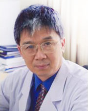 Xiangen Shi (石祥恩)合作论文数首都医科大学三博脑科医院69
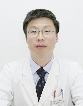 Yuming Sun (孙玉明)合作论文数首医大三博脑科医院(北京)有限公司65
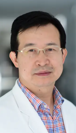 Zhongqing Zhou (周忠清)合作论文数Sanbo Brain Hospital, Capital Medical University30
 Yongli Zhang (张永力)合作论文数Sanbo Brain Hospital,Capital Medical University29
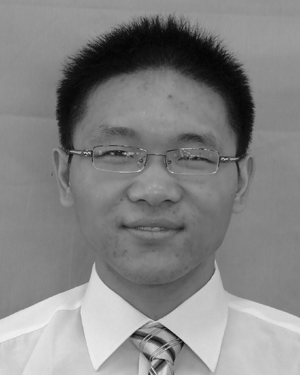 Hai Qian合作论文数Google Inc.20
 Lei Ting合作论文数19MengQing Hu (胡孟庆)合作论文数16Xin Xiang合作论文数11
 Long Wang (王龙)合作论文数SanBo Brain Hospital, Capital Medical University8Zaitao Yu合作论文数7JiQiang Wang (王计强)合作论文数5
 Chuzhong Li (李储忠)合作论文数Capital Medical University5
 Chengyin Liu (柳成荫)合作论文数Harbin Institute of Technology, Shenzhen4
 Songbai Gui (桂松柏)合作论文数Beijing Tiantan Hospital, Capital Medical University4
 Xingang Li (李新钢)合作论文数Qilu Hospital, Shandong University;Institute of Brain and Brain-Like Science, Shandong University4
 Yazhuo Zhang (张亚卓)合作论文数Beijing Institute of Neurosurgery;Beijing Tiantan Hospital, Capital Medical University4Huifu Zhang合作论文数4Xiaoling Ruan合作论文数3Zelin Sun合作论文数3YeShuai Hu (胡业帅)合作论文数3
 Yan Li (李燕)合作论文数Institute of Materia Medica, Chinese Academy of Medical Sciences3Yang Yang合作论文数3Heju Li合作论文数2
 Yaoling Liu合作论文数2Jin WANG (王瑾)合作论文数2xiaochun zhao (赵晓春)合作论文数2Pengbo Li (李鹏波)合作论文数2YuanPeng Zhang (张元鹏)合作论文数2DongHai Wang (王东海)合作论文数2
 Jie Zhang (张杰)合作论文数Beijing Tiantan Hospital, Capital Medical University2
 Peng Zhao (赵鹏)合作论文数山东省立医院2Qian Jia合作论文数2
 Wei Zhang (张伟)合作论文数Department of Neurosurgery, Beijing Tiantan Hospital, Capital Medical University;Graduate School of Capital Medical University2
 Fuzheng Zhang (张福征)合作论文数Beijing Pinggu Hospital2
 Bin Wu (吴斌)合作论文数Sanbo Brain Hospital Capital Medical University2
 Siqi Liu (刘斯奇)合作论文数BGI2Lujun Jing合作论文数1
 Hao Yang (杨浩)合作论文数Research Center for Social Computing and Information Retrieval, Harbin Institute of Technology1Gang Hu合作论文数1Jun Yang合作论文数1tao wenyan合作论文数1Haidong Deng合作论文数1Xiaocui Shi合作论文数1Yong-li ZHANG (张永力)合作论文数1Xin Li合作论文数1




