Chemoresistance driven by pro-survival autophagy remains a major obstacle in hepatocellular carcinoma (HCC) treatment. Berbamine (BBM), a natural alkaloid with a favorable clinical safety profile, shows potential as an autophagy inhibitor, yet its precise mechanism in HCC remains unclear. Using CCK-8, colony formation, and apoptosis assays, we first demonstrated that BBM synergistically enhanced the efficacy of multiple chemotherapeutic agents (5-FU, Sorafenib, Paclitaxel) against HCC cells in vitro. This synergistic effect was confirmed in an H22 xenograft mouse model in vivo. To investigate the mechanism, we monitored autophagic flux and lysosomal function. Western blot and immunofluorescence analyses revealed that BBM treatment led to the concurrent accumulation of LC3-II and p62, indicating a blockade of late-stage autophagic flux. Further experiments, including LysoTracker staining and assessment of lysosomal protease levels, showed that BBM impaired both autophagosome-lysosome fusion and lysosomal acidification. Mechanistically, we found that BBM downregulated SIRT1 protein expression and reduced the intracellular NAD+/NADH ratio, thereby inhibiting SIRT1 deacetylase activity. This suppression impaired the nuclear translocation and function of the key autophagy transcription factor TFEB, leading to decreased levels of its downstream targets RAB7, CTSB, and CTSD. Crucially, rescue experiments using specific agonists revealed that SIRT1 activation completely reversed all BBM-induced effects, including autophagic flux blockade and downstream protein suppression, whereas TFEB activation only partially rescued the expression of RAB7, CTSB, and CTSD without restoring autophagic flux. This establishes SIRT1 as the primary upstream regulator in this pathway. Our study identifies BBM as a novel autophagy inhibitor that targets the SIRT1-TFEB axis to disrupt autolysosomal fusion and degradation, and nominates it as a promising combinational agent to overcome chemoresistance in HCC.
Major histocompatibility complex class I (MHC-I)-mediated antigen presentation plays a pivotal role in anti-tumor immunity by enabling CD8+ T cells to recognize and eliminate malignant cells. In melanoma, modulation of this pathway is critical for improving the efficacy of immunotherapies. Our study demonstrates that the natural compound Cepharanthine (CEP) exhibits notable antitumor activity by enhancing MHC-I-mediated antigen presentation. CEP treatment upregulated MHC-I expression (both membrane-bound and total levels) in melanoma cells in a concentration-dependent manner, thereby improving antigen-presenting capacity. Interestingly, when autophagy was pharmacologically blocked using Bafilomycin A1, co-treatment with CEP did not lead to further elevation of MHC-I expression, suggesting that CEP’s effect is mediated through disruption of the autophagic pathway. Mechanistically, CEP induced autophagosome accumulation, as evidenced by an increase in GFP-LC3 puncta. Fluorescence imaging further confirmed that CEP selectively impaired lysosomal acidification without affecting autophagosome–lysosome fusion, thereby inhibiting late-stage autophagic flux. Furthermore, CEP treatment promoted CD8+ T cell infiltration into tumor tissues and enhanced the antitumor efficacy of anti-PD-1 therapy, resulting in greater tumor suppression compared to either treatment alone. The study elucidates how CEP’s selective lysosomal inhibition creates a tumor microenvironment more susceptible to immune surveillance, primarily through preserved MHC-I surface expression and subsequent T cell recognition. This work highlights CEP as a promising immunomodulatory agent and provides a potential strategy for improving the outcomes of immune checkpoint blockade therapy.
BackgroundClinically, although chemotherapy is one of the most commonly used methods of treating tumors, chemotherapeutic drugs can induce autophagic flux and increase tumor cell resistance, leading to drug tolerance. Therefore, theoretically, inhibiting autophagy may improve the efficacy of chemotherapy. The discovery of autophagy regulators and their potential application as adjuvant anti-cancer drugs is of substantial importance. In this study, we clarified that Fangjihuangqi Decoction (FJHQ, traditional Chinese medicine) is an autophagy inhibitor, which can synergistically enhance the effect of cisplatin and paclitaxel on non-small cell lung cancer (NSCLC) cells.MethodsWe observed the changes of autophagy level in NSCLC cells under the effect of FJHQ, and verified the level of the autophagy marker protein and cathepsin. Apoptosis was detected after the combination of FJHQ with cisplatin or paclitaxel, and NAC (ROS scavenger) was further used to verify the activation of ROS-MAPK pathway by FJHQ.ResultsWe observed that FJHQ induced autophagosomes in NSCLC cells and increased the levels of P62 and LC3-II protein expression in a concentration- and time-gradient-dependent manner, indicating that autophagic flux was inhibited. Co-localization experiments further showed that while FJHQ did not inhibit autophagosome and lysosome fusion, it affected the maturation of cathepsin and thus inhibited the autophagic pathway. Finally, we found that the combination of FJHQ with cisplatin or paclitaxel increased the apoptosis rate of NSCLC cells, due to increased ROS accumulation and further activation of the ROS-MAPK pathway. This synergistic effect could be reversed by NAC.ConclusionCollectively, these results demonstrate that FJHQ is a novel late-stage autophagy inhibitor that can amplify the anti-tumor effect of cisplatin and paclitaxel against NSCLC cells.
Autophagy is typically activated in cancer cells as a rescue strategy in response to cellular stress (e.g., chemotherapy). Herein, we found that Berbamine Hydrochloride (Ber) can act as an effective inhibitor of the late stage of autophagic flux, thereby potentiating the killing effect of chemotherapy agents. Lung carcinoma cells exposed to Ber exhibited increased autophagosomes, marked by LC3-II upregulation. The increased level of p62 after Ber treatment indicated that the autophagic flux was blocked at the late stage. The lysosome staining assay and cathepsin maturation detection indicated impaired lysosomal acidification. We found that Nox2 exhibited intensified co-localization with lysosomes in Ber-treated cells. Nox2 is a key enzyme for superoxide anion production capable of transferring electrons into the lysosomal lumen, thereby neutralizing the inner protons; this might explain the aberrant acidification. This hypothesis is further supported by the observed reversal of lysosomal cathepsin maturation by Nox2 inhibitors. Finally, Ber combined with cisplatin exhibited a synergistic killing effect on lung carcinoma cells. Further data suggested that lung carcinoma cells co-treated with Ber and cisplatin accumulated excessive reactive oxygen species (ROS), which typically activated MAPK-mediated mitochondria-dependent apoptosis. The enhanced anti-cancer effect of Ber combined with cisplatin was also confirmed in an in vivo xenograft mouse model. These findings indicate that Ber might be a promising adjuvant for enhancing the cancer cell killing effect of chemotherapy via the inhibition of autophagy. In this process, Nox2 might be a significant mediator of Ber-induced aberrant lysosomal acidification.
Purpose: To explore whether EVO affects metabolism of liver tumor, and how EVO exert its anti-tumor effect by inhibiting cancer metabolism.Method: CCK8 assay, plate colony forming experiment, transcriptomics and metabolomics assays, flow cytometry, Western Blot analysis, q-PCR technique, lentiviral overexpression gain-of-function experiments were used in vitro experiment, and tumor cells transplantation mice model was used in vivo experiments.Results: EVO markedly inhibited tumor cell proliferation and promoted apoptosis. In the transcriptomic gene chip analysis, EVO suppressed cancer metabolism by down-regulating phosphofructokinase (PFK), PRPP synthase (PRS) and NDP reducase (RR),showed it could restrained glycolysis, PPP and nucleotides synthesis, regulated cell cycle genes transcription and promoted the expression of apoptosis-related genes, and inhibited Met/EGFR, p53-GADD45and HIF pathway, etc. Those results of gene chip were verified by q-PCR and Western Blot. In the result of metabonomics, EVO inhibited pyrimidine nucleotide synthesis, fructose metabolism, active hyaluronic acid, phospholipids, sphingolipid, purine nucleotides synthesis, aspartate, arginine and some other amino acids metabolism. The result of metabonomics assay was consist with Genechip technology. We also used recombinated lentivirus transfection (over-express Met gene) to verify that EVO inhibited the cancer cells growth, RAS-MAPK, PI3K-AKT pathways and cancer metabolism above through inhibiting Met expression, but Met overexpression could not reverse the inhibition of EVO on HIF pathway, indicated that EVO inhibited HIF pathway directly, not through Met. In the experiment of molecular docking, the results showed that EVO had a certain binding energy and a high affinity with Met and EGFR, the intervention of EVO may be through its binding with Met and EGFR. NTP/dNTP could reverse the growth inhibition of EVO, indicated that EVO inhibited the cancer cell growth via inhibiting NTP/dNTP synthesis. But NTP/dNTP could not reverse the growth inhibition of 0.4μM EVO, probably because Evo induced irreversible apoptosis. In vivo experiments, EVO (intragastric administration 50mg/ Kg.d) could significantly reduce the volume and weight of transplanted liver tumor in mice after intragastric administration two weeks, prolonged the survival significantly compared with the control group.Conclusion: EVO may be a specific inhibitor of Met and EGFR. EVO inhibited liver cancer cell proliferation, induced apoptosis and prolonging the survival time of transplanted liver tumor model mice through binding with Met and EGFR so as to inhibit their downstream RAS-MAPK, PI3K-AKT pathways, meanwhile repressing HIF pathways, and then restraining cancer metabolism.Funding Information: This work was financially supported by the National Nature Science Foundation of China (Nos.81774028).Declaration of Interests: The authors declare no conflict of interest.Ethics Approval Statement: Animal experiments were approved by the Animal Ethics Committee of Guangzhou University of Chinese Medicine.
Berberine (BBR), a natural alkaloid derived from Coptis, has anticancer activity. Some researchers have found that it could restrain epithelial-mesenchymal transition (EMT) of melanoma, neuroblastoma, and other tumor cells. However, it is unclear whether BBR can reverse EMT in hepatocellular carcinoma (HCC) and gastric carcinoma (GC). In our study, BBR inhibited the migration and invasion of HepG2, MGC803, and SGC7901 cells in a dose-dependent manner. Transcription sequencing assays showed that Vimentin, MMP, and Smad3 were downregulated, but Smad2, Smad6, TAB2, ZO-1, and claudin 7 were upregulated when treated with BBR. GO Enrichment analysis of KEGG pathway showed that BBR significantly inhibited TGF-β/Smad at 12 h, then, PI3K/Akt and Wnt/β-catenin signaling pathways at 24 h, which were closely related to the proliferation, migration, and EMT. The results of the transcriptome sequencing analysis were verified by Western Blot. It showed that the expression of epithelial marker E-cadherin and ZO-1 remarkably augmented with BBR treatment, as well as declined mesenchymal markers, including N-cadherin and Vimentin, decreased transcription factor Snail and Slug. The effects of BBR were similar to those of the PI3K inhibitor LY294002 and TGF-β receptor inhibitor SB431542. Furthermore, β-catenin and phosphorylation of AKT, Smad2, and Smad3 were changed dose-dependently by BBR treatment, which upregulated p-Smad2 and downregulated the others. Combined with LY or SB, respectively, BBR could enhance the effects of the two inhibitors. Simultaneously, IGF-1 and TGF-β, which is the activator of PI3K/AKT and TGF-β/Smad, respectively, could reverse the anti-EMT effect of BBR. The Molecular Docking results showed BBR had a high affinity with the TGF-β receptor I (TGFβR1), and the binding energy was -7.5 kcal/mol, which is better than the original ligand of TGFβR1. Although the affinity of BBR with TGF-β receptor II (TGFβR2) was lower than the original ligand of TGFβR2, the more considerable negative binding energy (−8.54 kcal/mol) was obtained. BBR upregulated p-Smad2, which was different from other reports, indicating that the function of Smad2 was relatively complex. Combination BBR with SB could enhance the effect of the inhibitor on EMT, and the results indicated that BBR binding to TGFβR was not competitive with SB to TGFβR since different binding amino acid sites. Our experiments demonstrated BBR increased p-Smad2 and decreased p-Smad3 by binding to TGFβR1 and TGβFR2 inhibiting TGF-β/Smad, then, PI3K/AKT and other signaling pathways to restrain EMT, metastasis, and invasion in tumor cells. The effect of BBR was similar on the three tumor cells.
Atherosclerosis (AS), the most common underlying pathology for coronary artery disease, is a chronic inflammatory, proliferative disease in large- and medium-sized arteries. The vascular endothelium is important for maintaining vascular health. Endothelial dysfunction is a critical early event leading to AS, which is a major risk factor for stroke and myocardial infarction. Accumulating evidence has suggested the critical roles of histone deacetylases (HDACs) in regulating vascular cell homeostasis and AS. The purpose of this review is to present an updated view on the roles of HDACs (Class I, Class II, Class IV) and HDAC inhibitors in vascular dysfunction and AS. We also elaborate on the novel therapeutic targets and agents in atherosclerotic cardiovascular diseases.
Autophagy inhibition has been demonstrated to increase the efficacy of conventional chemotherapy. In this study, we identified hederagenin, a triterpenoid derived from Hedera helix, as a potent inhibitor of autophagy and then hypothesized that hederagenin might synergize with chemotherapeutic drugs (e.g., cisplatin and paclitaxel) to kill lung cancer cells. Firstly, we observed that hederagenin induced the increased autophagosomes in lung cancer cells concomitantly with the upregulation of LC3-II and p62, which indicated the impairment of autophagic flux. The colocalization assay indicated hederagenin could not block the fusion of lysosomes and autophagosomes, whereas the lysosomal acidification might be inhibited by hederagenin as revealed by the reduced staining of acidity-sensitive reagents (i.e., Lysotracker and acridine orange). The aberrant acidic environment then impaired the function of lysosome, which was evidenced by the decrease of mature cathepsin B and cathepsin D. Lastly, hederagenin, in agree with our hypothesis, promoted pro-apoptotic effect of cisplatin and paclitaxel with the accumulation of reactive oxygen species (ROS); while the synergistic effect could be abolished by the ROS scavenger, N-acetyl-L-cysteine. These data summarily demonstrated hederagenin-induced accumulation of ROS by blocking autophagic flux potentiated the cytotoxicity of cisplatin and paclitaxel in lung cancer cells.
Purpose: Dioscin, a natural glycoside derived from many plants, has been proved to exert anti-cancer activity. Several studies have found that it reverses TGF- β 1-induced epithelial – mesenchymal transition (EMT). Whether dioscin can reverse EMT by pathways other than TGF- β is still unknown. Methods: We used network-based pharmacological methods to systematically explore the potential mechanisms by which dioscin acts on lung cancer. Cell Counting Kit-8 assay, scratch healing, Transwell assay, Matrigel invasion assay, immuno fl uorescence assay, and Western blotting were employed to con fi rm the prediction of key targets and the effects of dioscin on EMT. Results: Here, using network-based pharmacological methods, we found 42 possible lung cancer-related targets of dioscin, which were assigned to 98 KEGG pathways. Among the 20 with the lowest p-values, the PI3K-AKT signaling pathway is involved and signi fi cantly related to EMT. AKT1 and mTOR, with high degrees (re fl ecting higher connectivity) in the compound-target analysis, participate in the PI3K-AKT signaling pathway. Molecular docking indicated the occur-rence of dioscin-AKT1 and dioscin-mTOR binding. Functional experiments demonstrated that dioscin suppressed the proliferation, migration, invasion, and EMTof human lung adenocarcinoma cells in a dose-dependent manner, without TGF- β stimulation. Furthermore, we determined that dioscin downregulated p-AKT, p-mTOR and p-GSK3 β in human lung adenocarcinoma cells without affecting their total protein levels. The PI3KinhibitorLY294002 augmented these changes. Conclusion: Dioscin suppressed proliferation, invasion and EMT of lung adenocarcinoma cells via the inactivation of AKT/mTOR/GSK3 β signaling, probably by binding to AKT and mTOR, and inhibiting their phosphorylation.
PURPOSE:To investigate the synergistic effect of resveratrol on the bystander effect of TK/GCV suicide gene system in melanoma cells. METHODS:The effect of resveratrol on the growth of B16 cells and the synergistic effect of resveratrol with or without GCV were detected by MTT assay and high content screening assay. The effect of resveratrol on GJIC function was detected by flow cytometry combined with fluorescence tracer and fluorescence microscope, and the expression of gap junction protein was detected by western blotting. Synergistic killing effect of resveratrol plus TK/GCV was tested in vivo using transplanted melanoma model. RESULTS:In vitro, resveratrol can enhanced GJ function and upregulated Cx32 and Cx43 protein expression in B16 cells. Resveratrol synergized with GCV to kill mixed B16 melanoma cells (20% TK+ cells and 80% TK- cells) and to improve apoptosis rate of TK- cells (the bystander effect of TK system), and the synergistic action was reversed by the GJ inhibitor AGA. In vivo, when B16 cells were mixed with 30% TK+ B16 cells, significantly reduced tumor weight and volume were observed after combinational treatment with resveratrol plus GCV as compared with GCV or resveratrol treatment alone. CONCLUSIONS:Resveratrol could synergistically enhance the killing effect of TK/GCV suicide gene system in melanoma B16 cells and transplanted melanoma. It might be a promising adjuvant of TK/GCV therapy.
The main mechanistic function of most chemotherapeutic drugs is mediated by inducing mitochondria-dependent apoptosis. Tumor cells usually respond to upregulate autophagy to eliminate impaired mitochondria for survival. Hypothetically, inhibiting autophagy might promote mitochondria-dependent apoptosis, thus enhancing the efficacy of chemotherapeutic therapies. We previously identified N-methylparoxetine (NMP) as an inducer of mitochondrial fragmentation with subsequent apoptosis in non-small cell lung cancer (NSCLC) cells. We discovered that ROS was accumulated in NMP-treated NSCLC cells, followed by c-Jun N-terminal kinase (JNK) and p38 MAP kinase (p38) activation. This was reversed by the application of a reactive oxygen species (ROS) scavenger, N-acetylcysteine (NAC), leading to a reduction in apoptosis. Our data suggested that NMP induced apoptosis in NSCLC cells by activating mitogen-activated protein kinase (MAPK) pathway. We further speculated that the remarkable increase of ROS in NMP-treated NSCLC cells might result from an inhibition of autophagy. Our current data confirmed that NMP blocked autophagy flux at late stage wherein lysosomal acidification was inhibited. Taken together, this study demonstrated that NMP could exert dual apoptotic functions—mitochondria impairment and, concomitantly, autophagy inhibition. NMP-related excessive ROS accumulation induced apoptosis by activating the MAPK pathway in NSCLC cells.
Melanoma is a global concern and accounts for the major mortality of skin cancers. Herpes simplex virus thymidine kinase gene with ganciclovir (HSV-TK/GCV) is a promising gene therapy for melanoma. Despite its low efficiency, it is well known for its bystander effect which is mainly mediated by gap junction. In this study, we found that curcumin reduced B16 melanoma cell viability in both time- and dose-dependent manner. Further study showed that curcumin improved the gap junction intercellular communication (GJIC) function, and upregulated the proteins essential to gap junction, such as connexin 32 and connexin 43, indicating the potential role in enhancing the bystander effect of HSV-TK/GCV. By co-culturing the B16TK cells, which stably expressed TK gene, with wildtype B16 (B16WT) cells, we found that co-treatment of curcumin and GCV synergistically inhibited B16 cell proliferation, but the effect could be eliminated by the gap junction inhibitor AGA. Moreover, curcumin markedly increased apoptosis rate of B16WT cells, suggesting its effect in enhancing the bystander effect of HSV-TK/GCV. In the in-vivo study, we established the xenografted melanoma model in 14 days by injecting mixture of B16TK and B16WT cell in a ratio of 3:7. The result demonstrated that, co-administration of curcumin and GCV significantly inhibited the xenograft growth, as indicated by the smaller size and less weight. The combinational effect was further confirmed as a synergistic effect. In conclusion, the results demonstrated that curcumin could enhance the killing effect and the bystander effect of HSV-TK/GCV in treating melanoma, which might be mediated by improved gap junction. Our data suggested that combination of HSV-TK/GCV with curcumin could be a potential chemosensitization strategy for cancer treatment.
The protein EPHB4 plays a vital role in various tumor types. However, few studies into the function of circ‐EPHB4 (hsa_circ_0001730) in tumors have been conducted. This study aimed to investigate the functions of circ‐EPHB4 and the underlying mechanism of circ‐EPHB4 in regulating hepatocellular carcinoma (HCC). The expression of circ‐EPHB4 was found to be downregulated in HCC tumor tissues, whereas circ‐EPHB4 overexpression suppressed cell viability, induced apoptosis, and inhibited cell migration and invasion in Huh7 and HepG2 cells. circ‐EPHB4 levels were negatively correlated with tumor weight, size, and metastasis foci in nude mouse models, suggesting circ‐EPHB4 inhibits tumorigenesis, tumor development, and metastasis. In addition, HIF‐1α and PI3K–AKT pathways were markedly affected by circ‐EPHB4 overexpression. HIF‐1α could potentially be the target of circ‐EPHB4. By overexpressing both HIF‐1α and circ‐EPHB4, the antitumor effect of circ‐EPHB4 should be most probably correlated with HIF‐1α. In conclusion, circ‐EPHB4 is a tumor inhibitor in HCC and functions by inhibiting HIF‐1α expression.
本科生科研创新能力的培养是高校建设一流大学和一流学科的重要手段,而科研实验室是高校科研成果的重要产地,对创新型科研人才的培养具有重要作用.本文结合各高校本科生积极申请进入科研实验室的现状,探讨医学院校科研实验室在招收本科生及培养方面的对策.
Chemoresistance is a major limiting factor that impairs the outcome of non-small cell lung cancer (NSCLC) chemotherapy. Paclitaxel (Tax) induces protective autophagy in NSCLC cells, leading to the development of drug resistance. We recently identified a new autophagy inhibitor (alpha-hederin) and hypothesized that it may promote the killing effect of Tax on NSCLC cells. We found that alpha-hederin (α-Hed) could block late autophagic flux in NSCLC cells by altering lysosomal pH and inhibiting lysosomal cathepsin D maturation. Combination treatment of α-Hed and Tax synergistically reduced NSCLC cell proliferation and increased NSCLC cell apoptosis compared with treatment with α-Hed or Tax alone. Furthermore, α-Hed plus Tax enhanced the accumulation of intracellular reactive oxygen species (ROS) in NSCLC cells, while the ROS inhibitor N-acetylcysteine reversed the inhibitory effect of the combination treatment. Our findings suggest that α-Hed can increase the killing effect of Tax on NSCLC cells by promoting ROS accumulation, and that combining α-Hed with classical Tax represents a novel strategy for treating NSCLC.
为响应广东省提出的创新强校工程,2014年我院首次开办了生物技术本科专业,在实验教学过程中采用开放和自主相结合的实验教学模式,建立形式多样、过程测试的考核机制,为培养具有实践能力和科研能力较强的高素质创新性的生物技术专业人才打下坚实基础.
Objective: To study the inhibitory effect of Zuojinfang and its main ingredient berberine on epithelial-mesenchymal transition of human gastric cancer cell line SGC7901. Method: Zuojinfang freeze-dried powder was prepared. High-performance liquid chromatography (HPLC ) was adopted to define the purity and content of freeze-dried powder. Berberine standards were compared, and single drug content of freeze-dried powder was detected. Methylthiazolyldiphenyl-tetrazolium bromide (MTT ) assay was used to detect the effects of Zuojinfang and berberine on the growth of SGC7901 cells, and the concentration of the drug was determined. transwell chamber was used to detect the migration and invasion of cells. The expressions of E-cadherin, N-cadherin were detected by Western blot. Result: Prepared Zuojinfang freeze-dried powder contained 9.85% berberine. The median inhibitory concentration (IC50) of Zuojinfang was 165 mg·L-1. Zuojinfang (165 mg·L-1 ) and berberine (16.3 mg·L -1 ) were adopted for the subsequent experiment. Scratch and transwell experiments showed that Zuojinfang and berberine could significantly inhibit the migration and invasion of SGC7901 cells. Western blot analysis showed that Zuojinfang and berberine could significantly up-regulate the expression of E-cadherin in SGC7901 cells and down-regulate the expression of N-cadherin. The expressions of E-cadherin and N-cadherin were statistically significant different (P < 0.05). Conclusion: Zuojinfang and berberine could inhibit the growth, migration and invasion of SGC7901 cells and inhibit the epithelial mesenchymal transition. Berberine is the main effective component in compound prescription.
近年来,我校新设立了医学生物技术专业,然而,目前传统生物技术毕业生就业难,而创新性人才又匮乏.这种结构性过剩的根本原因是专业课程设计不合理.本文针对医药生物技术新专业的课程建设和教学进行了改革,对生物技术人才进行四个层次进阶式培养,包括基础理论的学习,基本实验动手能力培养,生物化学和分子生物学大实验的设计,加入科研团队进行创新性课题研究.从毕业班实习单位的反馈来看,改革的效果良好.
传统医学检验毕业生存在创新性人才匮乏,就业前景差的问题,其根本原因是专业课程设计和教学模式相对陈旧落后.广州中医药大学在2014年新设立了医学检验专业,我们作为课程负责人对新设医学检验进行了创新改革,对人才进行四个层次进阶式培养,包括基础理论的学习及基本实验操作能力培养,临床化学检验和分子检验的技能训练,实体与虚拟教学的结合促进自主能力培养,加入科研团队进行创新性课题研究.本文阐述的培养新模式旨在为培养适合现代医学检验需求的人才提供思路.
Despite its low transfer efficiency, suicide gene therapy with HSV-TK is known for its bystander killing effect. The connexin-based gap junction is believed to mediate the bystander effect. Recently, we found that resveratrol, a polyphenol compound, increased the expression of Cx26 and Cx43, which are connexins and important constituents of gap junctions, in murine hepatoma cells. Hypothetically, the resveratrol-induced upregulation of gap junctions may improve the bystander effect that HSV-TK/GCV has on hepatoma cells. Our present investigation revealed that resveratrol could enhance intercellular communication at the gap junctions in CBRH7919 hepatoma cells and thereby enhance the bystander killing effect of GCV on CBRH7919TK cells. However, inhibition of gap junction using its long-term inhibitor alpha-glycyrrhetinic acid had a negative influence on the bystander effect of gene therapy with HSV-TK/GCV. In addition, combined resveratrol and GCV treatment in tumor-bearing mice with CBRH7919TK and CBRH7919WT cells at a ratio of 2:3 resulted in a significant decrease in the volume and weight of the tumor in comparison to GCV or only resveratrol. The present results demonstrate that resveratrol can enhance the bystander effect exerted by the HSV-TK/GCV system by enhancing connexin-mediated gap junctional communication.
 Biaoyan Du (杜标炎)合作论文数Guangzhou University of Chinese Medicine68
 YingYa Wu (吴映雅)合作论文数58
 Jianyong Xiao (肖建勇)合作论文数Guangzhou University of Chinese Medicine24JieFen Li (李杰芬)合作论文数17
 Hua Yi (易华)合作论文数Guangzhou University of Chinese Medicine15Pengxiang Qiu合作论文数11
 Kun Wang (王坤)合作论文数Guangzhou University of Chinese Medicine10Yizhen Ning (宁异真)合作论文数9Yujuan Zhan合作论文数7Hui Luo (罗惠)合作论文数7Yueshan Hu (胡岳山)合作论文数7Xiao-lan WANG (王小兰)合作论文数6YuRong Guo (郭玉荣)合作论文数6Xiaodong Liu合作论文数Chinese University of Hong Kong, The Chinese University of Hong Kong6Weiwen Chen (陈蔚文)合作论文数The First Affiliated Hospital of Guangzhou University of Chinese Medicine6
 Bonan Chen合作论文数Department of Anatomical and Cellular Pathology, Faculty of Medicine, The Chinese University of Hong Kong5Yanni JIANG (江燕妮)合作论文数5FuYou Zhong (钟富有)合作论文数5Yao Zhou (周瑶)合作论文数5
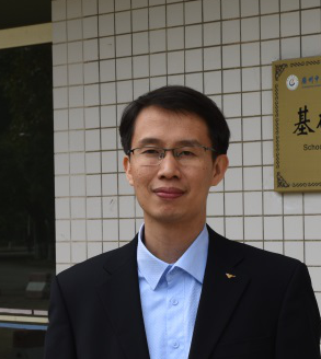 Ren Zhang (张韧)合作论文数Guangzhou University of Chinese Medicine5
 Hongwei Shao合作论文数4Xianli Wei合作论文数4Hui-feng WANG (王慧峰)合作论文数4LIU Jingjing (刘晶晶)合作论文数4Xiao-Dong Liu (刘晓东)合作论文数4GuanLin Chen (陈冠林)合作论文数4ZaoYuan Kuang (邝枣园)合作论文数4
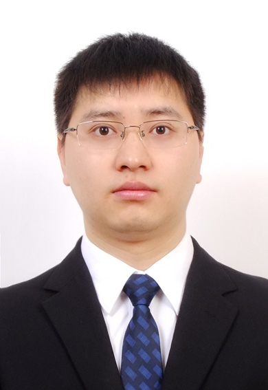 Jiangyong Gu (古江勇)合作论文数Guangzhou University of Chinese Medicine4
 Hong Li (李红)合作论文数Guangzhou University of Chinese Medicine4Peng Zhao (赵鹏)合作论文数4Xijuan Liu合作论文数4
 Qiaohong Yang (杨巧红)合作论文数Guangzhou University of Chinese Medicine4Wenyin Zhang (张文银)合作论文数3Idy H T Ho合作论文数3LIU Juan (刘娟)合作论文数3Ting Yin合作论文数3Haiyan Du合作论文数3Hai-yan DU (杜海燕)合作论文数3JiSheng Huang (黄暨生)合作论文数3RuLiu Li (李茹柳)合作论文数3Tingxiu Zhao (赵婷秀)合作论文数Guangzhou University of Chinese Medicine3Jian Wang (王剑)合作论文数Guangzhou University of Chinese Medicine3
 Benjie Zhou (周本杰)合作论文数School of Pharmaceutical Sciences (Shenzhen), Sun Yat-sen University;The Seventh Affiliated Hospital, Sun Yat-sen University3
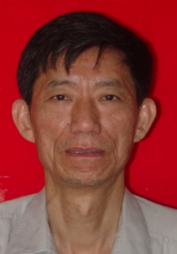 Zhicheng Lian (连至诚)合作论文数Guangzhou University of Chinese Medicine3Zengqiang Zhao (赵增强)合作论文数3Yaoju Li (李药菊)合作论文数3
 Yanli He (何彦丽)合作论文数Guangzhou University of Chinese Medicine3Wenling Yue (岳文玲)合作论文数3




