This study investigated the influence of cement type and water content on Er: YAG laser debonding efficiency and residual bond strength between zirconia restorations and titanium abutments with varying zirconia thicknesses. One hundred and sixty titanium-zirconia specimens were fabricated with zirconia thicknesses of 1 mm, 2 mm, and 3 mm. Specimens were divided into four groups (n = 40 per group) based on cement type: glass ionomer cement (Ketac Cem Easymix, KCE), modified glass ionomer (KCE with 35 µL water, KCEW), resin cement (Clearfil SA Luting, CSAL), and modified resin cement (CSAL with 35 µL water, CSALW). Each group was further subdivided by zirconia thickness (n = 10 per subgroup). After 48 h of storage at 100% relative humidity, Er: YAG laser irradiation (2940 nm) was applied for debonding, with the debonding time recorded. Specimens not debonded within 2 min underwent shear bond strength testing. A non laser irradiated 2 mm zirconia group served as the control. Statistical analysis employed Kruskal-Wallis and Wilcoxon tests with Bonferroni correction. Interfacial morphology was examined using scanning electron microscopy (SEM). The KCE and KCEW groups demonstrated significantly higher debonding rates at 1 mm and 2 mm thicknesses (> 50%), while the CSAL and CSALW groups showed minimal debonding(< 40%), particularly at 3 mm. The Kruskal-Wallis test indicated significant variations in both debonding time (χ² = 72.9, p < 0.001) and shear bond strength (χ² =107.8, p < 0.001) across groups. Debonding time varied significantly among groups, with the shortest times observed for KCE/KCEW at thinner thicknesses. Residual bond strength was comparable among KCE, KCEW, and CSALW groups; however, CSAL at 1 mm and 2 mm exhibited significantly lower strength versus 3 mm and control (p < 0.01). SEM analysis revealed microstructural alterations at zirconia-cement interfaces post-irradiation. Cement type, water content and restoration thickness significantly influence the debonding efficiency of zirconia crowns on titanium abutments using Er: YAG laser. Specifically, glass ionomer cements facilitate more efficient debonding, whereas for resin cements, water modification introduces a trade-off between bond strength and debonding performance. Debonding efficiency decreases substantially as restoration thickness increases. Clinically, these parameters are essential for optimizing the feasibility of laser-assisted crown retrieval.
To explore the influence of different inlay materials, depth, and width variations on stress distribution using the finite element method, and to provide a quantitative theoretical basis for the selection of clinical inlay restoration schemes. A three-dimensional finite element model of the mandibular first molar was established. The models were organized into four main groups based on the restorative material: Zirconia, Lithium Disilicate (LD), Gold Alloy (GA), and Resin-Based Ceramics (RBCs). Each material group was further divided into nine subgroups, simulating MOD inlay restorations with different preparation parameters: inlay depth (d = 2 mm, 4 mm, or 6 mm) and width (w = 2 mm, 4 mm, or 6 mm). A total of 36 groups of models were constructed. A vertical and oblique static load of 100 N was applied to the occlusal surface, and the distribution characteristics of the maximum principal stress (MPS) under different variable combinations were analyzed. The MPS of the four types of inlays were all concentrated on the stress-bearing contact surface and the bottom of the inlays. In terms of materials, under vertical loading, when d = 2 mm and w = 2 mm, the Zirconia group had the highest MPS (143.2 MPa), the RBCs group had the lowest (115.5 MPa), and the values of the LD group (127.9 MPa) and the GA group (128.3 MPa) were between the two. According to this finite element study, the stress distribution is influenced by the complex interaction between the material properties and the geometry of the preparation. Moreover, the stress distribution of RBCs is more uniform than that of high-modulus materials. In clinical applications, the restorative materials and preparation parameters should be comprehensively selected according to the conditions of the affected teeth, and the long-term effects still need in-depth research.
With the rapid advancement of digital technologies, digital intraoral scanning offers potential advantages for post-and-core impressions. However, its accuracy in relation to the complete spectrum of clinically relevant post space diameters, particularly smaller conservative preparations, requires comprehensive evaluation. This in vitro study aimed to systematically assess the accuracy of direct digital impressions compared to conventional impressions across four diameters and varying depths. Forty extracted single-rooted teeth were divided into four groups (n = 10/group) based on post space diameter: 1.0, 1.5, 2.0, and 2.5 mm. Each specimen was scanned using an intraoral scanner (TRIOS 5). Silicone impressions scanned with an extraoral scanner served as the reference. Root mean square (RMS) deviations were analyzed at four depths (1.65, 5.0, 7.0, 9.0 mm). Statistical analysis employed linear mixed-effects models. The scanner completely failed to capture data for the 1.0 mm diameter at depths ≥ 7 mm and for the 1.5 mm diameter at 9 mm. A significant diameter-by-depth interaction was found (p < 0.001). For diameters ≥ 1.5 mm and depths ≤ 7 mm, accuracy was clinically acceptable. At the 9 mm depth, the 2.0 mm and diameter group showed significantly higher deviation and critically high variability (135.0 ± 79.5 μm, CV 59.0
Aim or purpose: The long - term success of implant restoration and related complications are closely related to the stress on implants. Natural teeth with a periodontal ligament are superior to implants in buffering occlusal stress on alveolar bone. Elastic implant crown materials have been reported to relieve stress around implants. This study focuses on comparing displacement values of single implant crowns with diverse materials and abutment designs, exploring if elastic crown materials can imitate the periodontal ligament's cushioning function. Materials and methods: Personalized and prefabricated abutments, along with zirconia, resin - based ceramic, and PEEK crown materials, were used. CAD/CAM - made implant crowns with different central fossa thicknesses and corresponding abutments formed 12 groups (120 samples). A testing machine measured displacement, analyzed by ANOVA and Tukey's test. Results: ANOVA indicated that both crown material (F = 284.88, P = 1.39×10⁻⁴³) and abutment design (F = 48.63, P = 6.56×10⁻²⁰) significantly influenced implant displacement. The 3mm PEEK crown group had the highest displacement (0.331 ± 0.024 mm), and the 3mm zirconia group the lowest (0.143 ± 0.002 mm). The finished - abutment group's displacement was similar to that of 1 - mm zirconia and resin - based ceramic crown groups. Conclusions: For PEEK and resin - based ceramic, the thicker they are, the more they deform; for zirconia, it's reversed. The 3mm PEEK crown's displacement nears the periodontal ligament's thickness (0.38 mm), suggesting elastic crowns might replicate its cushioning effect.
Introduction: A 25-year-old female with posterior occlusal dysfunction, orthodontic/TMD history, and dental anxiety received minimally invasive occlusal veneers. The treatment preserved tooth structure, restored functional occlusion in two visits, and reduced anxiety. CBCT-confirmed TMJ health despite subjective symptoms. This demonstrates psychologically-informed adhesive dentistry for complex cases. Case description: A 25-year-old female patient presented with a chief complaint of posterior occlusal dysfunction persisting for over 1 year. The patient had a history of orthodontic treatment and temporomandibular disorder (TMD) management, along with significant dental anxiety. Clinical examination revealed inadequate posterior occlusal contacts without evident TMJ pathology on CBCT imaging. The treatment involved minimally invasive occlusal veneer restorations, preserving maximum tooth structure while reestablishing functional occlusion. The two-visit protocol significantly reduced treatment time and patient stress. At the two-week follow-up, the patient expressed dissatisfaction with minor occlusal interferences and requested selective adjustment. the patient reported no discomfort after adjustment. Discussion: This case demonstrates how minimally invasive veneers can effectively restore occlusion while addressing psychological needs in complex patients. The success highlights three key factors: (1) adhesive techniques preserving tooth structure, (2) shortened treatment time reducing anxiety triggers, and (3) the importance of correlating subjective symptoms with objective findings. Conclusion/clinical significance: This case highlights the successful integration of adhesive dentistry principles with psychological considerations for managing complex occlusal cases in anxious patients. The conservative approach achieved both biological preservation and functional rehabilitation while addressing the patient's emotional needs through tailored communication strategies and efficient treatment delivery.
Purpose: Elastic dental implant crown material may be able to reduce the impact force that is transmitted to the implant and the surrounding alveolar bone during the occlusal process. This study aims to compare the displacement values of implant-supported single crowns with different crown materials and abutment designs. The hypothesis (T0) of this study was that different crown materials and abutment designs have no effect on the displacement values of dental implant-supported restorations. Methods: Crowns were fabricated from three different materials: Aidite zirconia, Vita Enamic resin-matrix ceramics, and breCAM PEEK. These crowns were paired with custom abutments featuring different clearance values (1 mm, 2 mm, and 3 mm), as well as a prefabricated abutment group. This resulted in a total of 12 experimental groups, formed by combining the three materials with the four types of abutments. Each group consisted of 10 samples, for a total of 120 samples. Displacement values were measured using a universal material testing machine. Data were analysed using analysis of variance with Tukey's post hoc test. Results: One-way ANOVA with Tukey's post hoc test and Levene's test for homogeneity of variance were employed. Additionally, the two-way ANOVA revealed that both crown materials and abutment designs significantly affected the displacement values of implant-supported restorations. The polyetheretherketone 3-mm group exhibited the highest displacement value. The polyetheretherketone 3-mm group exhibited the highest displacement value (0.331 +/- 0.024 mm, P < .001), while the calcium oxide 3-mm group showed the lowest displacement value (0.143 +/- 0.002 mm, P < .001). Conclusion: For elastic materials, an increase in material thickness corresponded with increased deformation. For zirconia, increased material thickness led to decreased deformation. Therefore, their buffering effects on implant restorations are completely different.
PURPOSE:With the advancement in digital technology, post-and-cores can be produced using full-digital techniques. This study aimed to evaluate the accuracy of two full-digital impression techniques for scanning the post space with different root canal entrance diameters using an intraoral scanner (IOS) (TRIOS 3) with and without a scan post, compared to the half-digital impression technique. METHODS:Standard models of three intact teeth with varying root canal entrance diameters were prepared. Fifteen post-and-core designs per tooth were created using direct IOS and IOS with scan posts, with the half-digital technique as the control. All files were exported as standard tessellation language (STL) files, and the root mean square (RMS) deviations were analyzed between the experimental and control groups. Deviations were measured at standardized points across four regions: the occlusal external, occlusal internal, middle third, and apical third. RESULTS:No significant differences in the overall RMS were observed among the different root canal entrance diameters (3 mm, 4 mm, and 5 mm) for direct IOS and IOS with scan posts (P > 0.05). However, an independent samples t-test revealed a significant difference (P < 0.05) in both trueness and precision between the two experimental groups, with IOS with scan posts consistently showing lower RMS values. CONCLUSIONS:For post space lengths less than 7.4 mm in single-rooted teeth, both direct IOS and IOS with scan posts can serve as viable alternatives to half-digital techniques. However, in deeper areas, IOS with scan posts are recommended as alternatives to the half-digital technique.
Background: With the rapid advancement of digital impressions technologies, digital post-and-cores impressions can be applied in restorative dentistry. This study compared direct digital impressions with conventional silicone impressions to evaluate the influence of different post space diameters on the accuracy of digital post-and-core impressions at various depths. Methods: A total of three extracted teeth with different post space diameters were used to obtain three datasets s using an intraoral scanner (IOS) (TRIOS 5), with ten scans per group (1.5 mm, 2.0 mm, and 2.5 mm). An extraoral scanner (inEos X5) was used to scan the silicone impression as the control. All files were exported as standard tessellation language (STL) files.the root mean square (RMS) deviations were analyzed between the experimental and control groups. Deviations were measured at standardized points across four regions at various depths (1.65 mm, 5.0 mm, 7.0 mm, and 9.0 mm). Results: In the evaluation of trueness, the mean RMS values at all post space diameters (1.5 mm, 2.0 mm, and 2.5 mm) and depths (1.65 mm, 5.0 mm, 7.0 mm, and 9.0 mm) were below 100 μm, indicating clinically acceptable accuracy (RMS < 100 μm). There were no significant differences in mean RMS values among the different diameters (1.5 mm, 2.0 mm, and 2.5 mm) (P > 0.05) At depths of 1.65 mm, 5.0 mm, 7.0 mm. The mean RMS values at 9.0 mm were significantly higher than those at 1.65 mm, 5.0 mm, and 7.0 mm (P < 0.05), indicating a marked decrease in scanning accuracy at this depth. Conclusions: For the post space diameter more than 1.5 mm and the depth is within 7 mm in single-rooted teeth, the direct intraoral scanning method using TRIOS 5 can be considered a reliable clinical option. For post space diameters of 2.0 mm and 2.5 mm, the scanning accuracy of the direct intraoral scanning method significantly decreases at a depth of 9 mm, but it can still be considered a viable option in clinical practice. A wider post space diameter (≥2 mm) can improve the intraoral scanning accuracy of deeper post spaces (≥7 mm).
Background:Traditional impression techniques for customized post-and-core restorations often encounter challenges such as bubble and void formation, requiring a high level of skill from practitioners. Recent advancements in digital technology, particularly intraoral scanners (IOSs), have improved restoration processes, though capturing post spaces with IOS remains problematic due to their deep and narrow nature. Case Presentation:This report details the use of digital impressions for post-and-core restorations in both anterior and posterior teeth. By designing a scan post that conforms to the shape of the post space, accurate data capture of the post space is achieved. The integration of IOS with computer-aided design/computer-aided manufacturing (CAD/CAM) technology enables the creation of post-and-core restorations that precisely fit the root canal morphology. This approach results in a thin and evenly distributed cement layer, thereby reducing the risk of tooth fracture. Conclusion:This technique effectively overcomes the limitations of traditional silicone impressions for customized post-and-core restorations. It offers a viable restoration method for single-rooted teeth and addresses challenges related to acquiring impressions for split cast post-and-cores. However, further laboratory research and clinical trials are necessary to thoroughly evaluate the accuracy and reliability of this method.
Objective To compare the effects of different crown materials on the stress distribution of dental implants by finite element method. Methods A three-dimensional finite element model of implant-supported crown restoration of the right mandibular first molar was established. Four kinds of crown restoration materials were used, including polymer infiltrated ceramic network Vita Enamic, resin nanoceramic Lava Ultimate, zirconia ceramic Cercon, and polyether-ether-ketone breCAM. A displacement load of 0.01 mm or 200 N was applied to the occlusal surface of the right mandibular first molar, and the analysis of reaction force, the maximum variability of shape, the maximum principal stress, the maximum equivalent stress was performed. Results The results of stress analysis under 0.01 mm displacement load showed that the maximum equivalent stress of Cercon was 937.30 MPa, and that of breCAM was 67.09 MPa. The stress concentration of crowns and implants was obvious. The maximum equivalent stress of the surrounding alveolar bone of Cercon was the highest(26.61 MPa), and that of breCAM was the lowest(3.87 MPa), which was about 1/7 of that of maximum group. The stress concentration range of resin-matrix ceramics and polyether-ether-ketone was relatively small, and the distribution was more uniform. When 200 N was applied, the maximum principal stress and the maximum equivalent stress distribution of the four models were similar, and the maximum variability of shape of breCAM was larger at the loading site. Conclusion Under the same displacement load, the stress concentration of polyether-ether-ketone and resin-matrix ceramics crown was lower than that of zirconia. There was no significant difference in stress concentration in the implant and surrounding alveolar bone among polyetheretherketone, resin-ceramic composite, and zirconia crown restorations under the same load. Further research on long-term durability of PEEK and resin-matrix ceramicsis needed through practical and clinical trials.
The retention force of a realistic clasp is influenced by multiple, interrelated factors, which complicates the identification of the fundamental relationship between clasp geometry and retention force. While realistic clasps exhibit various shapes, they share basic geometric elements such as length, diameter, and curvature. Simpler geometries are often more conducive to identifying the underlying issues. The aim is to investigate the relationship between clasp geometry and retention force using finite element analysis. A three-dimensional clasp model was created in ANSYS 19.0 (ANSYS, USA). Two types of models were analyzed: rod-shaped clasps with varying lengths (1–15 mm) and diameters (0.6–1.6 mm), and bending clasps with different base widths (6–12 mm) and heights (0.5–5 mm), all made from cobalt-chromium alloys. For the rod models, stress and retention force were assessed by applying displacement loads and analyzing data with nonlinear regression. For the bending models, a similar analysis was conducted for varying base widths and heights. Maximum stress consistently concentrated at the clasp base. In rod models, retention force decreased with the third power of length and increased with the fourth power of diameter. For bent specimens, the retention force was approximately inversely proportional to the cube of the base width and inversely proportional to the first power of the height. Finite element analysis revealed distinct functional relationships between clasp geometry and retention force. Further laboratory validation is required.
全瓷修复体在口腔临床特别是种植修复中应用广泛,但临床上粘接固位的种植冠部修复体拆除较为困难.传统拆除采用磨除、敲击等机械破坏的方法,不仅增加临床诊疗时间和费用,同时给患者带来恐惧和不安.近年来有学者着手于研究使用Er:YAG激光作为替代传统机械拆除种植全瓷修复体的新方法,全瓷修复体的材质和厚度、水门汀类型、激光参数设置等多种因素均对拆除结果存在影响.该文对Er:YAG激光拆除种植牙全瓷冠修复体的应用及研究进展作一综述,为医师的临床操作提供相关参考.
Customized posts-and-cores have been widely used for improved fitness within a prepared post space. However, in comparison to direct restoration, they necessitate an increased number of appointments for patients. A 24-year-old man presented with a maxillary left canine that had fractured due to trauma 10 months previously. For this case, a digital process was used for simultaneous restoration with a personalized titanium post-and-core and a zirconia crown achieved with an intraoral scanner (IOS) and computer-aided design/computer-aided manufacturing (CAD-CAM). This workflow allowed the restoration to be completed in 2 visits, facilitating more effective and predictable treatment, with reduced time and cost for the patient.
近年来,随着种植修复技术的成熟和相关材料的不断发展,如何选择合适的种植义齿粘接水门汀引起了不少关注.口腔医生在选择种植义齿和牙体缺损全冠修复2种固定修复的粘接水门汀时,考虑的因素存在一定的区别.目前市售的粘接水门汀多以配合牙体粘接为主,牙体缺损固定修复通过水门汀将牙体组织与冠修复体进行粘接,其粘接界面与种植义齿的基台-水门汀-冠的粘接界面不同,适用于牙体组织与冠的粘接水门汀不一定能用于基台与冠的粘接.同时因牙体缺损固定修复粘接时,通常要考虑水门汀对牙釉质的粘接强度、对牙髓的刺激性以及释氟性能等,而种植修复则要考虑并发症.如何平衡水门汀的粘接力和可拆卸性[1],从而在拆卸牙冠的同时又不破坏种植体的各部分?选用种植冠粘接固位时的水门汀要考虑的因素有哪些?现就种植牙冠修复粘接水门汀的分类、固位性和可拆卸性、生物相容性、残留的影响、多余水门汀的检测和去除方法等进行综述,为临床工作中种植修复用水门汀的选择提供参考.
Background The purpose of this study is to investigate the performance and fracture resistance of different resin-matrix ceramic materials for use in implant-supported single crowns with respect to the abutment design (crown thickness: 1 mm, 2 and 3 mm). Methods Forty-eight abutments and crowns were fabricated on implants in the right lower first molar. Two resin-matrix ceramic materials for dental crowns were selected for study: (1) a glass-ceramic in a resin interpenetrating matrix (Vita Enamic, Vita, Germany) and (2) a resin-based composite with nanoparticle ceramic filler (Lava Ultimate, 3 M ESPE, USA). Three types of abutments were designed: 1 mm thick crown + custom titanium abutment, 2 mm thick crown + custom titanium abutment and 3 mm thick crown + prefabricated titanium abutment. The experiment was divided into 6 groups (n = 8) according to the crown materials and abutment designs. After 10,000 thermocycles, fracture resistance was measured using a universal testing machine. The statistical significance of differences between various groups were analysed with ANOVA followed by a post hoc Tukey’s honestly significant difference test. The surfaces of the fractured specimens were examined with scanning electron microscopy (SEM). Results Two-way ANOVA revealed that the abutment design (F = 28.44, P = 1.52 × 10 − 8 <0.001) and the crown materials (F = 4.37, P = 0.043 < 0.05) had a significant effect on the fracture resistance of implant crown restoration. The Lava Ultimate-2 mm group showed the highest fracture resistance of 2222.74 ± 320.36 N, and the Vita Enamic-3 mm group showed the lowest fracture resistance of 1204.96 ± 130.50 N. Most of the 1 and 2 mm groups had partial crown fractures that could be repaired directly with resin, while the 3 mm group had longitudinal fracture of the crown, and the crowns were detached from the abutments. Conclusion Based on the in vitro data of this study, the fracture resistance of the 2 mm thick resin-matrix ceramic crown design was higher than that of the 1 and 3 mm groups. The 2 mm thick resin-matrix ceramic crown and personalized abutment are an option to replace zirconia for implant crown restoration.
OBJECTIVES:This study compared the accuracy of the guide-supported and the microscope-assisted fiber post removal systems by using the extracted teeth. These new idea and theory can be used by clinicians to remove fiber posts. METHODS:Twenty-eight human extracted premolars were randomly divided into the guide and microscope groups. After root canal treatment and fiber post restoration, the fiber posts were removed by using a digital guide and via microscope-assisted ultrasonic instrument, respectively. Mimics 10.0 was used to measure the deviation, and the accuracy of the two fiber post removal systems were compared. RESULTS:In the guide group, the apical vertical deviation was 0.99 mm±0.52 mm, the apical horizontal deviation was 0.75 mm±0.19 mm, the angle deviation was 2.32°±0.64°, and the volume loss was 8.09 mm3±1.42 mm3. In the microscope group, the apical vertical deviation was 0.44 mm±0.23 mm, the apical horizontal deviation was 0.23 mm±0.07 mm, the angle deviation was 0.64°±0.31°, and the volume loss was 15.25 mm3±3.94 mm3. No significant difference was found in the apical vertical deviation between the two groups (P>0.05), whereas the apical horizontal deviation, the angle deviation, and the volume loss were significantly different between the two groups (P<0.05). CONCLUSIONS:The removal of fiber post supported by a digital guide helped reduce the volume loss of post-core restoration teeth, but its accuracy was lower than that of removal by using a microscope-assisted ultrasonic instrument.
随着"引导式牙髓治疗"在牙体牙髓领域的应用研究逐渐开展,动态导航系统和数字化导板技术也被应用于纤维桩拆除的临床操作中.相较于现有的纤维桩拆除系统,计算机辅助拆除具有准确、高效、微创等特点.本文通过介绍各种纤维桩拆除方式的临床应用和优缺点,分析计算机辅助技术在临床纤维桩拆除中的准确度及误差因素,以此作一综述,为医师的临床操作提供相关参考依据.
Background This research aimed to explore feasibility and the time required when erbium-doped yttrium aluminum garnet (Er:YAG) laser as a non-invasive treatment modality to retrieve different thicknesses of zirconia material bonded by two dental cements from titanium implant abutments. Methods Prepared 80 titanium blocks (length: 20 mm, width: 10 mm, height: 10 mm) and square zirconia sheets (length: 10 mm) with different thicknesses (1 mm, 2 mm, 3 mm, and 4 mm) were 20 pieces each. Resin modified glass ionomer cement (RelyX Luting 2; RXL) and resin cement (Clearfil SA luting; CSL) were used to bond zirconia sheet and titanium block. Specimens were kept in 100% humidity for 48 h. Er:YAG laser was used to retrieve the zirconia sheet and recorded the time. Universal testing machine was used to measure the residual adhesion of the samples that did not retrieve after 5 min of laser irradiation. Shear bond strength (MPa) and the time data (s) were analyzed using Kruskal–Wallis Test. The bonding surface and the irradiation surface of the zirconia sheet was examined with the scanning electron microscopy (SEM). Results Within 5 min of laser irradiation, RXL group: 1 mm group all fell off, 2 mm group had 3 specimens did not fall off, there was no statistical difference in the average time between the two groups; CSL group: half of the 1 mm group fell off. Shear bond strength test results: there was no statistical difference between 1 and 2 mm in RXL group and 1 mm in CSL group, there was no statistical difference between 3 mm in RXL group and 2 mm in CSL group, and there were significant differences statistically in comparison between any two groups in the rest. SEM inspection showed that the bonding surface and the irradiation surface of the zirconia sheet had changes. Conclusion In this vitro study, the following could be concluded: it is faster to remove zirconia crowns with thickness less than 2 mm from titanium abutment when luted with RelyX Luting 2 compared to Clearfil SA luting.
目的 探讨梨状窝甲状软骨颈部标记法应用于光棒盲探气管插管的指引效果及安全性.方法 选择我院收治的80例拟行全身麻醉患者,应用光棒指引盲探气管插管,随机分为两组,每组40例,分别采用传统正中直入法(对照组)和梨状窝及甲状软骨标记法(观察组)行光棒盲探气管插管,比较两组的气管插管首次成功率、总成功率、插管操作所用时间、术后并发症(咽痛、声音嘶哑)发生率、SpO2等.结果 两组气管插管的总成功率均为100%.观察组和对照组气管插管首次成功率分别为95.0%和62.5%;插管操作所用时间分别为(14.23±4.65)s、(35.72±9.68)s,差异均有统计学意义(P<0.05);插管过程中两组SpO2均保持在98%以上.术后并发症方面观察组1例出现咽喉部轻微疼痛;对照组6例出现咽喉部疼痛,2例出现声音嘶哑,两组并发症发生率差异有统计学意义(P<0.05).结论 梨状窝及甲状软骨颈部标记法光棒盲探气管插管操作直观快捷、成功率高、并发症较少,值得临床推广应用.
目的 评价两种不同粘接水门汀对树脂类冠修复材料和全瓷托槽剪切粘接力的影响.方法 选用2种水门汀Transbond XT和RelyX Ultimate,将上颌前磨牙全瓷托槽Inspire Ice与4种树脂类冠修复材料粘接.试件冷热循环5000次后测量断裂载荷(N),并观察破坏形态.结果 单因素方差分析结果F=4.52(P<0.001),显示粘接条件对陶瓷托槽和树脂类冠修复材料的剪切粘接力影响具有统计学意义.其中Lava Ultimate+Transbond XT组和Lava Ultimate+RelyX Ultimate+托槽喷砂组分别为(325.10±135.54)N和(304.71±35.71)N,较其他组高,Lava Ultimate+Transbond XT+托槽喷砂组最低,为(118.12±57.58)N.结论 树脂基陶瓷材料Lava Ultimate和全瓷托槽的剪切粘接力较暂时冠修复材料高.全瓷托槽未喷砂时,正畸全瓷托槽粘接水门汀Transbond XT和修复用水门汀RelyX Ultimate的粘接力未见明显区别,喷砂后全瓷托槽失去固位小球,RelyX Ultimate有更高的粘接力.
 Jie Lin (林捷)合作论文数28
 Hui Cheng (程辉)合作论文数Institute of Stomatology, Research Center of Dental Esthetics and Biomechanics, School and Hospital of Stomatology, Fujian Medical University15
 Pintong Huang (黄品同)合作论文数The Second Affiliated Hospital Zhejiang University School of Medicine12Zhenxiang Lin合作论文数11
 Ming Zheng (郑明)合作论文数Schoo and Hospital of Stomatology, FuUjian Medical UnfiversityFujian Stomatological Hospital11ZhaoJie Lu (卢兆杰)合作论文数10FuGuang Huang (黄福光)合作论文数9Pingping Cai合作论文数8Yingying Zhuo合作论文数7ShouZhi Ma (马守治)合作论文数6ZhiGang Hu (胡志刚)合作论文数6YuShuang Lin (林玉霜)合作论文数6YanPing Li (李艳萍)合作论文数6Nian-yu Xue合作论文数5ShaoPing Zhang (章少萍)合作论文数5ZongMin Wang (王宗敏)合作论文数5YaPing Zhao (赵雅萍)合作论文数5JiPing Deng (邓继平)合作论文数4ShiYan Li (李世岩)合作论文数4Zongyang Huang (黄宗阳)合作论文数3ChangWei Huang (黄常伟)合作论文数3Liang Wang (王亮)合作论文数3
 Houxuan Li (李厚轩)合作论文数Nanjing Stomatological Hospital, Medical School, Nanjing University3Xiangrong Cheng合作论文数3
 Akikazu Shinya合作论文数3Xue-li Zhu合作论文数2Jiesen LI (李杰森)合作论文数2Ying TANG (汤莹)合作论文数2honghui wang (汪宏晖)合作论文数2WenDuo Chen (陈文多)合作论文数2XinZhang Guo (郭心璋)合作论文数2YanJuan Tan (谭艳娟)合作论文数2ZaiXing Pan (潘在兴)合作论文数2Chao Zhang (张超)合作论文数2
 Michael George Botelho合作论文数Faculty of Dentistry, The University of Hong Kong;Prince Philip Dental Hospital, The University of Hong Kong2Sufeng Chen (陈素凤)合作论文数2
 Youguang Lu (卢友光)合作论文数Affiiated Stomatological Hospital, Fujian Medical University2
 Qun Chen (陈群)合作论文数福州中医药大学第二临床医学院2Akiyoshi Shinya合作论文数2
 Jukka Pekka Matinlinna (馬裕褀)合作论文数Faculty of Dentistry,The University of Hong Kong2
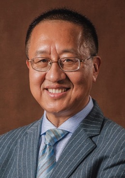 Fuhua Yan (闫福华)合作论文数Medical School of Nanjing University2Hui-liao He合作论文数1HUANG Feibin (黄飞彬)合作论文数1Xiaoyan HU (胡小艳)合作论文数1CHEN Rui-jie (陈瑞杰)合作论文数1l wang合作论文数1Jianmin Cheng合作论文数1Xuehui Long (龙学辉)合作论文数1




