Cerebral palsy (CP) is one of the most common disabling diseases of children’s nervous system,and its treatment is an obvious challenge in medical area.Spastic CP is the main type of CP,accounting for about 70%of CP patients,with increased muscle tone as its main characteristic.Spasticity has always been a major problem in the treatment of CP.
Objective To study the influence of different microsurgical methods on surgical outcomes and complications, and to improve the surgical outcomes for trigeminal neuralgia. Methods The clinical data of 109 patients with trigeminal neuralgia, who were treated with microsurgery, were analyzed retrospectively. All patients were divided into 3 groups according to surgical modality: the trigeminal neuralgia decompression group (TND group, 19 patients), the TND and rhizotomy group (rhizotomy group, 55 patients), and the TND and selective lesioning group (lesioning group, 35 patients). The mid-term and short-term effects of microsurgery, and the occurrences of complications, were compared between the 3 groups. Results There were no statistical differences in the frequency of complications between the 3 groups (P > 0.05). Eighty-four patients were followed up for 6 to 33 months. The rate of pain disappearance was found to be 94.4% in the TND group, and 100% in both the rhizotomy and lesioning groups; thus, no significant differences were found between these 3 groups (P > 0.05). Additionally, 50% of the patients in the rhizotomy group and 3.6% of the patients in the lesioning group had facial numbness while no patients were affected with facial numbness in the TND group, and the differences between these 3 groups were significant (P < 0.05). Conclusions Microsurgery is effective and safe for trigeminal neuralgia. The use of TND, in combination with selective lesioning, ensures therapeutic efficacy and improves the quality of life in postoperative patients.
OBJECTIVE To explore the outcomes of individualized neurosurgical treatments of spastic cerebral palsy. METHODS A total of 452 spastic cerebral palsy patients undergoing microneurosurgery during March 2006 and December 2011 were retrospectively analyzed. Performed on the basis of clinical manifestations, comprehensive procedures included selective cutting of neck or lumbosacral dorsal root ganglia (SPR, n = 182) for multiple muscle spasm, selective peripheral neurotomy (SPN, n = 270) for focal muscle spasm, line tendon lengthening or cutting and orthopedic surgery (n = 116) for tendon contracture plus limb deformities and carotid artery adventitia endarterectomy (n = 46) for salivation plus athetoid. RESULTS The average follow-up period was 18 (10-24) months. The limb spasm relief rate of SPR was 95.6% and that of SPN 98.3%. And the postoperative symptom improvement rate of common carotid arterial adventitia stripping was 91.3%. There were 8 cases (4.4%) of recurrent spasticity after SPR and 28 (10.4%) after SPN. CONCLUSION Offering individualized microsurgical treatments on the basis of clinical manifestations is essential for patients with spastic cerebral palsy.
Objective To analyze surface electromyography activities of spastic cerebral palsy,to explore the role of surface electromyography (sEMG) in evaluation effectiveness of peripheral nerve surgery.Methods Peripheral nerve surgery was carried out in 218 cases,including 171 selective peripheral neurotomy (SPN) and 47 selective posterior rhizotomy (SPR).The sEMG of patients was recorded before and after operations while passive and voluntary movements were performed during recording.The root mean square (RMS) were calculated by time domain analysis.Analysis of Variance was applied to compare RMS before and after operations.Modified Asworth Scale was emplvyed to estimated the patients' muscle tension.And Manual Muscle Testing was applied to assessed patients' muscle strength.Investigate the correlation among MAS,MMT and RMS.Results Two hundred and three cases were followed up for 22 to 30 months (mean 25.6 months).15 cases were lost.Among them,the cases were followed up with 350 sides of adductor,197 sides of quadriceps and 374 sides of triceps.In passive movements,the RMS of adductors,quadriceps and triceps post-surgery were significantly decreased comparing to values of pre-surgery (all P <0.05).And those values follow-up were significantly decreased than pre-surgery (all P < 0.05).In voluntary movements,the RMS of quadriceps post-surgery were significantly decreased than pre-surgery (all P < 0.05).However,no significant differences were observed in adductors and triceps between post-surgery and pre-surgery(all P >0.05).And there were no significant differences of adductors,quadriceps and triceps observed in follow-up and post-surgery (all P >0.05).A higher remission rate (91%) of adductor spasm was observed in 318 postoperative cases of 350 cases,and 98% (343/350) was observed during follow-up.The quadriceps spasm relief rate was 82% (161/197) postoperative and 95% (187/197) during follow-up.Besides the relief rate of triceps spasm was 93% (348/374) postoperative and 99% (370/374) during the follow-up.The muscle strength of adductor was reduced by 9% (31/350) postoperative,while it was increased by 23% (80/350) during follow-up.Moreover,there was a 21% (41/197) reduced rate of muscle strength of quadriceps,however,it was increased by 19% (37/197)during follow-up.The muscle strength of triceps was reduced by 18% (67/374) postoperative,but it was increased by 29% (108/374) follow-up.There are significant correlations between MAS,MMT and RMS(both P < 0.05).Conclusions sEMG can objectively evaluate the clinical effectiveness of spastic cerebral palsy as a non-invasive method.Muscle tension is obviously reduced after peripheral nerve surgery carried on spastic cerebral palsy,meanwhile the muscle strength is not significantly decreased.
目的 探讨Dellon三联周围神经减压术(腓总神经、腓深神经及胫后神经减压术)治疗糖尿病周围神经病(DPN)下肢病变的手术疗效.方法 分析2006年7月~2011年11月采用Dellon三联周围神经减压手术治疗的40例(DPN)患者的资料,观察他们的临床特点、手术方法、手术效果和随访结果.结果 临床表现以渐进性小腿和足底、足背部麻木起病者35例,以渐进性疼痛起病者5例.术后下肢麻木症状缓解率90%,术后下肢疼痛症状缓解率92%.术后平均随访12个月临床症状缓解的所有患者病情稳定,1例患者术后1个月出现足底部分区域疼痛缓解后再次出现疼痛,其余病人症状无复发或加重.结论 Dellon三联周围神经减压术对于(DPN)缓解疼痛、恢复感觉有较好疗效,是一种安全、有效、微创治疗新技术和新方法.
This study aimed to explore the role of surface electromyography (sEMG) on the changes of myoelectric activities of quadriceps femoris and the assessment of its clinical effect before and after selective femoral neurotomy on spastic cerebral palsy with stiff knee. Selective femoral neurotomy was carried out in 15 patients on 26 sides. The electromyography of quadriceps femoris was recorded before and after the operation. Passive and voluntary movements were performed during recording. The root mean square (RMS) and integrated electromyography (iEMG) was calculated by time domain analysis. Meanwhile, the range of the patients' knee joint motion (ROM) was measured by a joint goniometer. The RMS and iEMG of the quadriceps femoris during passive movement was significantly decreased post-operation when compared to those pre-operation (both P<0.05, n=26). Meanwhile, the RMS and iEMG of the quadriceps femoris during voluntary movement post-operation was significantly reduced than those pre-operation (both P<0.05, n=26). Additionally, total excursion on the sagittal plane and the peak knee flexion in the swing phase were significantly raised post-operation than those pre-operation (both P<0.05, n=26). The spasms in the quadriceps femoris in spastic cerebral palsy patients with stiff knee was clearly improved, and the ROM of the knee was significantly enhanced after the selective femoral neurotomy. Importantly, surface EMG can objectively evaluate the clinical therapeutic effect of spastic cerebral palsy stiff knee as a noninvasive detection method.
Objective To explore the methods and effectiveness of neurosurgical management of hereditary spastic paraplegia.Methods Selective peripheral neurotomy(SPN) was carried out in 17 patients,including 12 SPN and 15 SPN+orthomorphia.Modified Asworth scale was employed to estimated the patients' muscle tone.And the Fugl-Meyer scores was applied to assessed the motor function of lower limbs.Results All patients were followed up from 8 to 15 months(means 12 months).The postoperative spasticity relief rate is 98.5% and the follow-up is 92.7%.The improvement of patients' motor function in lower limbs is 80.5% after the operation and 90.8% by follow-up.Temporary sensory disturbance in lower limbs occurred in 2 cases postoperatively and disappeared by follow-up.Muscle weakness with severe spasm occurred in one case.The spasm recured in 2 cases by follow up.Conclusion With strictly control indications,the neurosurgery can effectively relief spasm,improve motor function and delay the development in lower limbs of hereditary spastic paraplegia patients.
目的研究神经片段结合神经导管治疗外周神经缺损,为周围神经损伤修复与重建的解决途径提供实验依据。方法 24只成年Wistar大鼠随机分为A、B、C组,分别为神经自体移植、与含有神经片段的神经导管和单纯导管移植桥接大鼠10mm神经缺损,各组间在形态学、电生理、组织学进行比较。结果 B组大鼠移植侧肢体足迹实验的坐骨神经功能指数(SFI)为-81.20±2.81,神经干传导速度为(28.93±5.07)m/s、动作电位的振幅为(5.46±2.76)mV,再生神经中段检查髓鞘化轴突密度为(3953±468)/视野,髓鞘面积为(2.77±0.83)μm,与C组比较差异有统计学意义(P<0.05)。结论含有神经片段的神经导管组织工程移植体桥接神经缺损有促进神经轴突再生与神经功能恢复的良好作用,效果优于单纯神经导管移植。
Autologous nerve grafts are widely used in bridging critical gaps of peripheral nerves, but they remain associated with high morbidity of the donor site and lack of full recovery. As an alternative, we have focused on chitosan nerve conduits filled with a heparin-incorporated fibrin–fibronectin matrix serving as delivery systems for basic fibroblast growth factor (bFGF). The artificial nerve conduits were used for repairing sciatic nerve defects of 10 mm in adult rats. Three months post-operation, the conduction velocity recovery index (CVRI) and the muscle restoration rate (MRR) in animals of the experimental group were 32 ± 4.1 and 77.4 ± 7.9%, respectively, which were significantly higher than those of the PBS control group (17.8 ± 1.9 and 66.7 ± 6.5%), and similar to those of the autograft group (38.4 ± 3.9 and 81.3 ± 7.8%). These results were also consistent with the densities of regenerated axons in the three groups, which were demonstrated by histomorphological analysis.
BACKGROUND: Peripheral nerve tissue engineering needs a large number of Schwann cells. In previous studies, lack of normal human nervous tissue, so animal (rat, rabbit, et al) nervous tissues are commonly used to isolate Schwann cells, but as xeno-cells it is limited in clinical application. OBJECTIVE: To investigate an effective technique for isolation, cultivation and purification of human Schwann cells of normal peripheral nerves cultured in vitro. METHODS: Normal peripheral nerves were obtained from the surgery of cerebral palsy patients. Schwann cells were cultured with enzymatic digestion culture method and differential attachment method. Tissues were cut into pieces and incubated in medium supplemented with fetal bovine serum, collagenase and Dispase enzyme, centrifuged. Tissue blocks were placed in the medium, triturated into monoplast suspension, and then moved into a DMEM Petri dish containing polylysine, supplemented with basic fibroblast growth factor. When adherent cells were confluent about 85%-90%, cells could subculture. Schwann cells were counted by Trypan Blue coloring method at 2, 3, 4, 5, 6, 7, 8, 9, 10. The purity of Schwann cells was identified through S-100 protein immunohistochemistry staining. RESULTS AND CONCLUSION: More than 0.5×10⁸/L Schwann cells were detected after four days under a microscope. Following the third passage, the number of Schwann cells was over 9×10⁸/L. The purity of Schwann cell population was up to 85%. Results suggested that plenty and purified human Schwann cells could be obtained by enzymatic digestion culture and differential attachment methods in a short time, which can be used for the source of peripheral nerve tissue engineering.
Instead of nerve autografts,tissue-engineered nerve conduits are being used for the treatment of peripheral nerve graft repair.These nerve tubes are made of different biomaterials or artificial synthetic materials with different physical properties.In order to increase the nerve gap that can be bridged,several modifications to the common hollow nerve tube(for example,the addition of Schwann cells,growth factors,and internal frameworks)are being investigated in laboratory.Some have been used clinically.
OBJECTIVE:To investigate the effects of topical fluorescein on the preoperative diagnosis of rhinorrhea of cerebrospinal fluid (CSF) and intraoperative localization of CSF fistula.METHODS:Cotton pads soaked with 5% fluorescein were placed in the middle turbinate meatus, roof of ethmoid plate, and sphenoethmoidalis recesses via endoscopic endonasal technique of 15 patients with CSF rhinorrhea, 5 caused by accidental trauma and 10 spontaneous. Change of the color of fluorescein from yellow to green fluorescence denoted the presence of CSF, and thus the site of the leak could be traced. The accuracy rates of diagnosis and leak site identification by this technique were compared with those by glucose analysis, intraoperative findings, and follow-up.RESULTS:The preoperative diagnosis rate of CSF rhinorrhea and the CSF fistula site localization rate by fluorescein-soaked cotton pad were both 100%. No recurrence was found during the follow-up for 2 - 24 months. No complication had been reported.CONCLUSIONS:Application of topical fluorescein is an easy, sensitive, safe, and highly accurate tool in the intraoperative localization of the site and extent of CSF fistulas and should be considered a viable noninvasive alternative to intrathecal fluorescein technique.
Background: The CSF rhinorrhea is a kind of common clinical disease. The preoperative diagnosis and intraoperative localization of CSF fistulas are critical to treatment of CSF rhinorrhea. At present, intrathecal fluorescein regarding endoscopic transnasal cerebrospinal leakage repair is a common method for localization of the fistula; however, it has some disadvantages because it needs a specific endoscope, and the trauma from lumbar Puncture as well as the potential complications of intrathecal fluorescein, as a result, widely limited its clinical use. Topical intranasal fluorescein can avoid the above-mentioned shortcoming. The aim of this work was to describe the use of topical fluorescein in the intraoperative localization of CSF fistulas and to screen its use in preoperative diagnosis of CSF rhinorrhea.Methods: Fifteen patients with CSF rhinorrhea were treated with an endoscopic endonasal technique. Topical intranasal 5% fluorescein for preoperative diagnosis and intraoperative localization of the site of the leak was placed in middle turbinate meatus, the roof of the ethmoid plate, and sphenoethmoidal recesses. A change in the color of the fluorescein from brown to green fluorescence denoted the presence of CSF, and the site of the leak could be traced. The accuracy rate of diagnosis and leak site identification was made by comparison with glucose analysis, intraoperative findings, and follow-up.Results: The cause of the leak was accidental trauma in 5 patients and spontaneous in 10 patients. The preoperative use of fluorescein-soaked cotton was 100% accurate in diagnosing CSF rhinorrhea. The intraoperative use of topical intranasal fluorescein was also 100% accurate in locating the site of the CSF fistula when compared with the surgical findings. It had achieved 100% success rate in sealing the CSF fistulas in our 15 patients with no recurrence detected during the follow-up period (mean, 2-24 months). No complications have been reported.Conclusion: In the presence of a clinically diagnosed CSF leakage and location of the leakage fistula, topical fluorescein is a very easy, sensitive, safe, and highly accurate tool in the intraoperative localization of the site and extent of CSF fistulas. It should be considered as a viable noninvasive alternative to intrathecal fluorescein. Crown Copyright (C) 2009 Published by Elsevier Inc. All rights reserved.
Objective To investigate the effect of extract of Ginkgo biloba (Egb) on apoptosis of nerve cells and its mechanism after spinal cord injury (SCI) in rats. Methods Forty eight adult SD rats weighing 200-230 g were divided equally and randomly into Egb group and normal saline (NS) group. After hemisectian of spinal cord at T9 vertebrae level, rats in Egb group were lavaged with 2 ml EGB (20 mg) daily and those in NS group with 2 ml NS daily. Tissue sections were collected and stained with Nissl's staining, myelin sheath staining, and inducible nitric oxide synthase (iNOS) immunohisto-chemistry as well as terminal deoxynueleotidyl transferase-mediated dUTP nick end lebeling (TUNEL) at days 1,7, 14 and 21 respectively to evaluate the injured spinal cord tissues after six rats from each group were sacrificed Results Nissl's staining manifested less swelling of the nerve cells near the injury epi-center ( rostral and caudal ), smaller cavity and demyelinated area and higher ratio of bilateral anterior horn neurons of transection side to normal side in Egb group, compared with NS group ( P <0.05). Ap-optotie index (AI) and expression of iNOS in NS group were higher than those in Egb group ( P <0.01 or P <0. 05). Furthermore, the rate of iNOS-positive cells was positively correlated with the AI (r = 0.729, P<0.01) after SCI. Conclusion Egb can prevent nerve cells from apoptosis after SCI in rats, as may be related with inhibition of expression of iNOS.
Objective To demonstrate the morphology of drainage of the cerebrospinal fluid through the olfactory nerves into the cervical lymphatics. Methods Microfil was injected into the cisterna magna of the rabbit, cervical lymphatics, the olfactory bulbs, olfactory tracts and the nasal mucosa were observed by macroscopic anatomy and light microscopy. Results Under the operative microscope, Microfil was found within the subarachnoid space and along the olfactory nerves. At the nasal mucosa, a lymphatic network stained in yellow was identified near the olfactory nerves, which cross nasopharyngeal region and finally emptied into the superficial and deep cervical lymph nodes. Light microscopically, Microfil was distributed around the olfactory nerves and within the lymphatic vessels. At the nasal mucosa, the lymphatics were frequently located close to the nerves. Conclusion These results indicate that the cerebrospinal fluid drains from the subarachnoid space along the olfactory nerves to the nasal Lymphatics, which in turn, empties into the cervical lymph nodes. This anatomical communication, thus, allows the central nervous system to connect with the lymphatic system. The presence of this route may play an important role in immune disease of the central nervous system and regulation of the cerebrospinal fluid circulation.
Objective To study the surgical technique of endoscopic transnasal biopsy for the diagnosis of cavernous sinus lesions.Methods Ten patients with cavernous sinus lesions were diagnosed by endoscopic transnasal biopsy.Retrospectively the clinical data of 10 patients were reviewed.Results The accuracy of diagnosis with this technique WSS 100%.Postoperative results included:5 patients with malignant tumors received radiotherapy,1 pauent with meningioma underwent yknife treatment,and 1 patient received antifungal therapy.2 patients with benign lesion and 1 patient underwent local radiotherapy were followed.Postoperative complications included:one patient with internal carotid artery injury was treated by interventional ballon occlusion and one with transient eerebrospinal leakage.Conclusion Endoscopic transrmsal biopsy is an effective.safe and mini-invssive approach for the diagnosis of cavernous sinus lesions.
Objective To demonstrate the connection between the subarachnoid space(SAS) and cervical lymphatic vessels in rabbit.Methods X-ray contrast examination was performed for cerebrospinal fluid(CSF) lymphatic drainage and the cervical lymphatic vessels in 10 New Zealand rabbit.Results X-ray contrast examination of CSF lymphatic drainage displayed the contrast medium reached the cribriform plate 7 min and distal the optic nerve 20 min after the contrast medium injection,and leaked into the nasal cavities 30 min later and the cervical lymphatic vessels were visualized at the same time.Cervical lymphangiography revealed contrast medium flowed slowly around foramen magnum at early stage after the injection,finally reached the chiasma via the optic nerve,and subsequently,the superficial lymphatic vessels in the nasal septum,ethmoid plate region,internal acoustic meatus,periphery of the superior sagittal sinus,and neck were visualized.Conclusion The intracranial subarachnoid space communicates with cervical lymphatic vessels.Under certain pressure,cervical lymph can retrogradely emptied into the cranial subarachnoid space.The pathophysiological significance of this phenomenon should be further researched.
Objective To describe the technique of endoscopic endonasal transsphenoidal surgery for treatment of pituitary adenoma.Method Clinical data of 100 patients with pituitary adenoma that underwent endoscopic endonasal transsphenoidal surgery between September 2003 and March 2006 was analyzed.30 patients had microadenomas,35 patients macroadenomas,and 35 patients gigantic adenomas.68 patients had hormone secreting adenomas.32 had non-functioning adenomas.All operations were performed through a nostril using pure rigid endoscopes.Results Among the 100 patients,total resection was in 93 patients,subtotal resection in 7 patients.32 patients developed postoperative diabetes insipidus,5 developed CSF leakage.All patients were followed by mean 28 months,residual tumor was found in 10 patients.Among 12 patients with tumor recurrence,10 performed second endoscopic endonasal surgery and 2 underwent transcranial surgery.18 patients were treated with gamma knife surgery and 11 patients were treated with a course of bromocriptine therapy.Conclusions Endoscopic endonasal transsphenoidal surgery is a mini-invasive surgical technique for treatment of pituitary adenomas.
Objective To discuss the clinical features of hemifacial spasm(HFS)due to tumors and the key points of microsurgical process to treat it.Methods The clinical data of a series of 48 patients with HFS due to cerebellopontine angle tumors surgically treated by the authors from Oct. 1984 to Mar.2007were reviewed retrospectively,and 41 cases of them had been followed-up for a long period.Results The tumors were found about 1.5cm to 5cm in diameter,and compressed the root exit zoon of the facial nerve in different extent.The tumors were totally resected in 43 patients,subtotally resected in 3 patients,and partially resected in 2 patients.After resecting the tumors the vascular were found compress the REZ accompanied with the tumor in 37 cases(77%),so it was push away from the REZ and fixed by Teflon.Intraoperative find and postoperative pathological examinnation disclosed the tumors as follows:epidermoid cysts in 37 cases,meningiomas in 4 cases,Schwannomas of the Ⅷ,Ⅸ and Ⅹ cranial nerve in 2,3,and 2cases respectively.41 cases had been followed-up for 4 to 216 months.The hemifacial spasm disappeared in 38 cases,improved in 2 case,and recurred in 1 case.8 patients suffered from tinnitus/decreased hearing ability and light hemifacial palsy respectively.Conclusions Most HFS secondery to CPA tumors are caused by compression at the REZ of facial nerve by tumors and vasculars.except for some HFS may due to epidermiod cysts only.The key points to success of the operation are exploring the REZ and decompression it after removing the tumor.Preoperative neuroimaging study conduce to judge whether HFS due to tumor or not.
Objective To summarize the surgical experience and intraoperative intrathecal fluorescein in repair of cerebrospinal fluid (CSF) rhinorrhea treated by endoscopic transnasal approaches.Methods Between Sep 2003 and May 2007,25 of 35 patients with CSF rhinorrhea were treated. Endoscopic endonasal approaches and surgical techniques in the repair of CSF leaks are reviewed in the series of 25 patients included 5 patients with traumatic CSF leaks,6 patients with spontaneous CSF leaks,and 14 patients with other reasons. 8 patients performed endoscopic endonasal surgery and received intrathecal fluorescein during operation. Results Follow-up 6 months to 3 yearn postoperative,achievement ratio at first attempt was 92%. 1 patient needed a second surgical repair and one patient a third surgical repair. The fistula of CSF leak can be detected in all of the 8 patients by intrathecal fluorescein examination. The complication included meningitis and obstruction hydrocephalus in two patients. Conclusions The endoscopic endonasal approaches is capital choice for repair of CSF leak for its minimal invasive injure and high achievement ratio. Leakage site identification, site preparation, accurate graft placement and postoperative management are critical to achieve an optimal prognosis. Injection of intrathecal fluorescein can improve identifying of leakage site.
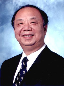 Huancong Zuo (左焕琮)合作论文数School of Medicine, Tsinghua University;Tsinghua University Yuquan Hospital15
 Chen Ye-Tao合作论文数10
 Haisheng Liu (刘海生)合作论文数清华大学第二附属医院7
 Qiang Ao (敖强)合作论文数College of Biomedical Engineering, Sichuan University;Institute of Regulatory Science for Medical Devices, Sichuan University4
 Qiuhang Zhang (张秋航)合作论文数,et al Department of Otorhinolaryngology Head-neck Skull Base Surgery,Xuanwu Hospital of Capital University of Medical Sciences, Beijing ,China4Zhao-Hui Sun合作论文数4Suhua Miao合作论文数3JiaPing Zheng (郑佳平)合作论文数3
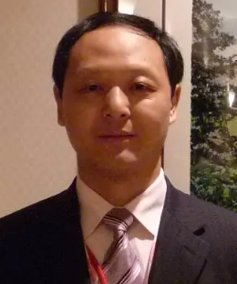 Hongyan Han (韩宏彦)合作论文数Aviation General Hospital, China Medical University;Chinese Academy of Sciences3
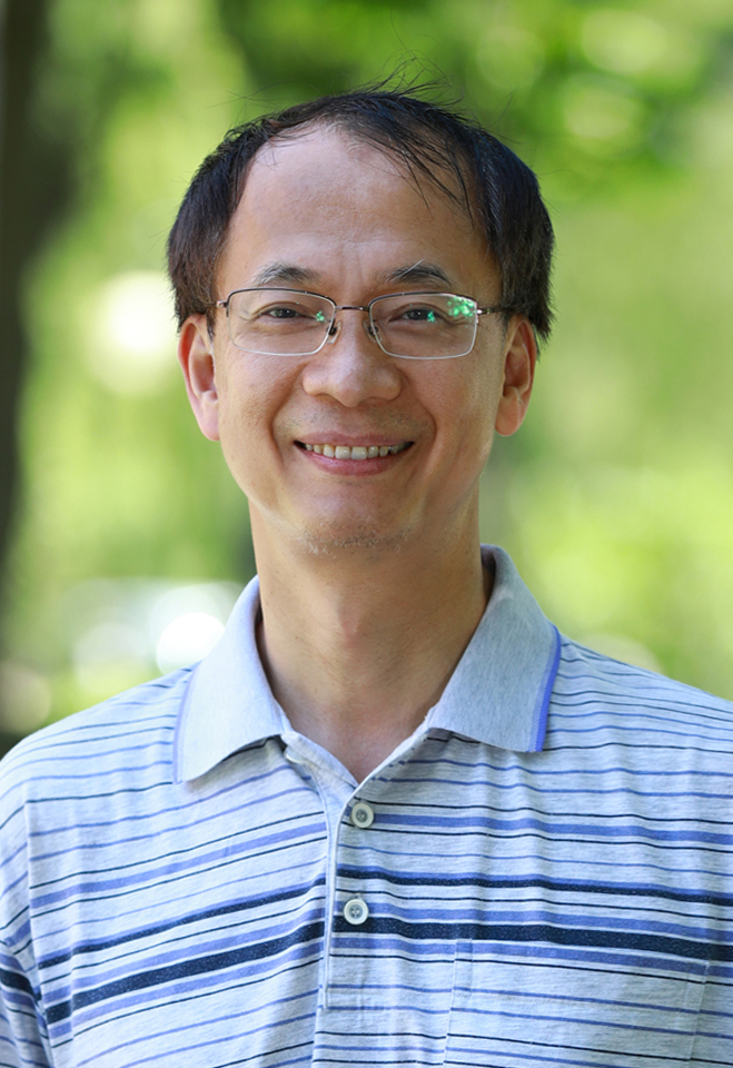 Guoqiang Chen (陈国强)合作论文数School of Life Sciences, Tsinghua University;Department of Chemical Engineering, Tsinghua University3Wang Yunpeng合作论文数3Yan LI (李妍)合作论文数2
 Bo Xiu (修波)合作论文数Beijing Chao-Yang Hospital2Yan Qi (齐岩)合作论文数2XiaoSong Wang (王晓松)合作论文数2YongGang Liu (刘勇刚)合作论文数2ZhiQiang Cui (崔志强)合作论文数2ZhiJun Zhang (张志军)合作论文数2Qiang Liu (刘强)合作论文数2Dong Wang合作论文数2
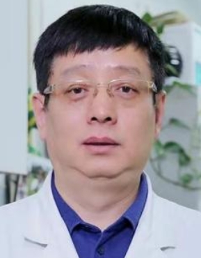 Yue Yuan (袁越)合作论文数China-Japan Friendship Hospital2Hui Liang (梁晖)合作论文数2luping li (李鲁平)合作论文数1Ying Chen (陈莹)合作论文数1LIU Qiang (刘强)合作论文数1Juan Fan合作论文数1Fucai XU (徐福财)合作论文数1YiBin Liu (刘义宾)合作论文数1JiZhi Zhang (张继志)合作论文数1YingBin Wang (王迎宾)合作论文数1YongFei Yang (杨永飞)合作论文数1JianShe Chen (陈建设)合作论文数1YunFu Ma (马云富)合作论文数1ZengLiang Liu (刘增良)合作论文数1Yan Wang (王岩)合作论文数1
 Ping Zhuang合作论文数1敖强合作论文数内蒙古民族大学1Zhijun Zhang (张志军)合作论文数1
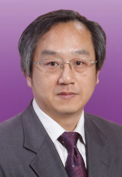 Yuqi Zhang (张玉琪)合作论文数Yuquan Hospital, Tsinghua University1
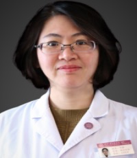 Yu Ma (马羽)合作论文数School of Clinical Medicine, Tsinghua University1
 Lin Xu (徐林)合作论文数Dongzhimen Hospital, Beijing University of Chinese Medicine1Lin Chen (陈琳)合作论文数1Tianqiao Yin (殷天樵)合作论文数1Zihang Xie合作论文数1
 Xiaohong Mu (穆晓红)合作论文数Dongzhimen Hospital, Beijing University of Chinese Medicine1




