Critical-sized bone defects, particularly segmental injuries such as radial defects, remain one of the most formidable challenges in orthopedic regeneration, as spontaneous healing is rarely achieved and existing grafts suffer from donor site morbidity, immune rejection, insufficient mechanical and biological performance. In this study, we present a biodegradable piezoelectric cryogel scaffold (PWH Gel) composed of a gelatin methacryloyl (GelMA) matrix embedded with piezoelectric whitlockite nanoparticles (PWH NP). Under physiological loading, PWH Gel generates localized electrical potentials and releases bioactive ions (Ca2+, Mg2+), creating a dynamic self-powered microenvironment that actively promotes bone regeneration. In vitro, the scaffold enhanced bone marrow mesenchymal stem cells (BMSCs) proliferation, migration, and osteogenic differentiation, facilitated endothelial tube formation, and triggered Piezo1-mediated Ca2+ influx and cytoskeletal remodeling. When implanted into a rat critical-size radial segmental defect, PWH Gel achieved complete bone bridging with markedly improved bone volume, trabecular organization, and vascularization. By coupling mechanical, electrical, and ionic stimulation within a single biodegradable system, this Mg2+-releasing piezoelectric cryogel establishes a new paradigm for functional reconstruction of segmental bone defects, representing a significant step toward clinically translatable electroactive biomaterials for large skeletal injuries.
Introduction: Three-dimensional (3D) printing technology has revolutionized the treatment of bone defects. However, medical education has lagged in integrating 3D printing into clinical training, limiting its widespread adoption. This study aimed to promote the dissemination of 3D printing technology through a novel, targeted teaching method. Methods: First, we implemented a bibliometrics aiming to identify core research forces and potential breakthrough directions. Then, from January 2022 to January 2025, a prospective cohort of 278 trainees (102 residents and 176 fellows) at a single center participated in a structured six-stage educational program encompassing: (1) foundational 3D printing knowledge, (2) video-assisted learning, (3) hands-on practice with 3D printed anatomical models, (4) virtual reality (VR)-based surgical simulation, (5) multidisciplinary treatment (MDT) discussions, and (6) supervised surgical operations. Teaching effectiveness was evaluated using pre- and posttraining theoretical exams, operational assessments, and comprehensive self-evaluation questionnaires. Results: Through the bibliometrics, 3D printing technology has been maintaining a high level of research hotspot, but the targeted education is still vacant. For the teaching study, both resident and fellowship doctors demonstrated statistically significant improvements in theoretical knowledge scores, rising from 14.3 to 32.7 in residents and from 19.1 to 33.3 in fellows (P < 0.001). Residents exhibited a greater magnitude of improvement compared to fellows. Operational metrics showed significant reductions in completion time and intraoperative blood loss (P < 0.001), alongside a marked increase in the proportion of "excellent" or "good" procedural quality ratings. Comprehensive ability assessment revealed significant gains across all evaluated domains (P < 0.001), except for self-study ability, which remained stable The total self-evaluation scores improved from 19.5 to 30.1 for residents and from 20.9 to 33.6 for fellows. Conclusion: This targeted, multimodal teaching method significantly enhances theoretical understanding, operative proficiency, and clinical competence in the use of 3D printing technology for bone defect repair.
BACKGROUND:This study aimed to identify the optimal method for detecting mild cognitive impairment (MCI) in elderly knee/hip osteoarthritis (OA) patients, a vulnerable group at high risk for postoperative delirium, by comparing neuropsychological tests, molecular biomarkers, and saccadic eye movements. METHODS:In this prospective cohort study, 316 patients (≥65 years) scheduled for knee/hip arthroplasty underwent preoperative assessments using neuropsychological tests (MMSE, MoCA, CDR), blood/CSF molecular analysis, and saccadic tasks. Participants were classified as MCI (n = 90) or non-MCI (n = 226) by geriatricians based on standard diagnostic criteria. RESULTS:No significant molecular differences were found between MCI and non-MCI patients. While MMSE, MoCA, and CDR scores were significantly lower in the MCI group, saccadic parameters showed superior diagnostic accuracy. A model based on saccades achieved an AUROC of 0.90, outperforming the MMSE model (AUROC = 0.85). CONCLUSIONS:Saccadic analysis outperformed standard cognitive screening and molecular tests in detecting MCI, indicating its potential as a superior, non-invasive preoperative assessment tool. TRIAL NUMBER:The study was registered with the Chinese Clinical Trial Registry (ChiCTR2200062483, August 8, 2022).
ABSTRACT Background Revision total knee arthroplasty (rTKA) poses substantial technical challenges. Although robotic assistance has demonstrated efficacy in primary TKA, its application in partial revision procedures remains poorly understood. This study presents a case of robotic‐assisted partial rTKA—specifically, isolated tibial component revision using the retained femoral component as a registration reference—and provides a comprehensive review of the current literature. Case Presentation A 66‐year‐old male presented with progressive knee pain 8 years after left TKA. Imaging revealed a well‐fixed femoral component but isolated aseptic loosening with posterior tilting of the tibial component. Given the stability of the femoral component, a robotic‐assisted partial revision targeting only the tibial component was performed. The robotic system was registered intraoperatively using the well‐fixed retained femoral component as the primary reference, enabling accurate spatial orientation despite the altered tibial anatomy. A critical prerequisite for this procedure was implant compatibility between the retained Triathlon femoral component and the planned Triathlon TS revision system. After removal of the loose tibial tray and debridement, robotic‐arm guidance was used for precise tibial osteotomy with a 10 mm augment. Postoperative imaging confirmed optimal component positioning. The patient achieved 0°–130° range of motion within 2 weeks, with favorable short‐term clinical and radiological outcomes at 1 year follow‐up. Conclusion This case demonstrates the technical feasibility of robotic‐assisted partial TKA revision, with favorable short‐term clinical and radiological outcomes at 1 year follow‐up. The technique enabled precise osteotomy, balanced gap restoration, and preservation of the well‐fixed femoral component. While these preliminary results are encouraging, larger studies with longer follow‐up are needed to establish comparative effectiveness and generalizability.
Surgical site swelling represents a prevalent complication following gluteal contracture release that adversely impacts functional recovery; while current literature predominantly focuses on rehabilitation outcomes, insufficient attention has been paid to swelling etiology. This review synthesizes causative evidence across three determinant categories: (1) preoperative factors—individual variations in coagulation profiles and symptom severity; (2) intraoperative variables—technical disparities in hemostasis, surgical trauma magnitude, and irrigation parameters; (3) postoperative divergences—inconsistent compression bandaging, exercise initiation timing, education quality, and compliance. Critical preventive strategies include implementing comprehensive preoperative education, standardizing surgical protocols, instituting graded rehabilitation, and establishing early detection-intervention frameworks, which collectively reduce swelling incidence and severity. These evidence-based approaches offer optimized clinical pathways while informing future research.
The clinical diagnosis of developmental dysplasia of the hip (DDH) typically involves manually measuring key radiological angles-Center-Edge (CE), Tönnis, and Sharp angles-from pelvic radiographs, a process that is time-consuming and susceptible to variability. This study aims to develop an automated system that integrates these measurements to enhance the accuracy and consistency of DDH diagnosis. We developed an end-to-end deep learning model for keypoint detection that accurately identifies eight anatomical keypoints from pelvic radiographs, enabling the automated calculation of CE, Tönnis, and Sharp angles. To support the diagnostic decision, we introduced a novel data-driven scoring system that combines the information from all three angles into a comprehensive and explainable diagnostic output. The system demonstrated superior consistency in angle measurements compared to a cohort of eight moderately experienced orthopedists. The intraclass correlation coefficients for the CE, Tönnis, and Sharp angles were 0.957 (95% CI: 0.952-0.962), 0.942 (95% CI: 0.937-0.947), and 0.966 (95% CI: 0.964-0.968), respectively. The system achieved a diagnostic F1 score of 0.863 (95% CI: 0.851-0.876), significantly outperforming the orthopedist group (0.777, 95% CI: 0.737-0.817, [Formula: see text]), as well as using clinical diagnostic criteria for each angle individually ([Formula: see text]). The proposed system provides reliable and consistent automated measurements of radiological angles and an explainable diagnostic output for DDH, outperforming moderately experienced clinicians.Clinical impact: This AI-powered solution reduces the variability and potential errors of manual measurements, offering clinicians a more consistent and interpretable tool for DDH diagnosis.
With the increasing incidence of joint diseases and the growing demand for surgical precision, robotic-assisted surgery has emerged as a promising advancement in orthopedic training. The Mako surgical robot, known for its precision in preoperative planning and intraoperative guidance, may offer significant educational benefits to joint surgery fellows. However, its potential impact on enhancing traditional surgical skills and research capabilities remains underexplored. To evaluate whether systematic training in Mako robotic-assisted hip replacement improves surgical performance and research skills among joint surgery fellows compared to traditional training. From October 2021 to May 2023, 40 joint surgery fellows were randomly divided into two groups, the experimental group received a comprehensive and systematic mako robot-assisted hip replacement training course. The control group received traditional training in hip replacement surgery within one month. The results of operation simulation and questionnaire were used to evaluate the training effect. The test results showed that in the simulation test of acetabular prosthesis placement, the overall pass rate of the experimental group was 90
Computer-assisted surgery (CAS) and patient-specific instrumentation (PSI) are digital techniques to improve the accuracy of implant positioning in total knee arthroplasty (TKA), but their effects on clinical outcomes are still in dispute. The objective of this trial is to evaluate the efficacy and safety of CAS and PSI compared to conventional instrumentation (CI) in TKA. A prospective randomized controlled trial was conducted. A total of 135 patients undergoing TKA were randomized into CAS group, PSI group and CI group with 45 patients in each group. Primary outcome is the coronal mechanical axis of lower extremity. Secondary outcomes include Femoral Rotation Angle (FRA) of the femoral prosthesis, operation time, perioperative blood loss, Western Ontario and McMaster Universities Osteoarthritis Index (WOMAC), Forgotten Joint Score (FJS) and complications. Outliers of Hip-Knee-Ankle angle (HKA) were 24.4
Anteromedial osteoarthritis (AMOA) is characterized by progressive and irreversible cartilage damage in anteromedial knee joint, and targeted therapies for it are lacking. To address this challenge, a magnetically controlled drug-loaded microrobot is developed based on Chlorella and incorporated quercetin, named M-CH@QUE, which is designed for precise drug delivery targeting AMOA. M-CH@QUE not only alleviated OA symptoms but also protected the cartilage and slowed the progression of AMOA. In vitro experiments demonstrated its excellent biocompatibility, drug-loading capacity, sustained-release properties, and precise control of flat surfaces and joint cartilage using a magnetic actuation system. In addition, the mechanisms are explored underlying the effects of quercetin. It is found that it promoted cell protection by enhancing insulin-like growth factor 1 receptor (IGF1R) phosphorylation and reducing intracellular oxidative stress. In vivo experiments revealed that M-CH@QUE accumulated in the medial knee joint of rats significantly slowed OA progression and effectively protected the cartilage. After 6 and 12 weeks of treatment, the M-CH@QUE significantly reduced both the Osteoarthritis Research Society International (OARIS) score and the Mankin score. In summary, the innovative M-CH@QUE design offers an effective strategy for alleviating AMOA symptoms and slowing the disease progression, thus presenting a highly promising option for treating AMOA.
Total knee arthroplasty (TKA) remains the gold-standard treatment for end-stage osteoarthritis, yet persistent challenges in prosthetic material performance limit its long-term clinical efficacy. CoCrMo alloy is commonly used material of femoral component in TKA due to its excellent mechanical durability. However, two critical limitations persist: (1) substantial elastic modulus mismatch inducing stress-shielding effects, and (2) bioinert surface impairing osseointegration. To address these dual challenges, we developed a synergistic surface engineering strategy combining 3D-printed porous architecture with Mg2+ functionalization via plasma immersion ion implantation (PIII). The porous structure significantly reduced the elastic modulus and achieve biomimetic mechanical compatibility. Mg2+-implanted scaffolds (CoCrMo-Mg) demonstrated multifunctional bioactivity through synergistic physicochemical interactions. Surface topography modification via 3D printing generated micro-scale features that enhanced osteoblast adhesion through mechanotransduction pathways, while the release of Mg2+ exerted immunomodulatory, pro-angiogenic and osteogenic effects. Mg2+-mediated downregulation of pro-inflammatory cytokines, established an anti-inflammatory microenvironment conducive to bone regeneration, while Mg2+ stimulation promoted substantial neovascularization - collectively creating an osteogenic niche favoring coupled angiogenesis-osteogenesis process. These findings were further validated in vivo, where the CoCrMo-Mg scaffolds showed improved anti-inflammation, neovascularization and bone ingrowth capacities, along with favorable biomechanical integration. Overall, this dual-modality approach combining structural optimization with bioactive ion engineering establishes a paradigm for developing mechanically compliant and biologically active orthopedic implants, with particular translational relevance for cementless TKA applications.
BACKGROUND:Postoperative delirium (POD) is a prevalent complication in elderly surgical patients. It is associated with long-term cognitive impairment and increased dementia risk. However, reliable tools to predict POD are currently lacking. METHODS:The study enrolled 316 arthroplasty patients (aged 65 yr or older) in this study. Preoperative assessments comprised neuropsychological tests ( i.e. , Mini-Mental State Examination [MMSE] and Montreal Cognitive Assessment [MoCA]), molecular biomarkers of serum/cerebrospinal fluid, and saccadic tasks. POD was diagnosed by expert persons based on the Confusion Assessment Method test. The effectiveness of abovementioned three types of assessments in predicting the occurrence of POD were compared. RESULTS:The incidence of POD was 8.2% (26 of 316). The MMSE and MoCA scales, serum neurofilament light chain levels, and five saccadic parameters values (reaction time, primary saccade error, saccadic gains in pro-saccades; peak velocity in anti-saccades and memory-guided saccades) differed significantly ( P < 0.05) between POD and non-POD participants. The logistic regression classifier model revealed higher predictive accuracy when using saccadic parameters (area under the receiver operating characteristic curve [AUROC], 0.81; 95% CI, 0.70 to 0.92) than when using MMSE and MoCA scores (AUROC, 0.64; 95% CI, 0.53 to 0.76) or neurofilament light chain levels (AUROC, 0.61; 95% CI, 0.50 to 0.72). The multilayer perceptron machine learning classifier model further increased the accuracy (AUROC, 0.89; 95% CI, 0.82 to 0.94) by using saccadic parameters to predict POD occurrence. CONCLUSIONS:Saccadic parameters exhibited higher accuracy in predicting the occurrence of POD than MMSE and MoCA scores and molecular test results. Therefore, saccadic parameters may serve as a complementary behavioral biomarker for predicting the occurrence of POD in elderly arthroplasty patients.
ABSTRACTObjectiveWith the global aging population, the incidence of OA is rising annually, and the number of TKA surgeries is rapidly increasing, placing a heavy economic and healthcare burden on society. As one of the key medications in the ERAS protocol, DXM can significantly reduce postoperative pain, suppress nausea and vomiting, and accelerate patient recovery. However, the safety of perioperative DXM use in patients with diabetes remains unclear. This study aims to clarify the safety of perioperative DXM application in diabetic patients.MethodsThis retrospective analysis involved 285 patients with type 2 diabetes and late‐stage knee osteoarthritis who underwent unilateral TKA at the Joint Surgery Center of Peking University Third Hospital from January 2019 to November 2022. After application of the inclusion and exclusion criteria, 161 patients were included in the study. The patients were divided into two groups according to whether they had received continuous intravenous administration of DXM for 3 days postoperatively: the DXM group (n = 66) and the non‐DXM group (n = 95). All other treatments and medications were the same in both groups. The patients' PBG, incidence of PONV, length of hospital stay, pain scores, and clinical data were collected and compared between the two groups.ResultsThere were no significant differences in the general preoperative data between the DXM and non‐DXM groups. The average PBG and the proportion of patients with levels exceeding 200 mg/dL were not significantly different between the two groups (10.84 mg/dL vs. 11.05 mg/dL and 43.2% vs. 43.9%). The postoperative visual analog scale scores (3.67 vs. 2.48) and the incidence of PONV were significantly lower in the DXM than non‐DXM group (40% vs. 16%). The preoperative level of glycated hemoglobin accurately predicted PBG. Furthermore, there were no statistically significant differences in the incidence of early postoperative complications between the groups.ConclusionsThe administration of DXM after unilateral TKA can effectively reduce postoperative pain and suppress the occurrence of PONV without affecting PBG in patients with type 2 diabetes. In addition, the preoperative level of glycated hemoglobin can accurately predict PBG.
OBJECTIVE:Postoperative nausea and vomiting (PONV) and abnormal inflammatory markers frequently complicate recovery following total knee arthroplasty (TKA), posing diagnostic challenges in distinguishing between transient inflammatory responses and early infections. This study aimed to evaluate the specific effects of dexamethasone (DXM) on PONV incidence and inflammatory markers after TKA. METHODS:This was a retrospective cohort study including 1853 patients who underwent unilateral TKA from January 2020 to August 2021. Patients were divided into a dexamethasone group (n = 756) and a control group (n = 1097) based on postoperative DXM use. All other perioperative management was consistent between groups. Data collected included PONV incidence, white blood cell count (WBC), C-reactive protein (CRP), and postoperative clinical outcomes. Statistical analysis included the χ2 test, t test, and multivariate logistic regression to control for confounding factors. RESULTS:The DXM group showed significantly lower incidences of PONV and lower postoperative CRP levels. WBC counts and neutrophil percentages were transiently elevated postoperatively in the DXM group without corresponding increases in postoperative complications. CONCLUSION:DXM administration after TKA reduces PONV and inflammatory markers but leads to transient leukocytosis, which is not associated with an increased incidence of complications.
BACKGROUND:Melorheostosis is a relatively rare disease, which may cause chronic pain, soft tissue mass and restricted range of motion. Diagnosis and treatment of melorheostosis remains challenging. CASE REPORT:We describe a patient with sclerotic bone involving the left acetabulum, femur, tibia, and talus. Over 19 years this patient showed slow progression of disease. The bone mass in the right popliteal region resulted in limitation of knee motion. Comprehensive management including physical therapy, medications and surgeries were adopted to obtain a full range of motion and no recurrence for 2 years. DISCUSSION:Accurate diagnosis and adequate treatments are critical to melorheostosis patients. Resection of redundant calcified mass is an effective method to treat severe limitation of range of motion associated with knee melorheostosis.
ObjectiveA robotic system was recently introduced to improve prosthetic alignment during total knee arthroplasty (TKA). The purpose of this multicenter, prospective, randomized controlled trial (RCT) was to determine whether robotic‐arm‐assisted TKA improves clinical and radiological outcomes when compared to conventional TKA.MethodsOne hundred and thirty patients who underwent primary TKA were enrolled in this prospective, randomized controlled trial, which was conducted at three hospitals. Five patients were lost to follow‐up 6 weeks after surgery. Therefore, 125 participants (63 in the intervention group and 62 in the control group) remained in the final analysis. The primary outcome was the rate at which the mechanical axis of the femur deviated by less than 3° from the mechanical axis of the tibia. This was evaluated by full‐length weight‐bearing X‐rays of the lower limb 6 weeks postoperatively. Secondary outcomes included operation times, 6‐week postoperative functional outcomes evaluated by the American Knee Society score (KSS) and the Western Ontario and McMaster Universities osteoarthritis index (WOMAC), short form‐36 (SF‐36) health survey results, and the occurrence of adverse events (AEs) and serious adverse events (SAEs).ResultsAt 6 weeks postoperatively, we found that the rate of radiographic inliers was significantly higher in the intervention group (78.7% vs 51.6%; p = 0.00; 95% confidence interval, 10.9% to 43.2%). The operation was significantly longer in the intervention group than in the control group (119.5 vs 85.0 min; p = 0.00). There were no significant differences in the 6‐week postoperative functional outcomes, SF‐36, AEs, and SAEs between the two groups. There were no AEs or SAEs that were determined to be “positively related” to the robotic system.ConclusionRobotic‐arm‐assisted TKA is safe and effective, as demonstrated in this trial.
OBJECTIVE:Total hip arthroplasty (THA) is currently one of the most effective treatment methods for end-stage hip joint disease, and its long-term effectiveness largely depends on the accurate placement of the acetabular prosthesis. In conventional surgery, the placement of the acetabular prosthesis mainly relies on the surgeon's clinical experience and surgical techniques. To further improve the accuracy of prosthesis placement, a new robotic system for THA is designed. The purpose of this study is to verify the effectiveness and safety of THA assisted by this robotic system. METHOD:A multicenter, prospective, randomized controlled, superiority study design was adopted with statistical methods of t test and Chi-squared test. Participants undergoing primary THA have been enrolled in three centers of joint surgery in China since July 17, 2023. Robotic THA was operated in the experimental group, and conventional instruments were used in the control group. The primary outcome is the proportion of anteversion and inclination angles in the safe zone. The secondary outcomes include operation time, WOMAC score, Harris score, SF-36 health questionnaire, dislocation rate of hip joint, and rates of adverse events and serious adverse events. RESULTS:A total of 138 patients were included in this study. The proportion of both anteversion and inclination angles in the safe zone was 92.2% in the experimental group and 50.8% in the control group, with significant difference (p < 0.01). The average operation time in the experimental group and control group was 116.4 and 80.5 min respectively, with significant difference (p < 0.01). There was no significant difference in WOMAC score, Harris score, and SF-36 between the two groups (6 ± 2) weeks after operation (p > 0.05). The dislocation rate of hip joint in the experimental group and control group were 3.0% and 1.5%, respectively, without significant difference (p > 0.05). The rate of adverse events and severe adverse events in the experimental group and control group also showed no significant difference (p > 0.05). No adverse events or serious adverse events were judged to be "definitely related" to the experimental instruments. CONCLUSION:Robotic THA could prolong the operation time within an acceptable range, but more precise acetabular prosthesis positioning could be obtained when compared with conventional surgery. Besides, no significant difference was found in function scores, dislocation rate or other adverse events, which indicates that this new robot system shows both good effectiveness and safety in THA. TRIAL REGISTRATION:Clinical Trials: NCT05947734.
BackgroundTourniquet is applied in Total Knee Arthroplasty (TKA) to reduce intraoperative blood loss and improve view of surgical field. However, tourniquet use in TKA is still in dispute. Some researchers find that tourniquet may lead to extra side effects such as aggravated pain and limb swelling, while others consider that tourniquet has no significant adverse impact on clinical outcomes of TKA patients. This trial is conducted to evaluate tourniquet effect on TKA patients.MethodsA prospective, single‐blind, randomized controlled trail is adopted with a sample size of 130 knees from August 2020 to February 2023. Patients undergoing TKA are randomly allocated to tourniquet group and non‐tourniquet group. Outcomes including quadriceps thickness and stiffness, operation time, total blood loss, intraoperative blood loss, postoperative blood loss, transfusion rate, thigh circumference, knee and thigh VAS, D‐Dimer and CRP level, knee function score, patient satisfaction, and complications are evaluated in this trial. Student's t‐test, Mann–Whitney U test, Pearson's chi‐square test, and Fisher's exact test are used in this study.ResultsNo significant difference in demographic information and baseline outcomes were found (p > 0.05). Participants in the tourniquet group had significantly less total blood loss and intraoperative blood loss, more postoperative blood loss, and higher D‐Dimer level on postoperative day 3 when compared with non‐tourniquet group (p < 0.05). Other outcomes including quadriceps thickness and stiffness, operation time, postoperative blood loss, transfusion rate, thigh circumference, knee and thigh VAS, D‐Dimer level on postoperative day 1, CRP level, knee function score, patient satisfaction, and complications showed no significant difference (p > 0.05).ConclusionTourniquet application can effectively reduce intraoperative blood loss and total blood loss, without significant side effects. Hence, we advocate the regular use of tourniquet in primary TKA.
Purpose:Various assistive techniques, such as conventional cutting instruments (CON), computer-assisted navigation systems (CAS), patient-specific instruments (PSI) and robot-assisted systems (RAS), have been developed and applied in primary total knee arthroplasty (TKA). In this study, we aimed to assess the relative accuracy and efficacy of several assistive techniques for TKA through a network meta-analysis (NMA) based on multiple published randomized controlled trials (RCTs). Methods:The PubMed, EMBASE and Cochrane databases were searched for RCTs to conduct this NMA from inception to 1 January 2024. We combined direct and indirect comparisons using a Bayesian NMA framework to assess and compare the effects of different assistive techniques on radiological and clinical outcomes. An NMA was conducted, and the study protocol was published online at PROSPERO (CRD42023402882). Results:One hundred and twelve RCTs involving 14,968 TKAs with four different assistive techniques (CON, CAS, PSI and RAS) were evaluated. Inconsistency and heterogeneity were acceptable for most outcomes. Based on the surface under the cumulative ranking curve, RAS could be the best technique for accurate mechanical axis alignment and component position, followed by CAS, PSI and CON. We observed no difference in clinical outcome scores. Additionally, CAS was the best intervention for visual analogue scale scores, and PSI had the shortest operative time. No significant differences were observed in postoperative complications, range of motion or total blood loss. Conclusion:RAS was most likely to achieve an accurate alignment, followed by CAS, PSI and CON. No differences were observed in clinical outcome scores and postoperative complications among the four assistive techniques. Level of Evidence:Level I (systematic review of Level-I randomized controlled studies).
Objective To identify prefrailty in patients undergoing total hip or knee replacement using preoperative ultrasound measurements of muscle dimensions and stiffness, with the goal of detecting high-risk prefrailty patients. Methods In this prospective cohort study, patients who underwent total hip/knee replacement were enrolled. Preoperative dimensions and stiffness of the biceps brachii (BB) were assessed using grey scale ultrasound (US) and shear wave elastography (SWE). Patients were preoperatively assessed for prefrailty based on FRAIL scale. Results In this study, a total of 121 consecutive patients [median age 71 years, (IQR 68–73 years), 94 women] were included. Sixty-five patients (53.7%) had prefrailty. The proportion of females is higher in the prefrail group compared to the non-frail group (86.2% vs. 67.9%, P = 0.023). The hemoglobin value in prefrail group was lower than that in non-frail group (130.2 ± 13.9 g/L vs. 136.2 ± 12.6 g/L, P = 0.031). US measurements revealed significant differences in BB thickness and area between prefrail and non-prefrail groups on both dominant and non-dominant sides. These differences were observed in both absolute values and relative values (normalized by BMI) (all p < 0.05). BB perimeter showed a significant difference between groups on the dominant side only (all p < 0.05). The AUC of BB thickness on dominant side (after normalization by dividing by BMI) was 0.664 (0.565–0.762), which was the largest among all US variables. Conclusion Preoperative US measurements of BB dimensions demonstrated its predictive value for prefrailty in patients undergoing total hip or knee replacement surgery. However, SWE was found to be insufficient in distinguishing between prefrail and non-frail patients.
Objective:To analyze the impact of robot assistance on the restoration of limb length and offset distance in total hip arthroplasty (THA). Methods:A retrospective analysis was conducted on the clinical data of 316 patients who underwent unilateral primary THA between September 2019 and August 2023. Among them, 117 patients underwent robot-assisted THA (group A), and 199 patients underwent conventional THA (group B). There was no significant difference between the two groups in the gender, age, or side of the hip replacement ( P>0.05); but there was a significant difference in the preoperative diagnosis ( P<0.05). The leg length discrepancy (LLD) and global offset (GO) dfference were measured on preoperative anteroposterior pelvic X-ray films, and absolute values were used for comparison between groups. Results:The operations in both groups were successfully completed. Postoperative imaging measurements showed that the LLD and GO dfference in group A were significantly lower than those in group B ( P<0.05). Among them, group A had 32 cases (27.4%), 5 cases (4.3%), and 0 case (0) of LLD>3 mm, >5 mm, and >10 mm, respectively, while group B had 115 cases (57.8%), 75 cases (37.7%), and 22 cases (11.1%), respectively; and the differences in above indicators between groups were significant ( P<0.05). Group A had 40 cases (34.2%), 3 cases (2.6%), and 0 case (0) of GO dfference>5 mm, >10 mm, and >20 mm, respectively; group B had 103 cases (51.8%), 54 cases (27.1%), and 7 cases (3.5%), respectively. There was no significant difference in the proportion of patients with GO>20 mm between groups ( P>0.05), while there were significant differences in other indicators between groups ( P<0.05). Conclusion:Compared with traditional THA, robot assisted THA has more advantages in restoration of limb length and offset distance.
 Hua Tian (田华)合作论文数Peking University Third Hospital39
 Xinguang Wang (王鑫光)合作论文数16
 Xiao Geng合作论文数12
 Minwei Zhao (赵旻暐)合作论文数Department of Orthopedics, Peking University Third Hospital12
 Feng Li (李锋)合作论文数Department of Orthopaedics, Peking University Third Hospital11
 Zijian Li (李子剑)合作论文数Orthopaedic Department, Peking University Third Hospital9
 Ke Zhang (张克)合作论文数Peking University International Hospital;Peking University Third Hospital8Ziyang Dong合作论文数6H Tian合作论文数6
 Tao Liyuan (陶立元)合作论文数北京大学第三医院6
 Cheng Wang (王成)合作论文数Department of Sports Medicine, Peking University Third Hospital6
 Hong Cai (蔡宏)合作论文数Department of Orthopedic, Peking University Third Hospital6X G Wang合作论文数4
 Xiangyang Guo (郭向阳)合作论文数Beijing Haidian Hospital4
 Yongzheng Han (韩永正)合作论文数Department of Anesthesiology, Peking University Third Hospital3Yi-Zhen HE (何宜蓁)合作论文数3
 Yanqing Liu (刘延青)合作论文数Peking University Third Hospital3
 Zhongjun Liu (刘忠军)合作论文数Peking University Third Hospital3DONG Ziyang (董子漾)合作论文数2WANG Cheng (王程)合作论文数2ge zhou (周歌)合作论文数2Qingqing Liu (刘青青)合作论文数2JianMing Chen (陈剑铭)合作论文数2
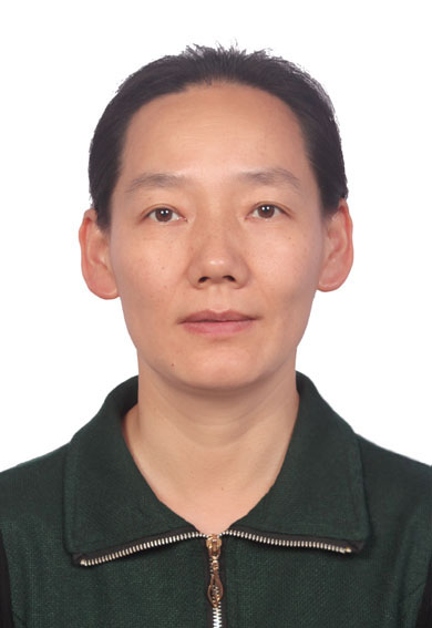 Qing Cai (蔡晴)合作论文数College of Materials Science and Engineering, Beijing University of Chemical Technology2Xinfeng Yan合作论文数2
 Zhiwei Xia (夏志伟)合作论文数Peking University Third Hospital2XU Chao-nan (徐超楠)合作论文数1Lianzhou Zhu合作论文数1Hongling Chu合作论文数1Hanjuan Cui合作论文数1Zhengkai Wang合作论文数1Qian Xiang合作论文数1KANG Meng (康猛)合作论文数1Chaodong Wang合作论文数1Ran Zhao合作论文数1LI Jian-guo合作论文数1
 Heng Xue (薛恒)合作论文数Department of Ultrasound, Peking University Third Hospital1T C Wu合作论文数1Meng Lu (卢猛)合作论文数1yuan gao (高源)合作论文数1Jiansuo Zhou合作论文数1
 Ji Tan合作论文数1Qirui Guo合作论文数1Yunke Liu合作论文数1
 Xiaoxiao Wang合作论文数1




