Background and purpose:In 2016 the National Cancer Institute(NCI)decided stopping to use NCI-60 cell lines for drug screening,suggesting that tumor cell lines were losing their value as a tool for drug discovery and basic research.The reason for NCI-60 cells'retirement'was that the preclinical studies based on traditional cellular and animal models did not obtain the corresponding expected efficacy in clinical trials.Since the major cancer behaviors,such as proliferation and metastasis,are fundamentally altered with long-term culture,the tumor cell lines are not representative of the characteristics of cancer in patients.Currently,scientists hope to create a new cancer model that are derived from fresh patient samples and tagged with details about their clinical past.Our purpose was to create patient-derived breast cancer primary cell lines as new cancer model for drug screening and basic research.Methods:Breast cancer tissues were collected in the Department of Breast Surgery,Affiliated Hospital of Guizhou Medical University.The collection of tumor tissue samples was approved by the Ethics Committee of the Affiliated Hospital of Guizhou Medical University(approval number:2022 ethics No.313),and the collection and use of tumor tissues complied with the Declaration of Helsinki.The primary breast cancer cell lines were isolated from the patient's breast cancer tissues and cultured in BCMI medium.After the cells proliferated,the media were replaced with DEME medium.Cell line STR genotyping was done to determine cell-specific genetic markers and identification.Clone formation assay and transplantation assay were done to analyze the ability of breast cancer primary cell lines to form tumors.Results:We created 6 primary breast cancer cell lines.The 6 primary breast cancer cell lines from the patients were tagged with the definitively clinicopathological features,clinical diagnosis,therapeutic regimens,clinical effectiveness and prognostic outcomes.The STR genotyping assays identified the genetic markers and determined the identities of the 6 primary breast cancer cell lines.Clone formation assays and transplantation assay showed that the proliferative capacities of the patient-derived primary breast cancer cell lines were significantly greater compared with the conventional breast cancer cell lines.Conclusion:We created a panel of 6 patient-derived primary breast cancer cell lines as new cancer model for drug screening and basic research in breast cancer.
Objective To explore the effect of aryl hydrocarbon receptor(AHR) on the immunomodulatory function of human adipose derived mesenchymal stem cells (MSCs). Methods RT-qPCR and immunofluorescence staining were used to detect the expression levels of indoleamine 2,3-dioxygenase 1 (IDO1), interleukin-4-inducible gene 1 (IL4I1) and AHR in MSCs before and after inflammatory cytokine stimulation; siCtrl, siIDO1, siIL4I1 and siAHR were transfected into MSCs and blocked the AHR signaling pathway; conditioned medium from MSCs was co-incubated with macrophages, RT-qPCR and ELISA were performed to detect the level of IL-6, IL-1β, TNF-α. Results MSCs without cytokine stimulation highly expressed AHR and hardly expressed IDO1 and IL4I1 proteins; IDO1, IL4I1 and AHR were up-regulated after different cytokine stimulation(P<0.05), the expression of IDO1 and IL4I1 was most significantly up-regulated after stimulation by IFN-γ(P<0.001), AHR entering the nucleus was increased; down-regulation of AHR expression could inhibit the immunomodulatory effect of MSCs(P<0.05). Conclusions AHR regulates the immune function of MSCs, downregulates expression of AHR could inhibit the immunomodulatory effects of MSCs.
Objective Modifying 3D tubular DNA origami nanostructures (DONs) onto the mesenchymal stem cells (MSCs) membrane surface to obtain DONs@MSCs. Utilizing the characteristics of MSCs homing lung tissue to achieve targeted delivery of DONs to the lungs of mice. Methods The computer-aided tools for DNA sequence were used to design and obtain obtain pre-designed DONs. The morphology of assembled DONs was characterized by atomic force microscopy(AFM), and the upload of functional groups was verified by agarose gel electrophoresis (AGE). Fluorescent labeled single-strand DNA(ssDNA) was modified on the surface of MSCs using biological orthometabolic sugar engineering. According to the principle of complementary base pairing, DONs were modified on the MSCs surface. Using confocal microscopy and flow cytometry to detect the ssDNA and DONs, and using in vivo imaging to observe the distribution of DONs in lung tissue. Results DONs had been successfully assembled. MSCs membrane surface was successfully modified with ssDNA, and DONs were successfully hybridized to obtain DONs@MSCs. Compared to DONs, the enrichment of DONs@MSCs in the lungs of mice was significantly enhanced. Conclusions DONs@MSCs can be used for DONs targeted delivery to lung tissue of mice. This method provides a new strategy for drug delivery of DONs in lung tissues.
在祭祀、狩猎等动物解剖的基础上,通过对人体的解剖,中医积累了大量有关人体结构的初步认识,包括五脏、六腑、奇恒之腑、气、血、津液、皮、筋、肉、骨、脉等.又通过司外揣内、取类比象的方法,从生理、病理的角度以及经络络属联系等,构建了以五脏系统为中心的人体结构模式.从以人为本的人本思想出发,结合天人相应、形神合一观,拓展了五脏系统的内涵和外延,建立了以五脏系统为核心的人本体系.
Islets have a high demand for oxygen and most of them will die of hypoxia injury before and after transplantation. Hypoxic damage is one of the key factors associated with islet graft dysfunction. Mesenchymal stem cells (MSCs) have multiple functions and can enhance the therapeutic effect of islet transplantation. In this study, islets were cultured together with or without MSCs derived from umbilical cord (hUC-MSCs) under normal and hypoxic conditions. The effect of hUC-MSCs on the survival and function of isolated islets was detected by immunofluorescence and ELISA. Hypoxia-inducible factor 1 alpha (HIF-1α) and PFKFB3 mRNA and protein expression in different conditions were tested by real-time quantitative reverse transcription polymerase chain reaction (qRT-PCR) and Western Blot. The islets co-cultured with hUC-MSCs have improved viability and function compared with islets cultured alone. The mRNA transcription of HIF-1α in the co-cultured group increased. The protein expression of PFKFB3 increased with the increase of HIF-1α. This study found that hUC-MSCs could protect islets from dysfunction caused by hypoxia, and HIF-1α/PFKFB3 played an important role in hypoxic resistance, suggesting a potential strategy to improve the outcome of islet transplantation.
Applications of stem cells have been playing significant roles in scientific and clinical settings in the last few decades. The foundation of these approaches is successful cryopreservation of stem cells for future use. However, so far we can only cryopreserve stem cell suspension of small volumes in the order of 1 mL mostly due to the lack of an effective rewarming technique. Rapid and uniform rewarming has been approved to be beneficial, and sometimes, indispensable for the survival of cryopreserved stem cells, inhibiting ice recrystallization or devitrification. Unfortunately, the conventional water bath thawing method failed in providing the rapid and uniform rewarming. The conversion of electromagnetic (EM) energy into heat provides a possible solution to this problem. This chapter will focus on (1) analysis of the combined EM and heat transfer phenomenon in the rewarming of a biospecimen, (2) numerical investigation of the rewarming system, (3) practical setup of an EM resonance system, and (4) test of heating performance with large volume of cells.
目的 基于自噬相关基因(ATGs)构建肝细胞癌病人预后风险模型.方法 TCGA数据库下载374例肝细胞癌及50例正常肝组织的转录组数据和临床信息,首先筛选出差异表达基因(DEGs),然后从中筛选出差异表达的ATGs(DEATGs),最终利用单因素Cox回归分析、LASSO回归分析以及多因素Cox回归分析构建预后风险模型,并应用ROC曲线评估模型对于肝细胞癌病人预后的预测能力.结果 肝细胞癌组织与正常肝组织筛选得到12471个DEGs,其中包含62个DEATGs.单因素Cox回归分析得到40个预后相关DEATGs,LASSO回归分析以及多因素Cox回归分析得到7个风险自噬基因(RAB7A、FOS、ATG9A、HSPA8、PRKN、TUSC1和GAPDH)用于构建预后风险模型.Kaplan-Meier分析显示,高风险组病人的总体生存期显著低于低风险组(χ2=36.972,P<0.05).多因素Cox回归分析表明,预后风险模型可以作为一个独立预后因素(HR=2.574,P<0.01).结论 基于ATGs构建的预后风险模型是一个独立的预后因素,可有效预测肝细胞癌病人的预后.
目的 探讨使用不同方法分离提取的脂肪间充质干细胞(ADMSCs)是否存在细胞生物学活性的差异.方法 分别使用直接消化法和干细胞基质胶消化法分离提取ADMSCs,进行细胞表型鉴定、增殖活性检测,应用Real-time PCR方法检测诱导分化后成脂标志基因过氧化物酶体激活受体γ(PPAR-γ)和成骨标志基因runt转录因子2(RUNX-2)mRNA的表达,Western blot方法检测PPAR-γ和RUNX-2蛋白表达.结果 基质胶消化法得到的ADMSCs相较于直接消化法得到的ADMSCs在培养第3天的增殖活力更高(F=727.139,P<0.05).与直接消化法比较,成脂诱导分化6~12 d后基质胶消化法得到ADMSCs的PPAR-γmRNA表达增高(F=40.260~157.532,P<0.05),PPAR-γ蛋白表达在诱导分化6 d后增高(F=1501.475,P<0.05);成骨诱导分化3~12 d基质胶消化法得到ADMSCs的RUNX-2 mRNA表达较直接消化法增高,诱导分化3 d蛋白表达增高(F=58.018~2402.049,P<0.05).结论 使用基质胶消化法得到的ADMSCs成脂、成骨分化潜力优于直接消化法.
Objective To investigate the effects of exosome secreted by macrophage J774A.1 on the proliferation and migration of mouse colorectal cancer cell lines MC38 and CT26 and potential mechanism. Methods The phenotype of J774A.1 cells was identified by flow cytometry.The morphological characteristics, size and surface markers of the exosome of J774A.1 cells were detected by transmission electronic microscopy, nanoparticle tracking analysis(NTA) and Western blot.The uptake of exosome by MC38 and CT26 cells was observed by fluorescence microscopy. Proliferation of MC38 and CT26 was detected by CCK8 assay. Migration ability of MC38 and CT26 was detected by wound healing assay. The expression of phosphorylated proteins p-MEK1/2 and p-ERK1/2 in proliferation-related pathways was detected by Western blot. Results J774A.1 cells are M2 macrophages expressing the cell surface marker molecules CD68 and CD206. J774A.1 exosomes can be taken up by colorectal cancer cells MC38 and CT26, and can promote their proliferation(P<0.05), enhance their migration and activate MEK-ERK signaling pathway. Conclusions Macrophage J774A.1 exosomes can promote the proliferation and migration of colorectal cancer cell lines MC38 and CT26, which is potentially attributed to the increased phosphorylation of MEK-ERK signaling pathway.
Abstract. The coronavirus disease 2019 (COVID-19) can be caused by severe acute respiratory syndrome coronavirus 2 (SARS-CoV-2) infection, and has led to millions of deaths among more than 100 million infected people around the world according to the declaration from World Health Organization. Dysregulated immune response of both the innate and adaptive immune systems is subsistent on COVID-19 patients, of which the degree are associated with disease severity, lung damage and long term functional disability. Current treatment options have included antiretroviral drugs, anti-inflammatory factors, antibodies, immune checkpoint inhibitors, and convalescent plasma therapy. More recently, mesenchymal stem cell (MSC) therapy has been explored for the management and control of COVID-19, particularly with the aim of preventing or at least mitigating respiratory co-morbidities. Though the safety and efficacy of stem cell therapy have been validated in multiple phase I–III clinical trials, to date, no standardized stem cell preparation, administration dosage or interval, product QA/QC testing, storage, transportation, or disposal protocols have been established. The present paper proposes a systematic methodology that addresses all the foregoing process steps and evaluation criteria for the efficacious and safe administration of MSCs in the treatment of patients infected with COVID-19.
目的 探讨Poly(I:C)活化人脂肪间充质干细胞(hAMSCs)Toll样受体3(TLR3)后对hAMSCs成骨分化的影响.方法 胶原酶消化法分离提取hAMSCs,对其细胞形态、免疫学表型和成骨分化能力进行鉴定.将原代分离培养的hAMSCs随机分为对照组和TLR3组,对照组不做处理,TLR3组用含有20 mg/L Poly(I:C)的正常培养基处理6 h活化TLR3.CCK8法检测两组hAMSCs增殖能力变化,Real-time PCR方法检测hAMSCs成骨标志基因runt相关转录因子2(RUNX2)和骨钙素(OCN)mRNA表达,Western Blot方法检测RUNX2和OCN蛋白及NF-κB通路相关蛋白p-P65、P65表达.对照组和TLR3组均用间充质干细胞成骨诱导培养基诱导分化,12 d后用茜素红染色法检测两组hAMSCs成骨诱导后钙质沉积情况.结果 对照组和TLR3组hAMSCs的增殖能力差异无显著性(P>0.05).TLR3组hAMSCs成骨标志基因RUNX2及OCN的mRNA和蛋白表达均高于对照组(t=2.98~36.36,P<0.05),NF-κB通路相关蛋白P65及p-P65表达也高于对照组(t=3.52、13.85,P<0.05).茜素红染色结果显示,成骨诱导12 d后TLR3组钙质沉积程度高于对照组.结论 TLR3活化能够通过激活NF-κB通路促进hAMSCs成骨分化.
The stem cell products has been transforming from research into drug therapy, which will benefit from traditional pharmaceutical ideas. Taking mesenchymal stem cells(MSCs) products as an example, this paper discusses the problems restricting the development of stem cell products from the perspectives of various pharmaceutical disciplines, such as medicinal chemistry, pharmaceutics, pharmaceutical analysis and pharmacokinetics, so as to provide a new strategy and pathway for the development of stem cell products in the future.
Objective To characterize the biological features of CD146+ cells from umbilical cord mesenchymal stem cells(UCMSCs), adipose-derived mesenchymal stem cells(AMSCs) and bone marrow mesenchymal stem cells(BM-MSCs). Methods MSCs from three tissues were sorted by magnetically activated cell sorting(MACS) to obtain highly purified CD146+ subpopulations. The phenotypes and ultra-structures of these three MSC were analyzed by flow-cytometry and transmission electron microscopy. The differentiation abilities were tested by adipogenic and osteogenic inductions. The expression of genes relative to adipogenesis, osteogenesis, stemness or angiogenesis were determined by RT-qPCR. Results CD146+ cells derived from three kinds of tissues had similar ultra-structures and the ability of angiogenesis. CD146+ AMSCs expressed highest levels of the OCT-4, SOX2 and NANOG genes.Under the same induction conditions, CD146+ AMSCs showed higher potential for adipogenesis and CD146+BM-MSCs showed higher potential for osteogenesis.However, CD146+ UMSCs had little ability for adipogenesis and osteogenesis. All of CD146+ MSCs from 3 tissues expressed BFGF, VEGF, Ang-1 and EGF although CD146+ BM-MSCs had stronger ability of angiogenesis in vitro. Conclusions CD146+ MSCs derived from the 3 sources exhibit different biological characteristics.These results provide a useful biological basis for further studies relative to application of MSCs.
背景:间充质干细胞因其众多优点被应用于多种疾病治疗,线粒体转移是间充质干细胞疗法的重要机制之一.目的:综述间充质干细胞通过线粒体转移在各类组织损伤中发挥的作用.方法:由第一作者以"stem cels,mitochondrial transfer,cel communication,rescue and repair,tissue injury"为英文关键词,以"干细胞,线粒体转移,细胞通讯,修复,组织损伤"为中文关键词,检索2008至2019年期间收录在PubMed、中国知网、万方数据库中的文献,纳入间充质干细胞的线粒体转移作用和机制及治疗组织损伤相关的文献,排除重复与相关性弱的文献,对64篇文献进行总结分析.结果与结论:间充质干细胞被应用于如脑卒中、心肌梗死、急性肾功能衰竭等多种疾病治疗,线粒体转移作为干细胞治疗的一种新机制被人们广泛关注,已被认为是组织损伤的潜在疗法;线粒体转移已发现由隧道管形成、间隙连接、微囊泡、细胞融合和分离等不同模式介导,但线粒体转移在组织损伤中的具体机制还未完全阐明,其在组织损伤修复中的具体作用也有待进一步了解.
目的 研究射干苷(tectoridin)、桃叶珊瑚苷(aucubin)对脂多糖(LPS)诱导后人脂肪间充质干细胞(hAMSCs)免疫调控作用的影响.方法 通过CCK-8细胞增殖实验和细胞周期检测实验,筛选合适的药物作用浓度;筛选LPS作用浓度,建立炎性反应模型;用ELISA、RT-qPCR检测两者对LPS诱导后hAMSCs分泌促炎因子、抑炎因子的影响.结果 50μg/mL射干苷和100μg/mL桃叶珊瑚苷能明显促进hAMSCs增殖(P<0.05,P<0.01);两者均促进LPS诱导后hAMSCs增殖(P<0.001);并且可抑制hAMSCs分泌IL-6、TNF-α、IFN-γ等促炎因子(P<0.05,P<0.01).结论 射干苷、桃叶珊瑚苷可显著抑制LPS诱导的hAMSCs炎性反应.
目的 探讨activin A、WNT通路激活剂、BMP4以及bFGF信号对人脂肪间充质干细胞(hADSCs)向限定性内胚层(DE)分化的影响.方法 基于胚胎干细胞(ESCs)或多潜能干细胞(iPS)向DE分化的诱导体系,建立并优化hADSCs向DE细胞分化的诱导体系.在hADSCs向DE诱导体系中,分别比较不同浓度的activin A、Chir99021替代Wnt3a、添加/去除bFGF以及BMP4等对分化效率的影响.用RT-qPCR检测诱导前后限定性内胚层标志基因FOXA2和SOX17的表达;Western blot检测FOXA2和SOX17蛋白水平;用免疫荧光检测FOXA2和SOX17的表达,鉴定DE分化.结果 与ESCs或iPS向DE分化的诱导体系相比,低浓度的activin A,GSK3抑制剂Chir99021替代Wnt3a,去除BMP信号和bFGF信号的诱导方案可以明显促进hADSCs向DE分化的影响(P<0.01).结论 hADSCs向DE分化与hESC/hiPS相比,需要激活不同信号通路,因此最适诱导体系不同.
目的探讨miR-181b在正常对照和青少年特发性侧弯患者(AIS)骨髓间充质干细胞(BM-MSCs)之间的表达差异,以及其对BM-MSCs成骨分化能力的影响。方法 RT-qPCR检测对照和AIS患者MSCs中miR-181b的表达;将miR-181b模拟物、抑制剂以及相应对照瞬时转染BM-MSCs,随后更换为成骨诱导培养基,比较不同处理组BM-MSCs成骨能力的差异; RT-qPCR和Western blot检测成骨标志基因以及特异性转录因子的表达;碱性磷酸酶(ALP)染色法以及ALP活性分析法来鉴定成骨早期分化;茜素红染色检测细胞外基质的形成情况;慢病毒在BM-MSCs中过表达miR-181b的模拟物和抑制剂后,建立裸鼠异位骨生成模型,通过组织染色检测类骨质的形成。Western blot检测miR-181b水平的改变对ERK1/2磷酸化的影响。结果与正常对照比,AIS患者BM-MSCs中miR-181b表达显著升高(P<0.01)。过表达miR-181b明显抑制间充质干细胞体外向成骨分化,降低体内异位骨生成(P<0.05);阻断内源性miR-181b可以促进BM-MSCs体外向成骨分化,增强体内类骨质的形成(P<0.05)。其作用机制是,过表达miR-181b抑制ERK1/2的磷酸化,而抑制miR-181b促进ERK1/2的磷酸化(P<0.05)。结论 miR-181b抑制间充质干细胞成骨分化,可能与AIS患者骨密度降低有关。
剖宫产疤痕息室是因剖宫产手术后,创口愈合不良,子宫基层形成一通向宫腔的疤痕缺口,给患者带来痛苦.目前没有好的治疗方法.温灸法有活血通经,温阳补虚的效果,自古为治疗妇科疾病的良法[1];而亚全能干细胞,大量国内外的临床试验研究表明,具有良好的促进体内组织损伤修复治疗效果,且具有很高的安全性[2].另外,创口的不愈合与炎性反应相关,二者都具有调节免疫的作用,因此尝试应用温灸结合干细胞的方法治疗此疾病.
随着科技的进步,尤其是细胞生物学与医疗活体检测的发展,人们对人体有了更多的认识与了解,近代医学的一些科学发现与远古医学逐渐契合.本文从间充质干细胞及间充质组织系统概念阐述三焦器官的发育来源和功能及其实质,提出了三焦器官是联系人体各组织脏器、具有特殊组织结构的最大组织系统,分布于人体组织之间、器官系统之间,连接并濡养全部组织和细胞的重要场所,将人体组织器官有机合一,发挥着干细胞储备、水液代谢、养分运输、免疫调节、激素运输、信号传递的通道等功能.赵春华教授团队分选并鉴定了构成三焦结构主要功能细胞-间充质干细胞/周细胞群体,并从活体脑组织中分离到具有可收缩功能的三焦组织细胞.三焦器官联系脏腑及全身各部,通过运行气血、感应传导、相互协调,完成人体经络功能的调控.三焦器官/间充质组织系统主要负责干细胞多组织增殖分化、组织器官再生修复和功能重建,参与人体免疫监督、应答及复杂免疫网络调控作用等,发挥着神经、组织间激素、内分泌调节及组织代谢的系统性调节功能.
Objective To investigate the effect of adipose mesenchymal stem cells(AMSCs) on the peripheral blood lymphocyte(PBL) in psoriasis vulgaris(PV) patients and the expression and secretion profiles of related inflammatory cytokines in the PBL.Methods AMSCs from three PV patients were co-cultured with PBL. Peripheral blood regulatory cells(Treg) and T helper cell 17(Th17)ratio was measured by flow cytometry. The anti- and pro-inflammatory cytokines expressed and secreted by PBL were detected by quantitative real-time polymerase chain reaction(qRT-PCR) and enzyme-linked immunosorbent assay(ELISA).Results The Treg/total lymphocyte ratio was significantly higher in the healthy people AMSCs+PBL co-culture group[(3.2±0.5)%;P=0.001],but AMSCs in patients had a tendency to promote the proliferation of Treg cells [(1.3±0.2)%],with no significant difference(P=0.485) when compared with the PBL culture alone group[(1.0±0.1)%]. qRT-PCR showed that the ability of PBL in expressing Treg transcription factor forkhead box p3 and transforming growth factor(TGF)-Β mRNA was significantly lower in psoriasis AMSCs+PBL co-culture group than in the healthy people AMSCs+PBL co-culture group(P=0.00,P=0.03),AMSCs had a tendency to promote the expression of interlukin(IL)-10 in peripheral blood lymphocytes,but there was no significant difference(P=0.09).ELISA showed the PBL in healthy people AMSCs+PBL co-culture group secreted the anti-inflammatory cytokine IL-10[(156.9±41.8) ng/Μl] and TGF-Β[(2774.1 ± 526.4) ng/Μl];in contrast,the abilities of PBL in PV patient AMSCs+PBL co-culture group in secreting the anti-inflammatory cytokines has a downward trend:IL-10[(90.4±28.8) ng/Μl] and TGF-Β[(1597.9±55.7) ng/Μl],although the differences were not statistically significant. After the co-culture,the proportion of Th17 cells in the psoriasis AMSCs+PBL co-culture group[(0.8±0.3)%] showed a decreasing trend when compared with the PBL culture alone group[(1.1±0.1)%],although the results were not statistically significant. Also,the proportion of Th17 cells showed no significant difference between PV patient AMSCs+PBL co-culture group and healthy people AMSCs+PBL co-culture group. Finally,both the psoriasis AMSCs+PBL co-culture group and the healthy people AMSCs+PBL co-culture group showed no obvious inhibitory effect on the expression and secretion of Th17 transcription factor retinoid-related orphan nuclear receptor Γt and pro-inflammatory cytokines IL-17 and IL-23 in PBL,and there was no significant difference between these two groups.Conclusions AMSCs in PV patients have decreased ability in regulating the anti-inflammatory function of peripheral blood Treg lymphocytes. However,they have no effect on the proinflammatory effect of peripheral blood Th17 lymphocytes.
 Lianming Liao (廖联明)合作论文数福建中医药大学中西医结合研究院20
 Limei Yu (余丽梅)合作论文数遵义医学院附属医院19
 Daixiong Chen (陈代雄)合作论文数Hunan Nonferrous Metals Research Institute Co., Ltd.17
 Mingxia Shi (史明霞)合作论文数昆明医科大学17
 Ning Fang (方宁)合作论文数遵义医学院附属医院16Zhao Sun合作论文数16
 Jing Li (李静)合作论文数Cancer Hospital, Chinese Academy of Medical Sciences15XingXia Liu (刘星霞)合作论文数13
 Xishan Zhu (朱希山)合作论文数Peking University First Hospital13
 Qin Han (韩钦)合作论文数Peking Union Medical College13
 Baijun Fang (房佰俊)合作论文数Henan Cancer Hospital12
 Hua Zhu (朱华)合作论文数Institute of Laboratory Animal Sciences, Peking Union Medical College11
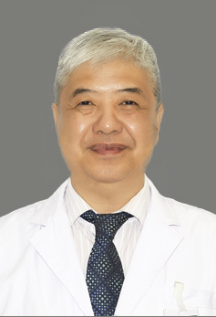 Renzhi Wang (王任直)合作论文数Peking Union Medical College Hospital;School of Medicine, The Chinese University of Hong Kong, Shenzhen10
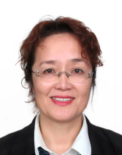 Chuan Qin (秦川)合作论文数Institute of Laboratory Animal Sciences, Chinese Academy of Medical Sciences & Peking Union Medical College9ShengGuo You (尤胜国)合作论文数8GuanJie Ma (马冠杰)合作论文数8KangHua Li (李康华)合作论文数8Xiaowei Dou合作论文数Laboratorium für Biochemie, Universität Bayreuth Universität8Bingzong Li合作论文数Soochow Univ, Affiliated Hosp 2, Suzhou, Peoples R China7Bin Zhang (张斌)合作论文数Institute of Molecular and Cell Biology, Agency for Science, Technology and Research7XueYing Jiang (姜学英)合作论文数7Tao Zhang (章涛)合作论文数7YuYing Wang (王钰莹)合作论文数7
 Yuhao Liu (刘煜昊)合作论文数Henan Provincial People's Hospital7
 Junji Wei (魏俊吉)合作论文数Peking Union Medical College Hospital7Lu Shan合作论文数7
 Changhong Li合作论文数7
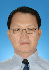 Yaojiong Wu (吴耀炯)合作论文数Shenzhen International Graduate School, Tsinghua University7HU Jian-Li合作论文数6Rongjia Zhu合作论文数6hong guo (郭虹)合作论文数6WeiMin Deng (邓为民)合作论文数6
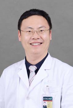 Ming Feng (冯铭)合作论文数Peking Union Medical College Hospital6Chunjing Bian合作论文数Chinese Academy of Medical Sciences, Peking Union Medical College6Chunzheng Yang合作论文数6
 Yaohong Tan合作论文数6Chunmei Ma合作论文数5Wei Ge合作论文数5LiHui Liu (刘丽辉)合作论文数5
 Huisheng Ai (艾辉胜)合作论文数Chinese PLA General Hospital5
 Lugui Qiu (邱录贵)合作论文数Institute of Hematology and Blood Diseases Hospital, Chinese Academy of Medical Sciences5
 Shihua Wang (王世华)合作论文数中国人民解放军海军总医院5
 Xi Yan (闫曦)合作论文数5
 Bin Chen合作论文数Chinese Academy of Medical Sciences, Peking Union Medical College5Qiyun Sun (孙琪云)合作论文数5Guo Ling合作论文数5Yanfeng Xu合作论文数5Hongye DENG (邓鸿业)合作论文数4Jie-wen Liu合作论文数4




