In pancreatic cancer, increased collagen I impairs the efficacy of gemcitabine; however, the role of gemcitabine itself in collagen I accumulation remains unclear. This study aims to explore the mechanism of gemcitabine-induced fibrosis and provide new insights to enhance its therapeutic efficacy. We analyzed COL1A1 expression in pancreatic cancer patient tumor tissues and found that gemcitabine treatment upregulated COL1A1 expression. Subsequently, cancer-associated fibroblasts (CAFs) were modeled by inducing human adipose-derived mesenchymal stem cells with tumor-derived exosomes. Using the autophagy inhibitor chloroquine (CQ) and the protein kinase B (AKT) activator SC79, we demonstrated that gemcitabine downregulated P62 expression and upregulated LC3BII, Beclin-1 expression, inducing autophagy in CAFs via decreasing AKT phosphorylation, which further led to collagen I accumulation. In addition, gemcitabine combined with CQ enhanced cell death in both CAFs and tumor cells, while inhibiting tumor cell proliferation and migration. In animal models, this combination therapy reduced gemcitabine-induced autophagy and collagen I deposition, contributing to delayed tumor growth. Collectively, gemcitabine upregulates collagen I by inducing CAF autophagy via reducing AKT phosphorylation. Targeting CAF autophagy can reduce collagen deposition, offering a promising strategy to improve the therapeutic efficacy of gemcitabine in pancreatic cancer.
Diabetic nephropathy (DN) is a leading cause of end-stage renal disease with limited therapeutic options. Ferroptosis contributes to renal tubular injury in DN. This study investigates whether mesenchymal stem cells (MSCs) ameliorate DN by inhibiting ferroptosis and elucidates the underlying mechanism. In a rat model of type 2 DN, MSCs transplantation improved renal function and histopathology, while reducing mitochondrial dysfunction, iron overload, and ROS-driven ferroptosis. In vitro, MSCs reversed high glucose-induced ferroptosis hallmarks in tubular epithelial cells. Mechanistically, RNA sequencing identified the MAPK/ERK pathway as key. MSCs suppressed the p-ERK/ERK-GPX4/ACSL4 axis, preventing glutathione depletion and lipid peroxidation. Activation of ERK abolished MSCs' protection, whereas ERK inhibition mimicked it. These findings reveal that targeting ERK-mediated ferroptosis in renal tubules offers a novel therapeutic strategy, with MSCs acting through this specific mechanism.
Traumatic brain injury (TBI) is a leading cause of disability in adults, significantly affecting patients’ quality of life. Extracellular vesicles (EVs) derived from human adipose-derived mesenchymal stem cells (hADSCs) have demonstrated therapeutic potential in TBI treatment. However, their limited targeting ability, short half-life, and low bioavailability present significant challenges for clinical application. In this study, we engineered extracellular vesicles (EEVs) by transfecting hADSCs with lentivirus and incorporating ultra-small paramagnetic nanoparticles (USPNs), resulting in EVs with enhanced miRNA expression and targeted delivery capabilities. These EEVs were administered intranasally to specifically target injury sites, effectively modulating the NF-κB signaling pathway to suppress neuroinflammation. In both in vitro and in vivo assessments, EEVs exhibited superior efficacy in promoting neurofunctional recovery and neurogenesis after brain injury compared to unmodified EVs. Furthermore, validation using human brain organoid models confirmed EEVs’ remarkable ability to suppress neuroinflammation, offering a promising strategy for TBI treatment.
BACKGROUND This study analyzed the dental follicle and alveolar bone of two patients with tooth eruption disorders, aiming to provide some reference for exploring the etiology and selecting treatment plans of this disease from the perspective of the influence of extracellular matrix on osteoclasts differentiation in dental follicle. CASE SUMMARY Collect dental follicle and alveolar bone tissue from one patient with single tooth eruption disorder and one patient with full permanent tooth eruption disorder, respectively. Simultaneously collect the dental follicle and alveolar bone tissue of obstructed teeth that need to be extracted due to orthodontic treatment as the control group. Hematoxylin and eosin (HE) staining was used to observe the morphology of dental follicle cells. Immunohistochemical staining was used to observe the expression of periostin, receptor activator of nuclear factor kappa B ligand (RANKL), and osteoprotegerin (OPG) protein in dental follicle and alveolar bone tissue. And observe the eruption of teeth after removing resistance from the crown of the permanent tooth germ. CONCLUSION HE staining of two cases of dental follicle tissues showed that the volume of dental follicle cells decreased, the nuclei were condensed, and there seemed to be cellular fibrosis. The immunohistochemical staining showed that both the dental follicle and alveolar bone tissue exhibited increased expression of periostin, decreased expression of RANKL and OPG proteins, and decreased RANKL/OPG ratio. After removing resistance, the permanent tooth germ often appears to have normal eruption. Tooth eruption disorders may be accompanied by abnormal remodeling of periostin, which affects the differentiation function of osteoclasts in the dental follicle and leads to metabolic imbalance of alveolar bone, resulting in tooth eruption disorders. Whether it is a single or full permanent tooth eruption disorder, once the coronal resistance is removed, the teeth can often erupt normally.
Background Cancer-associated fibroblasts (CAFs) are major non-tumor cellular components of the tumor microenvironment (TME) and are closely related to immune suppression. The enhancement of anti-programmed cell death protein-1 (PD-1) efficacy by antiangiogenic tyrosine kinase receptor inhibitors (TKIs) is partly due to the elimination of CAFs, which improves the immunosuppressive microenvironment. However, it remains unclear whether antiangiogenic TKIs regulate the autophagy of CAFs, thereby affecting the immunotherapy response.Methods We first examined the effects of autophagy inhibition and anlotinib on the TME and immunotherapy response via animal experiments. Then, CAF models were established in vitro. The effects and mechanisms of autophagy inhibition on the efficacy of anlotinib and CAFs were further explored in vitro. To specifically validate the role of CAF autophagy, we additionally constructed LLC tumor-bearing mouse models co-implanted with CAFs transfected with ATG5-targeting siRNA or non-targeting control siRNA.Results In anti-PD-1-resistant (LLC) and CAF-rich (LA795+MSC) non-small cell lung cancer models, the autophagy inhibitor chloroquine significantly enhanced anlotinib+anti-PD-1 efficacy—likely by inducing more apoptosis of CAFs and M2 macrophages, reshaping the TME to promote CD8+T cell infiltration. After ATG5 knockdown (autophagy inhibition) in CAFs, the efficacy-enhancing effect of anti-PD-1 therapy was significantly attenuated versus control, confirming CAF autophagy is core to TME regulation. In vitro, anlotinib induced CAF autophagy via AKT/mTOR inhibition; inhibiting CAF autophagy enhanced anlotinib-induced CAF apoptosis and impaired CAFs’ ability to recruit M2 macrophages.Conclusions Autophagy inhibition enhances the effects of antiangiogenic TKIs on tumor cells and CAFs and directly or indirectly regulates the TME, which might explain why autophagy inhibition enhances the efficacy of antiangiogenic TKIs combined with anti-PD-1 therapy.
OBJECTIVE:This study was conducted to assess the diagnostic accuracy of long non-coding RNAs (lncRNAs) in differentiating patients with oral squamous cell carcinoma (OSCC), and to explore their potential role in early detection. METHODS:A literature search was conducted in PubMed, Embase, Web of Science, and the Cochrane Library for studies published updated to 1 December 2024. Studies that explored the diagnostic accuracy of lncRNAs in OSCC were included. The risk of bias was assessed using the QUADAS-2. Diagnostic accuracy was calculated using indicators such as sensitivity, specificity, positive likelihood ratio (PLR), negative likelihood ratio (NLR), and diagnostic odds ratio (DOR) by random effects modeling. RESULTS:A total of eight studies were included in this meta-analysis, comprising 532 patients diagnosed with OSCC and 446 controls. The pooled sensitivity was 0.87 (95 % CI: 0.61 to 0.97), and the specificity was 0.87 (95 % CI: 0.76 to 0.93). The pooled PLR was 6.75 (95 % CI: 3.12 to 14.58), and the NLR was 0.14 (95 % CI: 0.04 to 0.57). The overall DOR was 46.87 (95 % CI: 6.20 to 354.18). A higher diagnostic value was found for blood and/or saliva lncRNAs (DOR: 123.22; 95 % CI: 9.79 to 1550.8) than for tissue lncRNAs (DOR: 6.24; 95 % CI: 3.29 to 11.86). CONCLUSIONS:This study has demonstrated that lncRNAs were found to achieve high diagnostic accuracy for OSCC. lncRNAs expressed in body fluids have higher diagnostic value than those in tissues, and can be effectively utilized as potential biomarkers for OSCC detection.
Metastasis is the primary cause of death in advanced/recurrent cancer patients. Cancer metastatic capability depends not only on cancer cells but also on the cancer microenvironment, particularly cancer-associated fibroblasts (CAFs), a highly heterogeneous population. Our prior work identified a POSTN-secreting CAFs subpopulation linked to gastric cancer (GC) invasion and poor survival. The Cancer Genome Atlas analysis in GC showed POSTN association with epithelial-mesenchymal transition and extracellular matrix degradation pathways. In vitro, GC exosomes induced adipose-derived mesenchymal stem cells (MSCs) into POSTN-expressing CAFs. Lentiviral POSTN overexpression in CAFs enhanced GC cell migration/invasion, while knockdown had the opposite effect. These results were validated in a nude mouse GC model. As POSTN is an integrin ligand, POSTN-positive CAFs (POSTN+ CAFs) activated integrin downstream AKT signaling. AKT inhibition significantly diminished the pro-migratory/invasive effect of POSTN-overexpressing CAFs. In summary, POSTN+ CAFs promote GC invasion via AKT pathway activation.
BackgroundGastric cancer is the fifth most common malignancy and third leading cause of cancer death in China, with advanced-stage five-year survival below 20%. βIII-tubulin (TUBB3) is overexpressed in cancers but its role in gastric cancer remains unclear.MethodsTUBB3 expression was analyzed using TCGA data and clinical samples. Knockdown models assessed its effects on proliferation, migration, and invasion in vitro and in vivo.ResultsTUBB3 was significantly upregulated in gastric cancer tissues versus normal mucosa. High TUBB3 correlated with poorer disease-free and overall survival but not other clinicopathological features. Functionally, TUBB3 knockdown inhibited proliferation via G2/M arrest and reduced migration/invasion by disrupting invadopodia, without affecting apoptosis, EMT, or ECM degradation. In vivo, TUBB3 depletion suppressed tumor growth and metastasis. Mechanistically, TUBB3 promoted G2/M transition via p21/Cyclin B1 and enhanced invasiveness through Cortactin/JNK activation.ConclusionTUBB3 overexpression predicts poor prognosis in gastric cancer. It drives proliferation via cell cycle regulation and metastasis through invadopodia formation, suggesting its potential as a therapeutic target.
Recently, cell therapies, including chimeric antigen receptor (CAR) modified T cell therapy and mesenchymal stem cell (MSC) therapy, have demonstrated considerable potential for systemic lupus erythematosus (SLE). In this study, a CAR-MSC model was constructed, combining two cell therapies. The structural domains of the CAR were designed by using the anti-CD19 scFv, targeting the CD19 antigen on the surface of B cells and the intracellular region of the interferon-gamma receptor, activating the JAK-STAT1 signaling pathway. Then we screened and identified the most effective structural domain of CAR as CAR1, as it facilitates MSCs to maintain significantly higher levels of JAK2 phosphorylation and IDO expression, as shown by western blot analysis. We also demonstrated CAR1 could be consistently and stably expressed at high levels in MSCs, and CAR1 transduction did not significantly affect the surface antigenic phenotypic criteria of MSCs via flow analysis. Furthermore, immunofluorescence results showed CAR1-MSCs could stably bind CD19 antigen, and they were activated by human CD19 antigen resulting in significantly high JAK2 phosphorylation and IDO expression via western blot analysis following co-culture. Besides, when activated peripheral blood mononuclear cells (PBMCs) were co-cultured with untransduced MSCs (UTD-MSCs) and CAR1-MSCs in vitro, respectively, the results showed that the percentage of activated CD3+ T cells and CD19+ B cells was both significantly lower after co-culturing. The percentage of activated CD19+ B cells was lower in the CAR1-MSCs co-culture group than in the UTD-MSCs co-culture group, whereas the percentage of activated CD3+ T cells was similar in the two co-culture groups. This suggests that CAR1 increased the inhibitory ability of MSCs on activated CD19+ B cells and had no significant effect on the ability of MSCs to inhibit activated CD3+ T cells. In conclusion, CAR1-MSCs were successfully constructed and demonstrated the ability to enhance the inhibitory effect of MSCs on activated human CD19+ B cells, facilitating SLE therapy.
The direct use of mesenchymal stem cells (MSCs) as therapeutics for skin injuries is a promising approach, yet it still faces several obstacles, including limited adhesion, retention, and engraftment of stem cells in the wound area, as well as impaired regenerative and healing functions. Here, DNA-based self-assembled composites are reported that can aid the adhesion of MSCs in skin wounds, enhance MSC viability, and accelerate wound closure and re-epithelialization. Rolling-circle amplification (RCA)-derived DNA flowers, equipped with multiple copies of cyclic Arg-Gly-Asp (cRGD) peptides and anti-von Willebrand factor (vWF) aptamers, act as robust scavengers of reactive oxygen species (ROS) and enable synergistic recognition and adhesion to stem cells and damaged vascular endothelial cells. These DNA structure-aided stem cells are retained at localized wound sites, maintain repair function, and promote angiogenesis and growth factor secretion. In both normal and diabetes-prone db/db mice models with excisional skin injuries, facile topical administration of DNA flower-MSCs elicits rapid blood vessel formation and enhances the sealing of the wound edges in a single dose. DNA composite-engineered stem cells warrant further exploration as a new strategy for the treatment of skin and tissue damage.
Although PD-1 inhibitors have revolutionized the treatment paradigm of non-small cell lung cancer (NSCLC), their efficacy in treating NSCLC has remained unsatisfactory. Targeting cancer-associated fibroblasts (CAFs) is a potential approach for improving the immunotherapy response. Multitarget antiangiogenic tyrosine kinase receptor inhibitors (TKIs) can enhance the efficacy of PD-1 inhibitors in NSCLC patients. However, the effects and mechanisms of antiangiogenic TKIs on CAFs have not been elucidated. In this study, we first compared anlotinib with other antiangiogenic TKIs and confirmed the superior efficacy of anlotinib. Furthermore, we established NSCLC-associated CAF models and found that anlotinib impaired CAF viability and migration capacity and contributed to CAF apoptosis and cell cycle arrest in the G2/M phase. Moreover, anlotinib treatment attenuated the capacity of CAFs to recruit lung cancer cells and macrophages. Experiments in animal models suggested that anlotinib could enhance the efficacy of anti-PD1 therapy in NSCLC and affect CAF proliferation and apoptosis. Anlotinib increased the abundance of tumor-infiltrating CD8 + T cells, and PD-1 inhibitor-induced cytotoxicity to tumor cells was achieved through the transformation of the tumor microenvironment (TME) caused by anlotinib, which may partly explain the synergistic antitumor effect of anlotinib and PD-1 inhibitors. Mechanistically, anlotinib affects CAF apoptosis and cell viability at least in part by inhibiting the AKT pathway. In conclusion, our study suggested that anlotinib could regulate the TME, inhibit the AKT pathway and promote CAF apoptosis, providing new insights into the antitumor effect of anlotinib and improving the efficacy of immunotherapy.
A major challenge for stem cell therapies, such as using mesenchymal stem cells to treat skin injuries, is the stable engraftment of exogenous cells and the maintenance of their regenerative capacities in the wound areas. DNA-based self-assembly strategies can be used for artificial and multifunctional cell surface engineering to stabilize and enhance their functions for therapeutic applications. Here, we developed DNA nanofiber-decorated stem cells, in which DNA-based, multivalent fiber-like structures were self-assembled in situ on the cell surfaces. These engineered stem cells have demonstrated robust reactive oxygen species (ROS) scavenging effects, specific adhesion to damaged vascular endothelial cells, and the ability to enhance angiogenesis, which were effective and safe for acute or chronic wound healing in a mouse model with excisional skin injury. This DNA nanostructure-engineered stem cell provides a novel therapeutic platform for the treatment of tissue damage.
BACKGROUND:Lower testosterone levels in men have been consistently associated with metabolic abnormalities, particularly obesity. This study aims to investigate the relationship between testosterone and obesity by analyzing the correlation between testosterone levels and body fat percentage using data from the NHANES (National Health and Nutrition Examination Survey) database. METHODS:The study included a total of 5959 participants from the NHANES 2011-2016. Multivariable linear regression models were used to assess the association between testosterone levels and body composition parameters, including total percent fat (TPF), android percent fat (APF), gynoid percent fat (GPF), android to gynoid ratio (A/G), and lean mass percent (LMP). Subgroup analyses stratified by sex were conducted using multivariable linear regression. To account for potential non-linear relationships, fitted smoothing curves and generalized additive models were utilized. A separate analysis of participants with a BMI ≥ 30 kg/m2 was conducted to validate the conclusions. RESULT:Among males, testosterone levels showed a significant negative correlation with TPF (β = -11.97, P <0.0001), APF (β = -9.36, P<0.0001), GPF (β = -10.29, P <0.0001), and A/G (β = -320.93, P<0.0001), while a positive correlation was observed between LMP and testosterone levels (β = 12.62, P<0.0001). In females, a contrasting pattern emerged in the relationship between testosterone and body fat, but no significant correlation was found between testosterone and body composition in obese women. CONCLUSIONS:The findings of this study support a negative association between body fat and testosterone levels in males.
OBJECTIVES:Type 2 diabetes (T2DM), a recognized risk factor for periodontitis, is characterized by insulin resistance. However, the molecular mechanisms concerning the role of insulin resistance in linking T2DM and periodontitis remain poorly elucidated due to the absence of an appropriate T2DM cell model. We aimed to explore an appropriate model of T2DM in human periodontal ligament stem cells (hPDLSCs) and uncover the involved mechanisms. MATERIALS AND METHODS:hPDLSCs were incubated with common reagents for recapitulating insulin resistance state including high glucose (HG) (15, 25, 35, 45 mM), glucosamine (0.8, 8, 18, 28, 38 mM), or palmitic acid (PA; 100, 200, 400, 800 μM), combined with LPS for 48 h. The insulin signaling pathway, inflammation, and pyroptosis were detected by western blots and quantitative real-time polymerase chain reaction (RT-qPCR). The effects on osteogenesis were evaluated by alkaline phosphatase staining, alizarin red S staining, RT-qPCR, and western blots. RESULTS:HG failed to recapitulate insulin resistance. Glucosamine was sufficient to induce insulin resistance but failed to trigger inflammation. In total, 100 and 200 μM PA exhibited the most proinflammatory, insulin resistance, and pyroptosis induced role, and inhibited the osteogenic differentiation of hPDLSCs. CONCLUSION:Palmitic acid is a promising candidate for developing T2DM model in hPDLSCs.
Lipotoxicity refers to the accumulation of lipids in tissues other than adipose tissue (body fat). It is one of the major pathophysiological mechanisms responsible for the progression of diabetes complications such as non-alcoholic fatty liver disease and diabetic nephropathy. Accumulating evidence indicates that lipotoxicity also contributes significantly to the toxic effects of diabetes on periodontitis. Therefore, we reviewed the current in vivo, in vitro, and clinical evidence of the detrimental effects of lipotoxicity on periodontitis, focusing on its molecular mechanisms, especially oxidative and endoplasmic reticulum stress, inflammation, ceramides, adipokines, and programmed cell death pathways. By elucidating potential therapeutic strategies targeting lipotoxicity and describing their associated mechanisms and clinical outcomes, including metformin, statins, liraglutide, adiponectin, and omega-3 PUFA, this review seeks to provide a more comprehensive and effective treatment framework against diabetes-associated periodontitis. Furthermore, the challenges and future research directions are proposed, aiming to contribute to a more profound understanding of the impact of lipotoxicity on periodontitis.
Aging -related hypogonadism involves complex mechanisms in humans, predominantly relating to the decline of multiple hormones and senile gonads. Late -onset hypogonadism (LOH) and erectile dysfunction (ED) are the main manifestations in men, while premature ovarian insufficiency (POI) and menopause are the main forms in women. Anti -aging measures include lifestyle modification and resistance training, hormonal supplementation, stem cell therapy, metformin, and rapamycin. In this expert consensus, the mechanisms, efficacy, and side effects of stem cell therapy on aging gonadal function are reviewed. Furthermore, various methods of stem cell therapy, administered intravenously, intracavernously, and intra-ovarially, are exemplified in detail. More clinical trials on aging -related gonadal dysfunction are required to solidify the foundation of this topic.
Remodeling the endogenous regenerative microenvironment in wounds is crucial for achieving scarless, functional tissue regeneration, especially the functional recovery of skin appendages such as sweat glands in burn patients. However, current approaches mostly rely on the use of exogenous materials or chemicals to stimulate cell proliferation and migration, while the remodeling of a pro-regenerative microenvironment remains challenging. Herein, we developed a flexible sono-piezo patch (fSPP) that aims to create an endogenous regenerative microenvironment to promote the repair of sweat glands in burn wounds. This patch, composed of multifunctional fibers with embedded piezoelectric nanoparticles, utilized low-intensity pulsed ultrasound (LIPUS) to activate electrical stimulation of the target tissue, resulting in enhanced pro-regenerative behaviors of niche tissues and cells, including peripheral nerves, fibroblasts, and vasculatures. We further demonstrated the effective wound healing and regeneration of functional sweat glands in burn injuries solely through such physical stimulation. This noninvasive and drug-free therapeutic approach holds significant potential for the clinical treatment of burn injuries.
Traumatic brain injury (TBI) is a significant contributor to global mortality and morbidity, with emerging evidence indicating a heightened risk of developing Alzheimer’s disease (AD) following TBI. This study aimed to explore the molecular intersections between TBI and AD, focusing on the role of adipose mesenchymal stem cell (ADMSC)-derived exosomes and hub genes involved in microglial polarization. Transcriptome profiles from TBI (GSE58485) and AD (GSE74614) datasets were analyzed to identify differentially expressed genes (DEGs). The hub genes were validated in independent datasets (GSE180811 for TBI and GSE135999 for AD) and localized to specific cell types using single-cell RNA (scRNA) sequencing data (GSE160763 for TBI and GSE224398 for AD). Experimental validation was conducted to investigate the role of these genes in microglial polarization using cell culture and ADMSC-derived exosomes interventions. Our results identified three hub genes—Bst2, B2m, and Lgals3bp—that were upregulated in both TBI and AD, with strong associations to inflammation, neuronal apoptosis, and tissue repair processes. scRNA sequencing revealed that these genes are predominantly expressed in microglia, with increased expression during M1 polarization. Knockdown of these genes reduced M1 polarization and promoted M2 phenotype in microglia. Additionally, ADMSC-derived exosomes attenuated M1 polarization and downregulated the expression of hub genes. This study provides novel insights into the shared molecular pathways between TBI and AD, highlighting potential therapeutic targets for mitigating neuroinflammation and promoting recovery in both conditions.
Psoriasis (Ps) is one of the most common chronic inflammatory skin disorders with its pathogenesis correlated with dysregulated innate and adaptive system. Even though biological agents have advanced the treatment of psoriasis, however, there are huge limitations, like high adverse reactions and relapse rate. Therefore, it is of great interest in searching clinical resolutions with better safety and efficacy. In the current study, we utilized the adipose-derived mesenchymal stem cell (AD-MSCs) to treat moderate/severe cases of psoriasis in a single-arm clinical study. This AD-MSC treatment has proven to be clinically safe and effective. Interestingly, a trend of adaptome improvement, including increased diversity, elevated uCDR3s and decreased large clone after AD-MSC treatment in a short (2 weeks) and long (12 weeks) terms. In conclusion, allogenic AD-MSC treatment has shown a good safety and efficacy in treating Ps and can effectively improve the compromised adaptive immune system of Ps patients.
Psoriasis is an immune-mediated, chronic, relapsing, inflammatory, systemic disease induced by individual-environmental interactions, and is often lifelong because of the difficulty of treatment. In recent years, a variety of targeted therapies, including biologics, have improved the lesions and quality of life of most psoriasis patients, but they still do not address the problem of relapse and may be associated with decreased efficacy or adverse events such as infections over time. Therefore, there is an urgent need for breakthroughs in psoriasis treatment and in relapse -delaying and non-pharmacologic strategies, and stem cell therapy for psoriasis has emerged. In recent years, research on stem cell therapy for psoriasis has received a lot of attention, however, there is no reference standard as well as consensus in this field of research. Therefore, according to the latest consensus and guidelines, combined with relevant literature reports, clinical practice experience and the results of discussions with experts, this consensus specifies the types of stem cells commonly used in the treatment of psoriasis, the methods, dosages, and routes of stem cell therapy for psoriasis, as well as the clinical evaluations (efficacy and safety) of stem cell therapy for psoriasis. In addition, this consensus also provides normative standards for the processes of collection, preparation, preservation and quality control of stem cells and their related products, as well as recommendations for the management of stem cells during infusion for the treatment of psoriasis. This consensus provides the latest specific reference standards and practice guidelines for the field of stem cell therapy for psoriasis.
 Robert Chunhua Zhao合作论文数70Zhao Sun合作论文数39
 Shihua Wang (王世华)合作论文数中国人民解放军海军总医院22Lianming Liao (廖联明)合作论文数福建中医药大学中西医结合研究院17
 Chunmei Bai (白春梅)合作论文数Peking Union Medical College Hospital15
 Jing Li合作论文数13
 Chunhua Zhao (赵春华)合作论文数Chinese Academy of Medical Sciences & Peking Union Medical College13
 Baijun Fang (房佰俊)合作论文数Henan Cancer Hospital13
 Xuechun Li合作论文数12
 Chunling Xue合作论文数11
 Junji Wei (魏俊吉)合作论文数Peking Union Medical College Hospital11Hongling Li合作论文数11
 Hongkui Deng (邓宏魁)合作论文数School of Life Sciences, Peking University9
 Yongping Song (宋永平)合作论文数Henan Cancer Hospital9WeiMin Deng (邓为民)合作论文数7
 Jinlin Song (宋锦璘)合作论文数The Affiliated Stomatological Hospital of Chongqing Medical University7
 Mingxiao Ding (丁明孝)合作论文数School of Life Sciences, Peking University7
 Ming Feng (冯铭)合作论文数Peking Union Medical College Hospital7
 Lin Zhao (赵林)合作论文数Peking Union Medical College Hospital7
 Yang Zhao (赵扬)合作论文数Institute of Molecular Medicine, College of Future Technology, Peking University7RC Zhao合作论文数6Xiaohui Xu合作论文数6Tingting You合作论文数6Yuanyuan Yin合作论文数6Ying Yang (杨瑛)合作论文数6
 Xiaojun Wang (王晓军)合作论文数 Department of Plastic and Reconstructive Surgery,PUMC Hospital,CAMS and PUMC,Beijing ,China6
 Xi Yan (闫曦)合作论文数6
 Bin Chen合作论文数Chinese Academy of Medical Sciences, Peking Union Medical College6
 Renzhi Wang (王任直)合作论文数Peking Union Medical College Hospital;School of Medicine, The Chinese University of Hong Kong, Shenzhen6Yanxia Liu合作论文数Shenzhen Graduate School, Peking University6Rch Zhao合作论文数5Li Ba合作论文数5Mingjia Zhang合作论文数5Junjie Gu合作论文数5
 Na Zhou合作论文数5
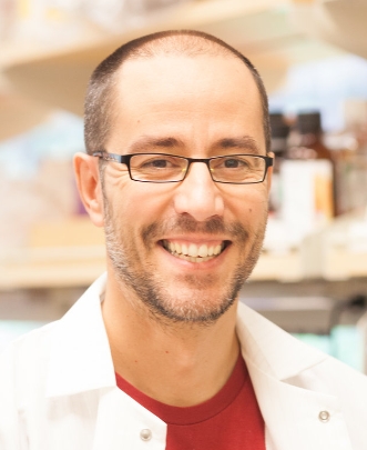 Miguel Ramalho-Santos合作论文数Department of Molecular Genetics, Temerty Faculty of Medicine, University of Toronto;Santos Lab, Lunenfeld-Tanenbaum Research Institute, University of Toronto5ShengGuo You (尤胜国)合作论文数5
 Xiao Long (龙笑)合作论文数Peking Union Medical College Hospital5
 Yang Zeng合作论文数School of Medicine, Tsinghua University;School of Medicine, Tsinghua University5Gao Yang合作论文数5Jun Cai合作论文数5Changhong Li合作论文数5Shengguo You合作论文数5Zhihua Song合作论文数5
 Laure Blouin合作论文数4Xuyun Xia合作论文数4Sihan Yang合作论文数4
 Xinglei Yao (姚醒蕾)合作论文数University of Chinese Academy of Sciences;Research Center for Eco-Environmental Sciences, Chinese Academy of Sciences4




