BackgroundHIV–syphilis coinfection poses a substantial challenge in the management of severe odontogenic infections, with few reports addressing maxillofacial multi-space infections in this setting.Case presentationA 50-year-old man developed severe left maxillofacial multi-space infection one week after molar extraction, presenting with respiratory distress, trismus, elevated C-reactive protein (235.20 mg/L), computed tomography evidence of gas-forming abscesses, reactive HIV antibody, positive syphilis serology, and a CD4+ T-cell count of 133.76 cells/μL. Emergency tracheostomy and incision and drainage were followed by multidisciplinary management comprising sequential antibacterial therapy, intravenous aqueous penicillin G, early antiretroviral therapy, and trimethoprim–sulfamethoxazole prophylaxis. The patient was decannulated on postoperative day 23 and discharged on day 25; the CD4+ T-cell count rose to 321.53 cells/μL within three weeks. At four-month follow-up, the infection had not recurred, the CD4+ T-cell count reached 415.36 cells/μL, and the RPR had reverted to negative.ConclusionIn patients with advanced immunosuppression, odontogenic multi-space infection mandates prompt airway control, adequate surgical drainage, sequenced pathogen-specific pharmacotherapy, and multidisciplinary collaboration.
BACKGROUND:Head and neck squamous cell carcinoma (HNSCC) carries a substantial mortality burden, driven largely by locoregional recurrence. Although immune checkpoint blockade (ICB) is now standard-of-care for recurrent/metastatic disease, primary resistance and limited durability remain major barriers. Clinical outcomes are not determined by PD-L1 status alone, but instead emerge from a multidimensional tumor ecosystem shaped by genomic alterations and epigenetic-metabolic reprogramming. KMT2D (MLL4), a frequently mutated histone methyltransferase in HNSCC, exemplifies this complexity: it is traditionally regarded as a differentiation-preserving tumor suppressor, yet accumulating data suggest context-dependent roles in sustaining oncogenic stemness and metabolic fitness. MAIN BODY:This review reconciles these seemingly discordant observations through an "Enhancer-Immune-Metabolic Framework," positioning KMT2D as an epigenetic rheostat rather than a binary determinant. We synthesize evidence that KMT2D alteration rewires enhancer landscapes, with particular emphasis on the PER2 axis, to promote a shift toward aerobic glycolysis. We further discuss how this metabolic state can potentiate immune exclusion by dampening antigen presentation programs and by supporting an immunosuppressive myeloid milieu. A central mechanistic link highlighted here is histone lactylation (e.g., H3K18la), proposed to couple glycolytic flux to chromatin remodeling and transcriptional outputs that favor immune evasion. Building on these concepts, we outline translationally actionable vulnerabilities in KMT2D-altered tumors, including: (i) synthetic lethal strategies with PARP inhibitors, (ii) metabolic blockade to disrupt glycolysis-associated immune suppression, and (iii) epigenetic "priming" approaches to re-open enhancers governing antigen presentation and T cell-inflamed states, thereby enhancing ICB responsiveness. CONCLUSIONS:KMT2D-driven enhancer remodeling provides a unifying lens to connect metabolic reprogramming with immune escape in HNSCC. Conceptualizing KMT2D as an epigenetic rheostat supports biomarker-informed combination strategies-integrating DNA damage repair targeting, metabolic interventions, and epigenetic priming-to overcome ICB resistance and improve durable disease control in KMT2D-altered HNSCC.
Adenoid cystic carcinoma (AdCC) is a malignancy that most frequently originates in the salivary glands of the head and neck, though it may also rarely occur in other sites such as the trachea or breast. It is characterized by its slow-growing nature in the early stages, along with distinctive features of perineural invasion and a tendency for late metastasis, particularly to the lungs. Recurrence or metastasis can occur even years after the initial treatment. Currently, no standardized clinical protocol exists for long-term follow-up. The disease can recur or metastasize even years after initial treatment. At present, no uniform clinical protocol exists for long-term follow-up. To our knowledge, no documented cases of retrograde metastasis from primary pulmonary AdCC to the tongue have been reported. We describe the case of a male patient in his 40s with primary low-grade pulmonary AdCC who developed lingual metastasis after aggressive treatment. This case highlights an unusual metastatic pattern and underscores the need for vigilant, long-term monitoring of patients with AdCC.
PURPOSE:This study aimed to investigate the real-world efficacy of combining Pembrolizumab with Nimotuzumab in patients with Recurrent/Unresectable/Metastatic HNSCC and to analyze the impact of p16 expression on treatment outcomes. METHODS:The study included 86 patients: 41 received Pembrolizumab monotherapy, and 45 received Pembrolizumab plus Nimotuzumab. Patients were divided into four groups based on treatment and p16 status, analyzing OS, PFS, and ORR. RESULTS:With a median follow-up of 30.2 months, the combination therapy showed a significantly higher 6-month ORR, longer 1-year PFS and OS compared to monotherapy. Additionally, the combination therapy group notably improved 6-month ORR, 1-year PFS rate, and 1-year OS rate in p16-negative patients when compared to monotherapy. However, there was no significant improvement in ORR, PFS, or OS in p16-positive patients. Adverse events occurred in 61 patients (70.93%), with incidences of 68.29% in the monotherapy and 73.33% in the combination group, showing no statistically significant difference (p > 0.05). CONCLUSION:The combination of Pembrolizumab and Nimotuzumab demonstrates a notable enhancement in ORR and OS, maintaining a dependable safety profile. Differential p16 expression in HNSCC patients may influence the efficacy of immune-combined targeted therapy, highlighting the importance of considering p16 in the formulation of therapeutic strategies.
Near-infrared (NIR) fluorescence imaging using cyanine dyes has gained significant attention for its applications in early tumor diagnosis and image-guided surgery. Despite advancements, many dyes suffer from poor targeting and low fluorescence intensity, limiting their clinical use. This study presents the synthesis of two novel NIR fluorescent contrast agents conjugated with HN-1 series peptides, aimed at enhancing DDR-1 targeting for improved tumor imaging. In comparison with ICG ((pf = 2.4 %), our fluorescent agent Cy756-CHN-1 showed higher fluorescent quantum yield ((pf = 16.3 %). Tail vein injection of Cy756-CHN-1 in tumor-bearing mice showed higher fluorescence intensity (over 8 times) than ICG. In vitro and in vivo imaging data confirmed that the Cy756-CHN-1 exhibited superior fluorescence brightness and enhanced tumor affinity. The DDR-1 targeting capability was validated through western blot analysis, molecular modeling, surface plasmon resonance (SPR) and colocalization studies. These results highlight its potential as a promising probe for NIR fluorescence-guided tumor imaging for future clinical applications.
[This retracts the article DOI: 10.21037/qims-23-1444.].
Histone modifications are critical determinants of chromatin accessibility and gene expression, both of which are intrinsically linked to human development and disease. Lysine methyltransferase 2D (KMT2D), a prominent member of the H3K4 methyltransferase family, is ubiquitously expressed across human tissues. Recent studies have found that it can regulate gene expression and signal pathway opening and closing in more than one way, playing an important role in cell proliferation and cell cycle homeostasis. Although previous studies have identified KMT2D as a potentially pivotal factor in the development and pathology of head and neck tissues, the regulatory networks associated with KMT2D in various complex head and neck diseases remain incompletely elucidated. This review seeks to consolidate recent findings on KMT2D's involvement in head and neck diseases, thereby laying the groundwork for future research into its mechanistic role in disease progression. A deeper understanding of KMT2D's functions and regulatory mechanisms is essential for advancing our comprehension of histone modifications and for the development of diagnostic tools and targeted therapeutic strategies for head and neck diseases.
Cryoablation therapy for tumors has a long history of clinical application. Its anti-tumor mechanisms and histopathological changes have been well established, with extensive clinical practice demonstrating its safety and efficacy, theoretically making it an ideal modality for tumor treatment. Historically constrained by limitations in cryogenic media and freezing equipment, its therapeutic effectiveness and clinical adoption were significantly restricted. The emergence of new-generation cryoablation systems represented by Argon-Helium cryosurgical systems has achieved substantial advancements in refrigeration efficiency, ablation range precision, and temperature monitoring accuracy, thereby greatly promoting the widespread adoption of tumor cryoablation technology. This consensus systematically summarizes the mechanisms of cryoablation technology, indications for cryotherapy in head and neck mucosal melanoma, standardized clinical treatment protocols, management of adverse reactions, and related principles. It aims to provide authoritative references for standardizing cryoablation therapy in the treatment of head and neck mucosal melanoma.
Tongue squamous cell carcinoma (TSCC) remains an unsolved medical problem due to its poor local recurrence rate and prognosis. The purpose of this study was to investigate the expression characteristics of RAC-related C3 botulinum toxin substrate 1(RAC1) in TSCCS and its role in tumor invasion and metastasis. In this study, immunohistochemical staining was used to detect RAC1 expression in 150 tongue cancer specimens. The correlation between RAC1 and clinical-pathological characteristics is analyzed, along with the association between protein levels and disease-specific survival, metastasis-free survival, and local recurrence-free survival. In vitro experiments, RAC1 was knocked down by short hairpin RNA transfection to reveal the role of RAC1 in the invasion and metastasis of TSCC. The regulatory effects of RAC1 on CAL27 cell migration and invasion were evaluated by scratch and invasion methods. The influence of RAC1 expression on tumor growth was scrutinized using a subcutaneous transplant model in nude mice. RAC1 is overexpressed in TSCC and positively correlates with tumor invasion and metastasis. Moreover, elevated RAC1 expression is associated with poorer differentiation. Survival analysis further indicates that high expression of RAC1 is linked to increased recurrence and metastasis rates, as well as poorer prognosis. Additionally, both in vivo and in vitro studies demonstrate that RAC1 may impact tumor progression by modulating the epithelial-mesenchymal transition process through the RAC1/PAK1/LIMK1 signaling pathways. RAC1 overexpression is closely related to the progression of TSCC, and may provide a new prognostic indicator and therapeutic target for tongue cancer.
OBJECTIVE:Betel-chewing-related oral squamous cell carcinoma (BCR-OSCC) has become a global health issue with increasing incidence year by year around the world. Active prevention of the occurrence of BCR-OSCC, monitoring the population exposed to Betel Nuts, and early diagnosis and treatment are very important to maintain and improve the quality of life of patients. However, there is currently no consensus or guideline that provides targeted guidance on the management of BCR-OSCC. SUBJECTS AND METHODS:A consensus panel consisting of 15 leading Chinese experts from multidisciplinary fields was convened, and a roundtable meeting was held to discuss the topics of BCR-OSCC. RESULTS:Based on existing research reports and the experts' clinical experiences, a consensus on staging, diagnosis, and treatment for BCR-OSCC was formed through extensive discussion. CONCLUSION:This manuscript presents consensus recommendations and a summary of evidence supporting each recommendation. This consensus may improve clinical practices about BCR-OSCC in China and propel more clinical trials to provide high-level evidence for BCR-OSCC management.
Concentrated growth factor (CGF) accelerates facial nerve rehabilitation in rabbits by enhancing Schwann cell activity, activating the PDGFRβ pathway, and facilitating axon and myelin regeneration.
Background: Cervical lymph node metastasis (LNM) is a well-established poor prognosticator of oral squamous cell carcinoma (OSCC), in which occult metastasis is a subtype that makes prediction challenging. Here, we developed and validated a deep learning (DL) model using magnetic resonance imaging (MRI) for the identification of LNM in OSCC patients. Methods: This retrospective diagnostic study developed a three-stage DL model by 45,664 preoperative MRI images from 723 patients in 10 Chinese hospitals between January 2015 and October 2020. It was comprehensively processed from training (8:2), multicenter external validation to reader study. The performance of the DL model was accessed and compared with general and specialized radiologists. Results: LNM was found in 36.51% of all patients, and the occult metastasis rate was 16.45%. The three-stage DL model together with a random forest classifier achieved the performance in identification of LNM with areas under curve (AUC) of 0.97 (0.93-0.99) in training cohort and AUC of 0.81 (0.74-0.86) in external validation cohorts. The models can reduce the occult metastasis rate up to 89.50% and add more benefit in guiding neck dissection in cN0 patients. DL models tied or exceeded average performance relative to both general and specialized radiologists. Conclusion: Our three-stage DL model based on MRI with three-dimensional sequences was beneficial in detecting LNM and reducing the occult metastasis rate of OSCC patients.
The aim of this study was to investigate the effect of inconsistent expression of p16 and HPV on the prognosis of patients with Oropharyngeal squamous cell carcinoma (OPSCC) in Chinese/Asian populations. The study included 130 patients. Inclusion criteria were primary OPSCC. The primary outcome was the proportion of cohort patients showing different combinations of p16 and HPV outcomes, as well as overall survival (OS) and progression-free survival (PFS). Patients with relapsed or metastatic disease or palliative care were excluded from the survival analysis. A multivariate analysis model was used to calculate the adjusted hazard ratio for overall survival for different p16 and HPV tests, adjusted for pre-specified confounders. Among the 130 patients, 25 (19.2
To investigate the targeting effect of toluidine blue-dextran-40 (TB-Dex-40) on the head and neck lymphatic system. Thirty healthy adult New Zealand white rabbits were randomly divided into two groups: the experimental group (TB-Dex-40 group, n = 15) and the control group (TB group, n = 15). In the experimental group, 1.0% TB-Dex-40 (0.14 mOsm/L) was submucosally injected at the lingual margin (1 cm from the tip of the tongue), while in the control group, 1.0% toluidine blue (32.60 mOsm/L) was administered under the same conditions. The time required for the dye to reach and stain the sentinel lymph node (SLN) was recorded, and the diffusion range of the dyes in the tongue was measured. SLN samples were collected at 30 min and 2 h post-injection for histopathological examination. SLN staining persistence was observed at 1 day, 2 days, and 4 weeks post-injection. Routine blood and biochemical tests were conducted before and 2 weeks after the experiment to evaluate systemic safety. Additionally, in two separate rabbits, the two dyes were injected into the common carotid artery to observe their effects on cervical lymph nodes, submandibular glands, and tongue tissue. A sucrose preference test was performed during animal rearing to assess potential neurotoxicity induced by the dyes. In the experimental group, it took (21.67 ± 0.19) seconds for the dye to reach the SLN and stain lymphatic vessels, which was significantly longer than that in the control group [(3.22 ± 0.34) seconds] (P < 0.01). The SLN stained in the experimental group remained clearly visible even after 4 weeks, whereas the SLN stained in the control group had completely faded by 2 days. The diffusion range of the dye in the tongue was significantly smaller in the experimental group [(10.53 ± 1.09) mm] compared with the control group [(20.04 ± 1.06) mm] (P < 0.01). No abnormalities were detected in the blood parameters of the experimental animals. Neither group exhibited neurological abnormalities. After injection via the common carotid artery, significant staining was observed in the lymph nodes of the TB group but not in the TB-Dex-40 group. TB-Dex-40 demonstrates superior targeting capabilities within the lymphatic system and holds substantial potential for clinical translation.Clinical relevance: TB-Dex-40 exhibits specificity for lymphatic vessels and serves as an effective tracer with significant clinical potential. Its molecular structure provides a robust theoretical foundation for the development of future imaging agents.
Anti-PD-1/PD-L1 has made breakthrough progress in the treatment of recurrent and metastatic head and neck squamous cell carcinoma (R/M HNSCC). However, the Asian population in KEYNOTE-048 only accounts for 13.2 % of the total population, and there is a lack of data on the mainland Chinese population. This multi-center trial (ChiCTR2400090060) evaluated the efficacy and safety of pembrolizumab in patients with R/M HNSCC. Between July 2020 and January 2024, 291 patients who received pembrolizumab-based therapy were enrolled from 20 hospitals across China. All patients were divided into two cohort: cohort 1 included patients unable surgery or radiotherapy (RT), who received first-line treatment of pembolizumab-based, and cohort 2 included patients eligible for surgery or RT, who received pembrolizumab-based treatment with or without local therapy. The primary endpoint was overall survival (OS), while secondary endpoints included time of pembrolizumab treatment (TOPT), immune-related adverse events (irAEs), and best overall response (BOR). With a median follow-up of 18.5 months, the mOS was not reached, with a 18mo-OS rate was 63.2 %. The mOS for cohort 1 was 21.2 months. Interestingly, mOS for cohort 2 was not reached. Patients who received local therapy had a significantly improvement on 18mo-OS rate compared to those who did not (86.1 % vs. 65.8 %, p = 0.021). A total of 24.7 % of patients experienced irAEs. The study was the first report that the patient who was eligible for local treatment had a benefit from pembrolizumab, especially the OS was significantly improved for pembrolizumab followed by surgery or RT.
ObjectiveThis study aimed to investigate the expression of serine protease inhibitor kazal type 1 (SPINK1) and its carcinogenic effect in oral tongue squamous cell carcinoma (OTSCC). Design: Initially, bioinformatics analysis was conducted using data from The Cancer Genome Atlas and Gene Expression Omnibus to compare SPINK1 mRNA expression between malignant and adjacent tissues. Subsequently, the impact of differential expression on survival and other clinical variables was examined. Additionally, histology microarray analysis was performed to assess SPINK1 protein expression in 35 cases of malignant and adjacent tissues. Finally, alterations in SPINK1 expression were evaluated to determine its biological phenotypes in OTSCC, including proliferation, apoptosis, invasion, and metastasis. Results: OTSCC tissues exhibit higher levels of SPINK1 compared to surrounding cancerous tissues. Notably, increased SPINK1 expression correlates with the pathological N stage and independently predicts overall survival among patients with OTSCC. Conclusion: Suppression of SPINK1 inhibited OTSCC cell proliferation, invasion, and motility while promoting apoptosis. These findings suggest that SPINK1 may serve as a prognostic biomarker as well as a potential therapeutic target for managing OTSCC.
Objectives:Chemerin, as a novel multifunctional adipokine, is proposed to be involved in high cancer risk and mortality. The present study was aimed to evaluate the prognostic value of serum Chemerin and neutrophils in patients with oral squamous cell carcinoma (OSCC).Materials and Methods:120 patients with OSCC were included in this prospective cohort study. The levels of serum Chemerin were measured by enzyme-linked immunosorbent assay (ELISA). We also explored the possible effects of Chemerin on neutrophils’ chemokines in OSCC using a real-time PCR, western blotting.Results:Levels of serum Chemerin, neutrophils and NLR were significantly higher among non-survivors compared to survivors of OSCC (both P < 0.05). Higher serum Chemerin levels were associated with advanced TNM stage, lymph node metastasis, differentiation and tumor recurrence (both P < 0.05). Serum Chemerin levels correlated with neutrophils and NLR levels (r = 0.708, r = 0.578, both P < 0.05). Based on ROC analysis, Chemerin + NLR predicted OSCC patient mortality with 81.54% sensitivity and 87.27% specificity, with an AUC of 0.8898. In a Kaplan-Meier analysis, high serum Chemerin levels, high neutrophil levels and high NLR levels were associated with shorter overall and disease-free survival (both P < 0.05). A univariate and multivariate Cox regression analysis showed that serum Chemerin and neutrophils were independent risk factors for OSCC. (both P < 0.05). QRT-PCR and western blotting results showed that Chemerin upregulated the expression of chemokines IL-17 and CXCL-5 in neutrophils (both P < 0.05).Conclusions:Our study suggests that measurement of serum Chemerin and neutrophils might be a useful diagnostic and prognostic biomarker for OSCC patients. Chemerin may promote neutrophils infiltration in OSCC through upregulation of chemokines IL17 and CXCL-5.
 Yuanyong Feng合作论文数37
 Kai Song (宋凯)合作论文数School of Stomatology , Qingdao University , Shandong Province , China; Department of Oral & Maxillofacial Surgery , the Affiliated Hospital of Qingdao University , Shandong Province , China.31
 Muyun Jia (贾暮云)合作论文数Department of Oral Maxillofacial Surgery,the Affiliated Hospital of Qingdao University Medical College25
 Ningyi Li (李宁毅)合作论文数Dept.of Oral and Maxillofacial Surgery, The Affiliated Hospital of Medical College, Qingdao University25
 Rongtao Yuan (袁荣涛)合作论文数Qingdao Municipal Hospital (Group)19Sheng-you GE (葛胜优)合作论文数18Xiao-chen YANG (杨小琛)合作论文数17ZongXuan He (何宗轩)合作论文数16
 Lingxue Bu (卜令学)合作论文数Department of Oral and Maxillofacial Surgery, The Affiliated Hospital of Qingdao University, Qingdao University15XiaoMing Jin (金晓明)合作论文数11Hao-yue XU (徐豪越)合作论文数8Yueqin Tao合作论文数8
 Moyi Sun (孙沫逸)合作论文数中国人民解放军第四军医大学8
 Jian Meng (孟箭)合作论文数Department of Stomatology, Xuzhou Central Hospital8
 Jiawei Zheng (郑家伟)合作论文数Shanghai Red Cross Ninth People's Hospital;College of Stomatology, Shanghai Jiao Tong University8XueCai Yang (杨学财)合作论文数7ShiEn Liu (刘世恩)合作论文数7Wei Li (李薇)合作论文数7
 Zhangui Tang (唐瞻贵)合作论文数Hunan Xiangya Stomatological Hospital, Central South University7
 Longjiang Li (李龙江)合作论文数West China School of Stomatology, Sichuan University7
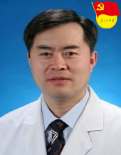 Wei Guo (郭伟)合作论文数Shanghai Ninth People’s Hospital, Shanghai Jiao Tong University School of Medicine7
 Guoxin Ren (任国欣)合作论文数Shanghai Ninth People's Hospital, School of Medicine, Shanghai Jiao Tong University7
 Zhijun Sun (孙志军)合作论文数Hospital of Stomatology, Wuhan University6
 Shengbiao Wan (万升标)合作论文数School of Medicine and Pharmacy, Ocean University of China6Baoxing Pang合作论文数5ZHOU Kai (周凯)合作论文数5Wen-lin Xiao (肖文林)合作论文数5HeMing Wu (武和明)合作论文数5ZhongCheng Gong (龚忠诚)合作论文数5
 Kai Yang (杨凯)合作论文数重庆医科大学附属第一医院口腔颌面外科5Lin Wang合作论文数4
 Ce Gao合作论文数4Yu KONG (孔钰)合作论文数4L Wang合作论文数4LI Heng-yong (李恒勇)合作论文数4QingYun Duan (段青云)合作论文数4Qi Wang (王琪)合作论文数4
 Jiang Tao (江涛)合作论文数School of Medicine and Pharmacy, Ocean University of China4
 Wei Ran (冉炜)合作论文数The First Affiliated Hospital, Sun Yat-sen University4
 Qing Xi (席庆)合作论文数Chinese People's Liberation Army General Hospital4Kexin Wang合作论文数3Jieying Li合作论文数3Houyu Ju合作论文数3LI Jie-ying (李洁莹)合作论文数3Qian-Qian Gao合作论文数3Jie Zhao合作论文数3ZHANG Zhuang (张壮)合作论文数3WU Jing (吴晶)合作论文数3HaiJun Lu (陆海军)合作论文数3




