To study the effects of enteral administration of carbachol on organ dysfunction induced by partial ischemia/reperfusion injury to the intestine.Seventy-five white rabbits were randomized into four groups: ischemia/reperfusion (I/R), carbachol+ I/R and sham operation. The superior mesenteric artery (SMA) was partially blocked with self-designed blocker, producing 50% decrease in SMA blood flow, lasting for 4 hours. One hour after SMA occlusion, carbachol was injected into gut in carbachol+I/R group. Sham group was treated as same as I/R group except without SMA occlusion. The levels of alanine aminotransferase (ALT), creatinine (Cr), MB isoenzyme of creatine kinase (CK-MB) and tumor-necrosis factor-alpha (TNF-alpha) were measured by automatic analyzer and with radio-immunology method before SMA occlusion, and at 2, 4, 6, 8, 24, 48 and 72 hours after occlusion. The pathological changes of the intestinal tissue were observed with hematoxylin and eosin stained sections.In I/R groups, the levels of TNF-alpha, Cr, ALT, CK-MB in plasma were increased dramatically after partial ischemia/reperfusion injury to the gut. Severe pathological changes were observed in the hearts, livers, and kidneys. While in carbachol treatment groups, the levels of TNF-alpha, Cr, ALT, CK-MB in plasma were decreased dramatically after enteral administration of carbachol during ischemia stage. The pathological injuries were alleviated in the heart, liver, and kidney.Enteral administration of carbachol may alleviate after the systemic inflammatory response and pathological changes in various organs, thus provide a protective effect on gut and remote organs partial ischemia/reperfusion of the intestine.
OBJECTIVETo investigate the effects of carbachol injection in intestine on plasma levels of tumor necrosis factor-alpha(TNF-alpha), interleukin-10 (IL-10) and cortisol in rats during gut ischemia/reperfusion.METHODSWistar rats were anaesthetized with soluble pentobarbitone, and subjected to superior mesenteric artery occlusion (SMAO) for 60 minutes, followed by reperfusion for 60 minutes. Animals were divided into three groups, pretreated group (carbachol injection in intestine at 30 minutes after SMAO; 0.1 mg/kg), treated group (carbachol injection at 30 minutes from onset of reperfusion), and controls (saline injection). Plasma TNF-alpha, IL-10 and cortisol levels were determined at 1.0, 2.5 and 6.0 hours after SMAO.RESULTSThe plasma levels of TNF-alpha significantly decreased in pretreated and treated groups than those in controls after carbachol injection (both P<0.01). However, the levels of IL-10 and cortisol didn't show significant differences among three groups.CONCLUSIONThe RESULTS suggest that carbachol can reduce the proinflammatory cytokine release and have a less inhibitory effect on the anti-inflammatory cytokine. It is indicated that carbachol play a potential role in alleviating systemic inflammatory response during splanchnic ischemia/reperfusion injury.
研究拟胆碱药卡巴胆碱对大鼠缺血再灌注损伤小肠组织髓过氧化物酶(MPO)和丙二醛(MDA)的影响及其与肠损伤相关指标变化的规律.Wistar大鼠被随机分为预防、治疗和对照三组.活杀后取小肠组织测MPO、MDA和肿瘤坏死因子(TNF-α)含量.结果显示,治疗组及预防组MPO活性、MDA和TNF-α含量均明显低于对照组,治疗组与预防组之间差异不明显.提示卡巴胆碱可抑制致炎因子TNF-α的释放,减少中性粒细胞在肠组织的聚集,从而使小肠MPO活性降低.
OBJECTIVETo study the barrier function, absorption, permeability and peristalsis of intestine in sepsis in rats.METHODSA Wistar rat model of sepsis was reproduced by ischemia/reperfusion (I/R) of the intestine combined with endotoxin challenge. Animal were randomly divided into normal, I/R 1 hours (I/R 1), I/R 2 hours (I/R 2), I/R 4 hours (I/R 4) and I/RL groups. The following parameters were measured in the experiments: (1) diamin oxidase activity (DAO), D-lactate and D-xylose levels in blood using spectrophotometry; (2) transit function of small intestine; (3) pathological examination of small intestine by light microscope.RESULTSThe results showed that plasma DAO activity was increased in I/R 1, I/R 4 and I/RL (all P<0.05), and small intestinal tissue DAO was decreased in I/R 2 and I/RL (both P<0.05). Negative correlations were found between plasma and intestinal DAO (r=-0.909, P<0.001). Plasma D-lactate was elevated significantly in I/R 1, I/R 2, and I/RL (all P<0.05). D-xylose content was increased at I/R1 and I/RL groups (both P<0.05), and it was significantly higher than controls at 3 hours. Similarly, a positive correlation was found between plasma DAO activity and plasma D-lactate level (r=0.559, P<0.05).CONCLUSIONThe intestinal barrier function, absorption function, permeability, and transit are impaired after gut ischemia/reperfusion combined with endotoxin challenge.
OBJECTIVE:To investigate the effects of carbachol on the levels of tumor necrosis factor-alpha (TNF-alpha), interleukin-10 (IL-10) and cortisol in plasma of rats during gut ischemia-reperfusion.METHODS:Wistar rats were anaesthetized with soluble pentobarbitone, and subjected to superior mesenteric artery occlusion (SMAO) for 60 minutes, followed by reperfusion for 60 minutes. Animals were divided into three groups, pretreated group (intramuscular injection carbachol at 30 minutes after SMAO, 0.1 mg/kg), treated group (intramuscular injection of carbachol at 30 minutes after reperfusion), and control group (saline injected). The levels of TNF-alpha, IL-10 and cortisol in plasma were determined at 0 hour, 1 hour, 2.5 hours and 6 hours after SMAO.RESULTS:The levels of TNF-alpha, IL-10 and cortisol significantly increased after SMAO (P<0.01 ). The levels of TNF-alpha significantly decreased in pretreated and treated groups than that in control after the intramuscular injection of carbachol (all P<0.01). However, the levels of IL-10 and cortisol did not show significant differences among three groups. It was also found that lower content of TNF-alpha in pretreated group than that in treated group, especially at 1 hour and 6 hours.CONCLUSION:The results suggest that carbachol reduce the proinflammatory cytokine releasing and has a less inhibiting effect on the anti-inflammatory cytokine. It is indicated that carbachol play a potential role in alleviating systemic inflammatory response during splanchnic ischemia-reperfusion injury.
Objective:To investigate the relationship between the electrical stimulation of vagus nerve and changes of liver malondialdehyde(MDA) and tumor necrosis factor-α(TNF-α) in rats with endotoxemia.Methods:Wistar rats were subjected to bilateral cervical vagotomy. The distal end of the vagus nerve trunk was placed across bipolar electrodes connected to a stimulation module with acquisition system control. Constant voltage stimuli (5 v, 2 ms, 1 hz) were applied to the nerve for 20 min immediately after administration of LPS, and then repeated twice at an interval of 10 min. The level of alanine aminotransferase(ALT) in plasma and the levels of TNF-α and MDA in liver were determined at 1 h, 1.5 h and 2 h after LPS challenge.Results:The hepatic TNF-α levels in endotoxin challenged rats were significantly higher than those in the sham controls. Compared to animals receiving no electrical stimulation, electrical stimulation of the efferent vagus nerve significantly decreased the hepatic contents of TNF-α, MDA and plasma ALT levels in endotoxin challenged rats.Conclusions:Excitation of the efferent vagus nerve could significantly reduce the production of the proinflammatory cytokine and alleviate the release of oxygen-derived free radicals after endotoxin challenge. The results suggested that excitation of vagus nerve might have potential protective effect on liver.
目的:探讨创伤并发感染时肝细胞线粒体的损伤及意义.方法:复制大鼠缺血-再灌注复合内毒素血症模型,测定血乙酰乙酸(AcAc)、β-羟丁酸(β-OHBA)及其比值,同时测定相关指标的变化,并进行肝组织光镜病理形态学检查.结果:休克加复苏组、休克复苏加内毒素Ⅰ组(输内毒素后2小时)以及休克复苏加内毒素Ⅱ组(输入内毒素后24小时)血AcAc均较健康对照组有不同程度升高,但无统计学差异;血β-OHBA在休克加复苏组、休克复苏加内毒素Ⅰ组和Ⅱ组均显著升高(P<0.05或P<0.01);AcAc/β-OHBA比值伤后各组均显著降低(P均<0.05);血丙氨酸转氨酶(ALT)和天冬氨酸转氨酶(AST)则均显著升高(P<0.05或P<0.01).结论:AcAc、β-OHBA及其比值在血中的变化能较敏感地反映创伤合并内毒素血症时肝细胞线粒体的损伤,可作为评价肝细胞代谢状况的指标.
目的:观察迷走神经兴奋对内毒素血症大鼠肝、肺组织中促炎因子(TNF-α)及抗炎因子皮质醇的影响,探讨胆碱能抗炎通路在炎症反应中的初步机制.方法:雄性Wistar大鼠,随机分为迷走神经刺激组、迷走神经切断后静注LPS组、假手术组和LPS组等4组.测定肝、肺TNF-α、血浆皮质醇和丙氨酸氨基转移酶(ALT)含量.结果:静注LPS后,组织中TNF的含量显著升高(最高可达15倍),其中肝组织中TNF含量在1.5h达到高峰,2h回落,而肺组织中TNF含量在1h达峰值;假手术组由于未注射LPS,TNF含量变化不明显,但肝、肺组织中TNF含量差异较大,肺组织中TNF含量明显低于肝组织,可能与TNF较多在肝脏合成有关.迷走神经切断后静注LPS组TNF水平较单纯LPS组有所增加;迷走神经刺激组肝组织TNF含量在1h和1.5h、肺组织在各时相点较单纯LPS组和迷走神经切断后静注LPS组显著降低.LPS组血浆皮质醇含量在2h与其余各组相比增高明显;迷走神经刺激组血浆ALT含量显著低于单纯LPS组.结论:迷走神经兴奋能显著降低内毒素血症大鼠肝、肺组织中促炎细胞因子的产生,有助于减轻其全身炎症反应.
目的:研究刺激传出迷走神经对内毒素[即脂多糖(LPS)]引起大鼠急性肺损伤的影响.方法:雄性Wistar大鼠70只,随机分为7组.切断大鼠双侧颈部迷走神经干或做假手术对照,将左侧迷走神经远端连接刺激电极,于静注LPS(10 mg/kg)即刻持续电刺激(5 V,2 ms,1 Hz)20分钟,每间隔10分钟后重复刺激,每次20分钟,共刺激3次.分别在1、1.5和2小时检测实验和对照各组肺组织中肿瘤坏死因子-α(TNF-α)、髓过氧化物酶(MPO)以及丙二醛(MDA)含量.结果:静注LPS后迷走神经刺激组肺组织中TNF-α、MPO 和MDA含量均显著降低,炎性病理改变减轻.结论:迷走神经兴奋能抑制LPS攻击大鼠肺组织炎症介质产生,减轻肺组织病理损害,对LPS所致急性肺损伤动物有潜在保护作用.
OBJECTIVE:To evaluate the effects of different enteral nutrients on intestinal absorptive capacity and ATP content of intestinal mucosa after ischemia-reperfusion (I/R) of the intestine.METHODS:A segment of jejunum was isolated to form a pouch in Sprague-Dawley rats. The jejunal pouches of animals were filled with either a mixture of nutrients (5 mmol/L alanine + 5 mmol/L glucose), glucose alone (10 mmol/L), alanine alone (10 mmol/L) or mannitol alone (10 mmol/L) as a nonabsorbable osmotic control. The superior mesenteric artery was then occluded for 60 minutes, followed by reperfusion for 60 minutes. Gut absorptive capacity, as measured by the changes in short circuit current (DeltaIsc), and mucosal ATP content were determined both at the end of 60 minutes ischemia and 60 minutes of reperfusion.RESULTS:DeltaIsc and ATP levels were decreased after I/R. Intraluminal alanine alone or alanine/glucose mixture further reduced DeltaIsc and ATP content. On the other hand, intraluminal glucose significantly increased DeltaIsc and ATP levels compared with intraluminal alanine, alanine/glucose mixture or I/R alone.CONCLUSION:Intraluminal alanine increases the absorptive capacity and energy metabolism of the gut caused by I/R, and indicated that intraluminal glucose improves gut absorptive capacity and energy metabolism and provides a protective effect on gut from ischemia and reperfusion injury.
AIM To investigate the mechanism underlying intestinal barrier function damage after severe trauma and the therapeutic effect of glutamine. METHODS Burned patients, and animal models of severe trauma replicated by hemorrhagic shock combined with endotoxin infusion and burn injury, were included in a serial experiment. Effects of oral glutamine on intestinal barrier function were observed in scalded rats. Parameters measured in these experiments were as follows: plasma levels of diamine oxidase (DAO), tumor necrosis factor (TNFalpha), endotoxin (LPS), and lactate as well as D-lactate by biochemical methods, lactose/mannitol (L/M) ratio in urine by SP-3400, and pathological examination of intestinal mucosa under light microscopy. RESULTS Plasma DAO activity was significantly increased after injury. There was a negative correlation between plasma DAO and intestinal mucosal DAO or pHi (r=-0.93, plasma 0.80+/-0.93,2.83+/-1.71, 1.14+/-0.64,2.36+/-2.06 and 2.49+/-1.67 vs intestinal 0.52+/-0.12,0.34+/-0.03,0.45+/-0.18,0.37+/-0.26 and 0.41+/-0.07 r=-0.533, plasma 0.87+/-0.75, 1.89+/-1.13,1.21+/-0.23,3.03+/-2.61 and 4.70+/-1.22 vs pHi 7.03+/-0.05,7.05+/-0.06,7.14+/-0.096, 7.20+/-0.08 and 7.05+/-0.07 P<0.01-0.05). Positive correlations were found between DAO activity and plasma TNFalpha, LPS, lactate, L/M and D-lactate (r=0.817, 0.842, 0.872, and 0.951 plasma DAO 0.87+/-0.75,1.89+/-1.13,1.21+/-0.23,3.03+/-2.61 and 4.70+/-1.22 vs TNF 0.08+/-0.02,0.03+/-0.25, 0.17+/-0.09,0.34+/-0.15 and 0.33+/-0.18 vs LPS 0.14+/-0.03,0.16+/-0.04,0.21+/-0.02,0.18+/-0.16 and 0.37+/-0.10 vs lactate 9.03+/-2.19,18.30+/-2.56, 9.81+/-2.83,12.01+/-6.83,12.01+/-6.84 and 43.61+/-11.27 vs L/M 0.03+/-0.01,0.41+/-0.27,0.62+/-0.20, 1.70+/-0.60 r=0.774, plasma DAO 1.25+/-0.41 2.17+/-0.71 2.29+/-0.87 1.23+/-0.55 and 1.11+/-0.47 vs D-lactate 8.37+/-2.48,18.25+/-6.18, 13.96+/-4.94,8.93+/-3.00 and 12.39+/-4.94 all P<0.01), respectively. Damage of intestinal mucosa was found by pathological examination. Intestinal barrier function was improved to a certain extent by oral glutamine in scalded rats. CONCLUSION Intestinal barrier function was damaged in the early stage after trauma. Plasma DAO activity, D-lactate content, intestinal pHi and urine L/M may be sensitive markers of intestinal mechanical injury, and glutamine may protect against intestinal barrier dysfunction after severe trauma.
 XiaoGuo Jiang (姜小国)合作论文数8Sen Hu (胡森)合作论文数Chinese Pla General Hospital8Junyou Li (黎君友)合作论文数The First Affiliated Hospital, PLA General Hospital7DeGuang Shi (石德光)合作论文数7Hua Jin (晋桦)合作论文数6
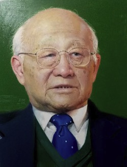 Zhiyong Sheng (盛志勇)合作论文数Chinese PLA General Hospital6
 Yi Lv (吕艺)合作论文数Fourth Medical Center PLA General Hospital3Yi Lu合作论文数2yi lv (吕艺)合作论文数2QiLin Liu (柳琪林)合作论文数2Shirong Sun (孙世荣)合作论文数1rong zhang (张蓉)合作论文数1Ming Yan (严鸣)合作论文数1Zhi-yong Sheng合作论文数1Han Cuihua (韩翠华)合作论文数1XiaoQing Sun (孙晓庆)合作论文数1
 Yongming Yao (姚咏明)合作论文数Chinese People's Liberation Army General Hospital;School of Medicine, Nankai University1




