BACKGROUNDIt is considered that the pathogenesis is closely related to an excessive production of pro-inflammatory cytokines caused by bacterial toxins and an imbalance between pro-inflammatory and anti-inflammatory mediators.MATERIALS AND METHODSThis work investigates the effect of electro-acupuncturing (EA), at Zusanli point (ST36) on plasma cytokine release and organ dysfunction and their mechanism in conscious rats with endotoxin challenge.RESULTSEA at Zusanli points obviously lowered the elevated levels of plasma TNF-α, and attenuated changes in parameters relevant to various organ functions at 2 h after LPS challenge. α-BGT injection or bilateral cervical vagotomy could weaken or eliminate the effects of EA, and further aggravated the elevated levels of pro-inflammatory cytokines and organ dysfunction.CONCLUSIONThe results suggested that EA at Zusanli points significantly reduced the release of pro-inflammatory cytokines and organ dysfunction after LPS challenge by activating cholinergic anti-inflammatory pathway.
目的 研究卡巴胆碱对H2O2引起体外培养的肠上皮细胞(intestinal epithelial cell,IEC)凋亡的影响.方法 采用H2O2损伤IEC模型,实验分为对照组、H2O2组、卡巴胆碱预防组和卡巴胆碱治疗组.H2O2组加入2.5 mmol/L的H2O2(溶于无血清培养基中);卡巴胆碱预防组预先30 min给予卡巴胆碱(卡巴胆碱100 μmol/L),然后加入2.5 mmol/L的H2O2;卡巴胆碱治疗组加入H2O2,同时给予卡巴胆碱(卡巴胆碱100 μmol/L);对照组只给予无血清培养液.于处理后4 h收集细胞.采用噻唑蓝法(MTF法)检测线粒体功能;硝酸酶还原法检测IEC中NO含量;用Annexin-V荧光染色;流式细胞仪检测凋亡细胞率.采用SPSS 11.5统计学软件处理数据.数据以均数±标准差(x±s)表示,比较以资料性质采用用t检验或χ2检验,P<0.05为差异有统计学意义.结果 H2O2组线粒体功能降低,细胞凋亡率显著增加.与H2O2组相比,卡巴胆碱预防和治疗组线粒体功能明显改善,细胞凋亡率显著降低.结论 线粒体参与了H2O2诱导的IEC凋亡,卡巴胆碱对H2O2引起的IEC凋亡有显著的抑制作用.
Aim To investigate the effects of carbachol on inflammatory cytokine releases from rat peritoneal macrophages induced by lipopolysaccharide and its receptor.Methods Rat peritoneal macrophages were collected and exposed to muscarinic cholinergic receptor antagonist atropine or α-bungarotoxin,an antagonist specifically binds the α7 subunit of nicotinic cholinergic receptor.15 min later carbachol or nicotine was added into cell culture.After another 15 min,cells were stimulated with LPS.Inflammatory cytokines TNF-α,IL-6,and IL-10 in the cultures media were assayed by ELISA.Rat peritoneal macrophages were exposed to nicotine and carbachol in high concentration for 15 minutes,and then stained with FITC-labeled α-Bungarotoxin and viewed with fluorescent confocal microscopy.Results Increases of TNF-α and IL-6 after LPS stimulation were significantly inhibited in the presence of carbachol or nicotine while the content of IL-10 was not apparently altered.Atropin did not down-regulate the inhibitive effects of both carbachol and nicotine.And α-bungarotoxin significantly down-regulated these effects.Fluorescent confocal microscopy showed that nicotine and carbachol pretreatment markedly reduced the intensity of binding between FITC-labeled α-Bgt and macrophages.Conclusion The results suggested that both carbachol and nicotine play a role in the anti-inflammatory process through the α7 subunit of nAChR.
OBJECTIVETo investigate the effects of carbachol on apoptosis of intestinal epithelial cells in rats after gut ischemia/reperfusion (I/R).METHODSA jejunal sac was formed in Wistar rats. The superior mesenteric artery (SMA) was occluded for 45 minutes followed by 240 minutes of reperfusion. Immediately after occlusion of SAM blood flow, either 0.1 mg/kg of carbachol or same amount of 0.9% saline normal was injected into the jejunal sac. Animals were randomized into three groups (each n=40): sham operation, I/R+normal saline injection (I/R model) and I/R+carbachol injection (0.1 mg/kg, Ca). The Pathological changes in gut epithelial cells were assessed by Chiu's scores. The apoptosis index of intestinal epithelial cell was determined with terminal deoxynucleotidyl transferase-mediated dUTP-biotin nick end labeling (TUNEL) staining. Expressions of caspase-3 and Bcl-2 in intestinal epithelial cells were assayed by immunohistochemistry method. All measurements were done at 0, 30, 60, 120 and 240 minutes after reperfusion.RESULTSThe Pathological injuries were less severe in Ca group than those in I/R model group. Apoptosis index of intestinal epithelial cell and expressions of caspase-3 were significantly decreased, while the expressions of Bcl-2 increased dramatically (all P<0.01) after I/R in Ca group compared with those in I/R model group, especially at 120 minutes after reperfusion.CONCLUSIONEnteral administration of carbachol can inhibit apoptosis of intestinal epithelial cells in rats after gut I/R injury.
Objective:To explore the protective effect of excitation of cholinergic receptor in different ways on the hepatic function of animals(rats and rabbits)challenged by endotoxemia or gut ischemia/reperfusion(GIR).Methods:The model of endotoxemia(n=10,in each group)was reproduced in rats by injecting endotoxin(LPS,10 mg/kg and 5 mg/kg)into the peritoneal cavity.Absolute GIR(n=5)was reproduced in rats by obstructing superior mesenteric artery(SMA)1 hour followed by recovering of blood perfusion,and partial GIR(n=6)rabbits model was established by partial SMA 4 hours followed by recovery of the blood perfusion.The endotoxemia animals were treated with electric stimulation of the vagas nerve or electroacupuncture(Housanli acupoint,后三里穴),while the sham operated group,model group,vagectomy group or sham acupoint electroacupuncture groups served as the controls.The GIR rats were injected carbachol(0.1 mg/kg)into the ileac sac(formed by ligating two ends of ileum to form a blind sac of 10 cm long with maintenance of blood supply)and the GIR rabbits were injected carbachol(3 μg/kg)into the lumen of superior segment of the jejunum,the model group and the sham operated group were the controls.Plasma ALT activity was determined in rats,and at 0,2,4,6,8 hours as well as 1,2,3 days after SMA obstruction in rabbits.Results:Compared with the model group,vagectomy group or sham acupoint electroacupuncture group,the plasma ALT activity in animals with endotoxemia treated by excitation of parasympathetic nerve was decreased dramatically(P<0.05 or P<0.01).ALT of GIR rats increased,and it was decreased after carbachol treatment.ALT of GIR rabbits was increased during ischemia and early stage of reperfusion,but lower than that of the untreated animals,and it recovered gradually and approached the normal level on the third day after injury.Conclusion:Stimulating parasympathetic nerve or administration of cholinergic drug locally could protect against hepatic injury induced by endotoxin or gut ischemia/reperfusion.
Objective To observe influence of enteral administration of carbachol on heart inflammation induced by partial ischemia/reperfusion injury to intestine.Methods Seventy five rabbits were randomized into three groups: intestine I/R group,carbachol group,sham operation group,the superior mesenteric artery(SMA) were occluded to 50% for 4h in intestine I/R group.In carbachol group,carbachol(3mg/kg) was injected into duodenum 1h after occlusion of SMA.Changes of TNF-α,IL-10 in heart and intestine were measured at 2,6h,1,2,3d after SMA occlusion.Results Levels of TNF-αin heart and intestine were increased dramatically after intestine I/R injury.Enteral administration of carbachol increased the blood flow of intestinal mucosa,decreased the level of TNF-αand inflammation in heart.Conclusion Partial ischemia/reperfusion injury to intestine could induce heart inflammation and dysfunction;Enteral administration of carbachol could inhibit the release of pro-inflammatory factors as TNF-α,but not increase the release of anti-inflammatory factors as IL-10.
AIM:To study the effects of carbachol on the H_2O_2-oxidative injury of intestinal epithelial cells (IEC). METHODS:IEC were cultured with the pres- ence of H_2O_2 in vitro to induce the oxidative injury.IEC were divided into 3 groups:control group,H_2O_2 (2.5 mmol/L) group and carbachol (100)μmol/L) group.The viability of IEC was measured by MTT assay.The level of lactate dehydrogenase (LDH) in culture media and the malondialdehyde (MDA) content in IEC were measured. RESULTS:In compared with those in control group,LDH level (7.40±2.10 vs 0.81±0.12,P<0.01) and MDA content in H_2O_2 group were sig- nificantly elevated,and the viability of IEC was obviously decreased (37.25%±0.80% vs 100%±0.13%,P<0.01).However,in carbachol group, LDH level (4.64±1.31 vs 7.40±2.10,P<0.01) and MDA content were lower while IEC viability was obviously higher than those in H_2O_2 group (78.70%±2.80% vs 37.25%±0.80%,P<0.01). CONCLUSION:Carbachol can protect rat IEC from the oxidative injury.
OBJECTIVE To explore the mechanism of the effect of carbachol in the treatment of sepsis at the angle of modulation of dendritic cell (DC) activity. METHODS Thirty male C57BL/6 mice were randomized into three groups: normal control, sepsis and carbachol. The sepsis model of mice was reproduced by the injection of lipopolysaccharides (LPS). The expression of interleukin-1 beta (IL-1 beta) and IL-12 p70 positive cells were measured by immunohistochemistry method. The changes in DC activity in the spleen of mice were studied using flow cytometry. RESULTS In LPS group, the number of splenic DC was increased, but it was not statistically significant. The expression of DC molecules including major histocompatibility complex-II (MHC-II), CD86 and the rate of positive cells containing IL-1 beta and IL-12 p70 were significantly higher than those in normal controls (all P<0.05). While in carbachol treatment group, the number of splenic DC had no significant change, the expression of IL-1 beta was down-regulated, dramatically. The expression of MHC-II, CD86 and IL-12 p70 were also lowered (all P<0.05). CONCLUSION Carbachol can lower the DC activity, suggesting that modulation of DC activity may lower extensive immune and inflammatory response in the early phases of sepsis.
OBJECTIVE:To evaluate the effect of cholinergic drug carbachol on apoptosis and expression of certain cytokines of peripheral blood lymphocytes and neutrophils in rats subjected to ischemia/reperfusion injury of the intestine.METHODS:Thirty-six Wistar male rats were randomly divided into groups as follows: sham operation, 1 and 2 hour-gut ischemia, 1 hour-gut ischemia followed by reperfusion for 1 hour and 2 hours, carbachol + 1 hour-gut ischemia, and carbachol + 1 hour-gut ischemia followed by reperfusion for 2 hours. Then the gut was subjected to ischemia/reperfusion. At different time points after the said injury, the total number and differential count of leukocytes, apoptosis rate of lymphocytes and neutrophils, and the mRNA expression levels of certain cytokines in peripheral blood leukocytes, were determined.RESULTS:At 1 hour after gut ischemia, the total number of leukocytes decreased (the ratio of lymphocytes increased and that of neutrophils decreased), but it increased after gut reperfusion. Carbachol could reduce the apoptosis rate of lymphocytes, but enhance that of neutrophils after gut ischemia. In leukocytes, mRNAs expression of inflammatory (TNF-alpha) and antiinflammatory (IL-10) cytokines were upregulated at 1 hour after gut ischemia, while that of IL-10, IL-4 and interferon-gamma were down-regulated distinctly at 2 hours following reperfusion. All of these phenomena were ameliorated by giving carbachol.CONCLUSION:Carbachol could reduce the peripheral lymphocytes apoptosis subsequent to gut ischemia, and regulate the balance between inflammatory and antiinflammatory cytokines expression in leukocytes during gut ischemia and reperfusion. The results suggest that carbachol might be a potential therapeutic agent in preventing uncontrolled inflammatory response as a result of ischemia/reperfusion injury of the intestine.
OBJECTIVE:To investigate the effect of burn injury on the expression of rat defensin-5 (RD-5) and Matrilysin mRNA in the intestine of scalded rats and to determine its relation to bacterial translocation.METHODS:Thirty-two Wistar rats were divided randomly into sham injury group (n=8) and burn group (n=24). Rats of the burn group were subjected to 30% total body surface area (TBSA) full-thickness scald injury. At 8, 24 and 72 hours after the injury, the animals (n=8 at each time point) were sacrificed and ileum was sampled for the determination of the expression of RD-5 mRNA and Matrilysin mRNA with reverse transcription-polymerase chain reaction (RT-PCR) technique. Cultures of mesenteric lymph nodes (MLN), liver, spleen and lung were carried out at 24 hours after the injury to assess the rate of bacterial translocation to visceral organs,and the morphological changes in ileal Paneth cells were observed.RESULTS:The expression of RD-5 mRNA was significantly up-regulated at 8 hours when compared with control group. It was down-regulated sharply at 24 hours but was still higher than control group, and at 72 hours it was down-regulated to a level lower than control. On the contrary, the expression of Matrilysin mRNA was up-regulated continuously to 72 hours, and it was significantly higher than control. Bacterial translocation rate was significantly higher in rats of the burn group at 24 hours (58.3%) than in control group (8.3%, P<0.01), but there were no obvious morphological changes in ileal Paneth cells at the same time point.CONCLUSION:The expressions of both RD-5 mRNA and Matrilysin Mrna are up-regulated in the early stage after the injury, but their time phases are different. The phenomenon might imply that there is a host protective response against bacterial translocation.
Aim: To investigate the effects of carbachol on the levels of the proinflammatory cytokines during partial ischemia-reperfusion(IR) of intestine injury in rabbits. Methods: White male big-ear rabbits(grade cleanness) were anaesthetized with soluble pentobarbitone via vein of ear margin, and then subjected to partial superior mesenteric artery occlusion(SMAO) (50%) for 4 hours, followed by reperfusion. The animals were randomly divided into three groups: carbachol group[carbachol (30 mg/kg) was injected into intestine 2 hours after partial SMAO], partial IR of intestine injury group, sham-operation control group. The levels of tumor necrosis factor-alfa (TNF-α) and interleukin-6(IL-6) in plasma were determined before partial SMAO and 2, 4, 6, 8 hours, 1 and 3 days after partial SMAO. The animals were sacrificed at 2, 6 hours, 1 and 3 days after partial SMAO, and then the pathological changes of the intestine tissue were fetched and observed. Results: The levels of TNF-α and IL-6 were significantly increased after partial IR injury. After injection with carbachol into intestine, the level of TNF-α was decreased significantly 1 day after operation, and the level of IL-6 was decreased significantly 4, 6 and 8 hours after SMAO in the carbachol group, compared with those in partial IR of intestine injury group. There were less inflammatory pathologic changes of the intestine issue after operation in the carbachol group than the partial IR of intestine injury group. Conclusion: Carbachol can inhibit the development and release of proinflammatory cytokines, and alleviate systemic inflammatory response during partial IR of intestine injury.
研究拟胆碱药卡巴胆碱对大鼠缺血再灌注损伤小肠组织髓过氧化物酶(MPO)和丙二醛(MDA)的影响及其与肠损伤相关指标变化的规律.Wistar大鼠被随机分为预防、治疗和对照三组.活杀后取小肠组织测MPO、MDA和肿瘤坏死因子(TNF-α)含量.结果显示,治疗组及预防组MPO活性、MDA和TNF-α含量均明显低于对照组,治疗组与预防组之间差异不明显.提示卡巴胆碱可抑制致炎因子TNF-α的释放,减少中性粒细胞在肠组织的聚集,从而使小肠MPO活性降低.
OBJECTIVE:To determine the serum level and mRNA expression of type-1/type-2 cytokines of T lymphocytes in spleens of rats after thermal injury and to investigate the effects of escharectomy during burn shock stage on IFN-gamma and IL-4.METHODS:One hundred and sixty male Wistar rats were randomized into four groups. In group A, animals were not subjected to escharectomy. In groups B, C and D, escharectomy and skin allograft were performed at 8, 24, 96 hours postburn (PB) respectively. At 4, 12, 24, 48, 96, 120 and 168 hours PB, animals were killed and blood and spleens samples were harvested. ELISA was applied to determine the concentration of IFN-gamma and IL-4 in serum. The expression pattern of IFN-gamma and IL-4 were observed at mRNA level in T lymphocytes isolated from spleen by RT-PCR.RESULTS:The serum level of IFN-gamma and IL-4 rose rapidly and significantly after scald injury, expression of IFN-gamma and IL-4 mRNA in rats' T lymphocyte were also up-regulated spontaneously. The serum level of IFN-gamma and its mRNA expression began to rise within 4 hours PB, peaking at 24 hours PB. Whereas IL-4 and its mRNA expression showed a persistent elevation. Thereby leading to a dominant tendency of Th2 cytokine response on 7 d PB. In group A all above parameters revealed most obvious changes compared with controls, then ranked in group D, B and C.CONCLUSION:Escharectomy during burn shock stage is helpful to decrease the harmful over expression of Th2-type lymphocyte after severe thermal injury.
目的:了解肠缺血-再灌流过程中不同脏器 ICAM-1表达的时间、空间规律。方法:用 RT-PCR 和免疫组化的方法检测肠缺血-再灌流大鼠肠、肝和肺组织中 ICAM-1mRNA 和蛋白表达的变化。结果:肠组织中的ICAM-1 mRNA 和蛋白在肠缺血期及再灌流1h 表达增加,肝脏和肺脏毛细血管内皮上的 ICAM-1表达增加在再灌流后2h 和6h,晚于肠组织,并且与组织中性白细胞的聚集增加一致。结论:肠缺血-再灌流大鼠不同组织的ICAM-1表达的变化呈现序贯性。
目的:探讨电刺激迷走神经对内毒素血症大鼠肠道炎症反应的影响.方法:雄性Wistar大鼠随机分为4组.实验组大鼠分离并切断双侧迷走神经,静注脂多糖(LPS)后持续刺激左侧迷走神经20min,间隔10min后重复刺激20min,共刺激3次,对照组无电刺激.分别于静注LPS后1h、1.5h和2h处死动物,测定肠组织TNF-α、髓过氧化物酶(MPO)含量,并观察病理形态学改变.结果:迷走神经刺激组肠组织TNF-α、MPO含量显著低于LPS组和迷走神经切断复合LPS组(P<0.05),组织炎性损害明显减轻.结论:迷走神经兴奋能显著减轻内毒素血症大鼠肠道局部炎症反应,其机制可能与对抗致炎因子有关.
目的探讨腹部外科手术病人全血细菌DNA检测结果与血细菌培养、术后全身炎症反应综合征(SIRS)及感染的关系.方法将2003年5~10月收治的63例腹部外科病人分为:胃肠手术组和非胃肠手术组;大手术组和中等手术组;术后发生SIRS组和未发生SIRS组.术前和术后2h、24h、48h采集外周血,利用聚合酶链反应(PCR)技术进行细菌DNA检测.结果术前PCR均为阴性,术后共有12例PCR阳性.术后2h、24h和48h PCR阳性率差异无显著性(P>0.05).胃肠和非胃肠手术组PCR阳性率分别为20.6%和17.2% (P>0.05).大手术组和中手术组PCR阳性率分别为36.0%和7.9%(P<0.01).术后出现SIRS的病人PCR阳性率为41.7%,高于无SIRS组的5.1%(P<0.01).PCR阳性组SIRS发生率为83.3%,阴性组为27.5% (P<0.01).PCR阳性者66.67%发生感染并发症,阴性者无一发生(P<0.01).术后血细菌培养阳性率4.8%,显著低于PCR的19.0%(P<0.01);培养阳性者,PCR均呈阳性.PCR阳性结果中大肠杆菌DNA阳性占66.7%.结论腹部外科术后早期(2h)即可发生肠道细菌易位(BT),以大肠杆菌为主.BT与手术时间长(>3h)和(或)失血量大(>500mL)有关,而与是否胃肠手术无关.术后SIRS和感染并发症与BT密切相关,PCR阳性对术后感染有良好的预测价值,有望为临床早期诊断提供指导.
目的:从不同角度探讨大鼠肠缺血再灌注对小肠屏障、吸收、通透和传输等功能的影响,为更深入地研究肠道损伤及其保护提供防治依据.方法:以Wistar大鼠肠缺血再灌注(I/R)作模型,将动物随机分为健康对照(C)、肠系膜上动脉夹闭1h(I)、夹闭后再灌1h(R 1h)、2h(R 2h)和4h(R 4h)共5个组.分别测血或小肠组织的二胺氧化酶(DAO)、D-乳酸、D-木糖、肠传输、脂质过氧化物(MDA)和髓过氧化物酶(MPO),并作小肠普通光镜检查.结果:R 1h和R 4h组的血浆DAO显著升高(P<0.05),小肠DAO各组有不同程度的降低,R 2h组降低显著,血浆和小肠DAO的变化呈负相关(r=-0.648,P<0.05).缺血和再灌注后各时相点血D-乳酸浓度升高,其中R 1h和R 2h升高显著(P<0.05).缺血再灌后肠道D-木糖的吸收增加,小肠的传输显著加快.结论:肠缺血和再灌注后小肠的屏障、吸收、通透和传输功能均显示不同程度的改变.
 Junyou Li (黎君友)合作论文数The First Affiliated Hospital, PLA General Hospital27
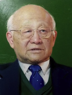 Zhiyong Sheng (盛志勇)合作论文数Chinese PLA General Hospital26Sen Hu (胡森)合作论文数Chinese Pla General Hospital25Hua Jin (晋桦)合作论文数15XiaoGuo Jiang (姜小国)合作论文数12Dan Sun (孙丹)合作论文数12XiaoQing Sun (孙晓庆)合作论文数8
 Jiake Chai (柴家科)合作论文数Chinese People's Liberation Arm General Hospital6ZhenRong Guo (郭振荣)合作论文数5
 Xiaofang Zou (邹晓防)合作论文数Air Force General Hospital5Shirong Sun (孙世荣)合作论文数4Li-xin HE (贺立新)合作论文数4Ming Yan (严鸣)合作论文数4DeGuang Shi (石德光)合作论文数4rong zhang (张蓉)合作论文数3cheng zhang (张诚)合作论文数3Ning Dong (董宁)合作论文数Burns Institute,First Hospital Affiliated to General Hospital of PLA3
 Jiangyang Lu (陆江阳)合作论文数Fourth Medical Center of PLA General Hospital3
 Yanqiu Wu (吴焱秋)合作论文数Chinese PLA General Hospital3Zhen-rong Guo合作论文数2Wei Pang合作论文数2yuqing zheng (郑玉清)合作论文数2Qi Song合作论文数2XiuRong Shuai (帅秀蓉)合作论文数2WeiHong Cao (曹卫红)合作论文数2GuoYong Zhou (周国勇)合作论文数2Yong Yu (于勇)合作论文数2
 Xiaobing Fu (付小兵)合作论文数Chinese PLA General Hospital;School of Medicine, Nankai University2
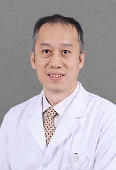 Baotong Zhou (周宝桐)合作论文数Peking Union Medical College Hospital2
 Hongming Yang (杨红明)合作论文数Chinese People's Liberation Army General Hospital2
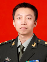 Daifeng Hao (郝岱峰)合作论文数中国人民解放军总医院第一附属(304)医院烧伤整形外科2zhiyong sheng (盛志勇)合作论文数1Lian-rong LU (陆连荣)合作论文数1Li-hong Yang合作论文数1jingjie liu (刘静杰)合作论文数1BAI Yu-mei (白玉梅)合作论文数1fei gao (高飞)合作论文数1Ai-hua MENG (孟爱华)合作论文数1Wei Sheng合作论文数1lihong yang (杨立红)合作论文数1Na SUN (孙娜)合作论文数1YuLei Gao (高玉雷)合作论文数1Jianchuan Gao (高建川)合作论文数People's Liberation Army General Hospital1DeXin Xiong (熊德鑫)合作论文数1ShiFan Zhang (张世范)合作论文数1




