ObjectivesScaphoid nonunion remains a challenging problem to manage with no general consensus on its treatment recommendations. We propose a novel minimally invasive (MIS) technique of arthroscopic bone grafting (ABG) with robot‐assisted fixation for the treatment of scaphoid nonunions.MethodsPatients with radiographically proven scaphoid nonunion treated by this novel surgical technique were included. Following arthroscopic debridement and iliac crest bone grafting, the scaphoid was fixed percutaneously using either multiple Kirschner (K)‐wires or a headless compression screw using a robotic navigation system.ResultsSix male patients with an average age of 29.2 years were enrolled. Four patients had scaphoid waist fractures, and the other two were proximal pole fractures. During wrist arthroscopy, punctate bleeding of the proximal scaphoid fragment was observed in four out of the six patients. Half of the patients were fixed using a headless compression screw and the other half using multiple K‐wires. All the guidewires were placed with a single‐attempt using the robotic navigation system. Postoperatively, all the scaphoid fractures had complete radiographic union by 16 weeks. At a mean follow‐up of 18.3 months, there were significant improvements in wrist range of motion, grip strength, and patient‐rated outcomes. No intraoperative or early postoperative complications were encountered in any of our patients.ConclusionArthroscopic bone grafting with robot‐assisted fixation is a feasible and promising therapeutic option for scaphoid nonunions, regardless of the vascularity of the proximal pole fragment. This novel technique allows for anatomic restoration of the scaphoid alignment and accurate, targeted placement of implants into the scaphoid nonunion site within a single‐attempt using a robotic navigation system.
Objective:To investigate the value of high-resolution ultrasound in the diagnosis of triangular fibrocartilage complex (TFCC) injuries.Methods:Forty-eight patients who underwent wrist arthroscopic surgery for wrist diseases in our hospital were studied and analyzed, including 34 cases of TFCC injury and 14 cases of other wrist diseases with normal TFCC. The TFCC high-resolution ultrasound was performed. Taking the results of arthroscopic exploration as the gold standard, the statistical research was carried out by comparing TFCC ultrasound imaging and arthroscopic manifestations. The sensitivity, specificity, positive predictive value, negative predictive value, positive likelihood ratio and negative likelihood ratio of high-resolution ultrasound in the diagnosis of TFCC injury and injury site were obtained.Results:The sensitivity, specificity, positive predictive value, negative predictive value, positive likelihood ratio and negative likelihood ratio of high-resolution ultrasound in the diagnosis of TFCC injury were 0.9375, 0.875, 0.9375, 0.875, 7.5, and 0.071 respectively. The sensitivity, specificity, positive predictive value, negative predictive value, positive likelihood ratio and negative likelihood ratio for the diagnosis of central degenerative perforation of TFCC were 0.9629, 0.9048, 0.9285, 0.95, 10.11 and 0.041 respectively. The sensitivity, specificity, positive predictive value, negative predictive value, positive likelihood ratio and negative likelihood ratio of high-resolution ultrasound in the diagnosis of TFCC ulnar insertion injuries were 0.95, 1, 1, 0.9655, + ∞ and 0.05 respectively.Conclusion:High resolution ultrasound has certain value in the diagnosis of TFCC injury, especially the common clinical TFCC central degenerative perforation and ulnar insertion injuries, which is worthy of further study on its method and diagnostic criteria.
Purpose To describe the clinical features, radiologic findings, differential diagnosis, and surgical treatment of a congenital flexion deformity of the middle, ring, and little fingers. The cause of the condition is the aberrant origin of the flexor digitorum profundus, leading to a congenital contracture of the ulnar digits. Methods We reviewed 8 patients with congenital contracture of the ulnar digits. The mean age at the time of surgery was 14 years. An examination revealed a flexion contracture of the middle, ring, and small fingers. Plain radiographs, 3-dimensional computed tomography, magnetic resonance imaging, and ultrasound were used to characterize bony and soft tissue pathology. Surgical treatments included resection of the aberrant origin and a muscle-sliding procedure. Results Bony prominence on the proximal ulna was seen in the plain radiographs and/or 3-dimensional computed tomography. A cord that extended from this bony prominence to the tendons of flexor digitorum profundus was revealed in the magnetic resonance imaging. The bony prominence and the cord were also seen using ultrasound. The median time of patient follow-up was 1.7 years. A simple resection of the tendinous origin only resulted in a release in 2 patients who were 4 years old. Older patients required a further muscle-sliding procedure. The average grip strength ratio on the contralateral side was 82%. Conclusions Congenital contracture of the ulnar digits is a new congenital flexion deformity involving the middle, ring, and small fingers. Bony prominence on the proximal ulna is the key finding for establishing its diagnosis and distinguishing it from an ischemic contracture. We recommend treating this surgically at 12 years of age or older after the phase of rapid growth of the extremities. We recommend the resection of the aberrant origin, combined with a muscle-sliding procedure, as the treatment of choice, even for young patients. (Copyright (C) 2022 by the American Society for Surgery of the Hand. All rights reserved.)
亚急性舟骨骨折的治疗迄今仍有争议。延迟的石膏固定使骨折不愈合的发生率较高;亚急性舟骨骨折也并不都需要植骨手术,稳定无移位的骨折可通过微创经皮螺钉固定实现愈合。但由于舟骨特殊的解剖形态,徒手拧入螺钉可能出现位置及长度不满意,这使得术后关节磨损、骨折不稳定等并发症的发生率较高。机器人辅助技术能大大提高螺钉置入的精确度及安全性,且目前尚无应用于稳定亚急性舟骨骨折的报道。笔者回顾性分析2例稳定无移位的亚急性舟骨骨折患者在导航机器人与腕关节镜辅助下行微创固定治疗的临床资料,探讨其可行性及初步临床疗效。
Objective:To investigate the value of high-resolution 3T MRI in the detection of triangular fibrocartilage complex (TFCC) injuries through a diagnostic test.Methods:A retrospective case series analysis was performed in 133 patients with ulnar wrist pain admitted in Beijing Jishuitan Hospital from January 2013 to April 2018. There were 68 males and 65 females, aged 14-69 years (mean 32.6 years). The patients was examined with the wrist MRI with consistent parameters and then received wrist arthroscopic detection or treatment. The TFCC injuries were classified as central injury, namely triangular fibrocartilage disc injury and peripheral injury, namely the tear of ulnar attachment, radial attachment, distal radioulnar ligaments and ulnocarpal ligaments. Take the results of wrist arthroscopy as the gold standard, the MRI and arthroscopic findings were compared and the sensitivity, specificity, positive/negative predictive value and positive/negative likelihood ratio were determined.Results:Wrist arthroscopy confirmed TFCC injuries in 122 patients, among which 72 patients were with central injury and 102 with peripheral injury. Meanwhile, TFCC injuries were diagnosed with MRI in 124 patients, among which 75 patients were central injury and 111 cases were peripheral injury. As for central injury, the sensitivity/specificity, positive/negative predictive value and positive/negative likelihood ratio of MRI were 0.972/0.918, 0.933/0.966 and 11.85/0.03, respectively; when the target pathology was peripheral injury, the sensitivity/specificity, positive/negative predictive value and positive/negative likelihood ratio of MRI were 0.882/0.323, 0.811/0.45 and 1.30/0.37, respectively.Conclusions:High-resolution 3T MRI is accurate in detection of central injury of TFCC, no matter the injury is traumatic or degenerated. In the diagnosis of peripheral injury of the TFCC, MRI can provide some help for its high sensitivity. However, the positive findings of MRI on peripheral structures should be cautious because of the poor specificity, and hence the results of history, physical examination and MRI should be considered together to attain an accurate diagnosis.
Objective To evaluate conventional wrist physical examination in detecting injury to the triangular fibrocartilage complex (TFCC).Methods A retrospective study was conducted of the 118 patients (119 wrists) who had been admitted from January 2013 to October 2017 to Hand Surgery Department,Beijing Jishuitan Hospital for wrist arthroscopic surgery.All of them underwent wrist physical examination preoperatively for conventional index tests for TFCC injury like ulnar fovea sign,piano-key test,distal ulnar ballottement test and ulnocarpal stress test.They were 68 males and 50 females with a mean age of 32 years.The results of physical examination were compared with the arthroscopic findings to calculate the sensitivity,specificity,positive/negative predictive value (PPV/NPV) and +/-likelihood ratio (LR) of each index test.In addition,we combined the results of any 2 tests to increase the capability of detecting peripheral TFCC injury.Results The diagnostic values of each index test were achieved by comparison between the results of physical examination and the arthroscopic findings:(1) ulnar fovea sign:sensitivity =0.648,specificity =0.742;(2) piano key test:sensitivity =0.817,specificity =0.735;(3) distal ulnar ballottement test:sensitivity =0.927,specificity =0.647;(4) ulnocarpal stress test:sensitivity =0.825,specificity =0.500.The combination of any 2 tests produced 3 new index tests,of which the most valuable combination was ulnar fovea sign & distal ulna ballottement test,with a sensitivity of 0.598,a specificity of 0.941,a PPV of 0.961and a + LR of 10.14,Conclusions The signs elicited in physical examination and special tests can be helpful in the diagnosis of TFCC injuries.However,as the value of any single test is not enough,we should clinically combine the results of ulna fovea sign and distal ulna ballottement test to increase the capability of detecting peripheral TFCC injury.
Objective To explore the effectiveness of wide awake local anesthesia no tourniquet (WALANT) in arthroscopy for diagnosis and treatment of wrist injuries and conditions.Methods Between January 2012 and January 2016,WALANT approach was used in wrist arthroscopy for 22 patients with wrist injury or condition at Department of Hand Surgery,Beijing Jishuitan Hospital.They were 10 males and 12 females,aged from 19 to 56 years(average,34 years).Arthroscopic exploration was conducted in 7 cases,arthroscopic synovectomy in 6,dorsal wrist ganglionectomy in 3,triangular fibrocartilage complex (TFCC) debridement in 5 and TFCC repair in one.All the patients were anesthetized using portal site local anesthesia (PSLA) technique.Additional local infiltration around ulnar-sided wrist incisions was performed in 2 TFCC repairs.Bupivacaine was used for both techniques.The effects were evaluated using a ten-point visual analogue scale (VAS).Results Desired objectives were achieved in all patients.No operative complication was observed.Mild pain was noted in all the patients when local anesthetic was injected to the portal sites,with an average VAS score of 2.5 points (from 1 to 3 points).In the following arthroscopic procedures,most of the patients did not report any discomfort.Only one patient undergoing TFCC repair reported mild pain (VAS score of 3 points) during ulnar-carpal suture procedure.Conclusion The new WALANT technique is safe,time-saving,economical and capable of pain control during arthroscopy for numerous wrist injuries and conditions.
目的 介绍小关节镜微创治疗第1腕掌关节疾病的常用手术方法及临床初步应用结果.方法 回顾分析2013年1月至2015年12月北京积水潭医院手外科采用1.9mm小关节镜及配套小关节镜手术器械行第1腕掌关节手术的11例患者的临床资料.对其中2例关节内游离体行游离体取出术,2例关节滑膜炎行滑膜切除术,5例骨关节炎行大多角骨部分切除关节成形术,2例韧带松弛行韧带皱缩术.术后随访以疼痛视觉模拟评分(VAS)、臂肩手功能障碍评分(DASH)、Kapandji评分以及拇指与示指侧方的侧捏力量等指标对效果进行评价.结果 通过小关节镜成功对所有患者第1腕掌关节的结构进行了观察,对病变进行了评估.2例接受关节内游离体取出的患者术后关节活动时的卡顿疼痛感消失.2例接受滑膜切除患者平均VAS评分从5.0分降至1.0分,平均Kapandji评分从9.0分增至10.0分.2例接受韧带皱缩患者术后平均DASH评分从23.9分降至0分.5例接受大多角骨部分切除关节成形术患者,术后平均VAS评分从6.8分降至0.4分,平均DASH评分从61.7分降至3.2分,平均Kapandji评分从6.8分提高至9.4分.患侧捏力占健侧捏力百分比从术前平均82.6%(60.0%~107.0%)增至术后96.0%(68.8%~111.0%).结论 小关节镜对适宜的第1腕掌关节疾病进行治疗的近期随访结果满意,是一种安全有效的微创治疗选择.
Triangular fibrocartilage complex (TFCC) plays an important role in maintaining stability of the distal radioulnar joint (DRUJ),buffering the ulnar carpal axial impaction and holding the proximal carpal bones.Patients with injured TFCC may suffer pain over the ulnar side of the wrist and DRUJ instability.Many of them may have difficulty in undertaking daily activities,leading to even psychological problems.With the development of arthroscopic technology and updated understanding of the mechanisms of TFCC injury,great progress has been made in the diagnosis and treatment of the traumatic injury,especially in the arthroscopy-assisted treatment.
Objective To introduce indication , surgical techniques and outcome of percutaneous screw fixation for acute and neglected scaphoid fractures .Methods We retrospectively reviewed 170 patients who had an acute or neglected scaphoid fracture treated with percutaneous screw fixation between January 2003 and January 2015 .Results One hundred and fourteen patients had full follow-up data , with 91 palmar approach and 23 dorsal approach procedures .All 77 acute fractures ( without wrist joint dislocation ) united at 8 or 12 weeks follow-up.The mean follow-up period was 32 months (range, 12~84 months).No sign of traumatic ar-thritis or other complications was observed .According to the Mayo Wrist Scores , overall functional outcomes were rated as excellent in all patients .The mean Patient-Rated Wrist Evaluation (PRWE) score was 1.8.For 26 transscaphoid perilunate dislocations , the mean follow-up period was 13 months (range,6-30 months). The Mayo wrist score were rated as excellent in 13 patients, good in 8, fair in 4 and poor in 1.The mean PRWE score was 11.For 11 neglected scaphoid fractures, the average follow-up period was 14 months (range, 6-36 months).Six of the 11 patients received arthroscopically assisted percutaneous fixation with arthroscopic debridement and bone grafting .Union was achieved in all 11 patients at a mean of 11 weeks ( range, 8-17 weeks ) .According to the Mayo Wrist Scores , overall functional outcomes were rated as excellent in 10 patients and good in 1.Conclusions Percutaneous screw fixation is a reliable and favorable approach in the treatment of acute scaphoid fractures and scaphoid fractures with delayed diagnosis and treatment according to our early follow-up results.
Objective To assess the effectiveness of arthroscopically assisted minimally invasive management for neglected scaphoid fractures in the subacute stage.Methods Between January 2012 and January 2015,ten patients who had a neglected scaphoid fracture were treated with arthroscopically assisted percutaneous fixation with or without arthroscopic debridement and bone grafting.Preoperatively fracture displacement,fracture sclerosis and bone absorption were evaluated on radiographic images.Fracture displacement and stability were further assessed intraoperatively via direct arthroscopic visualization through the mid-carpal portal.Based on the displacement and stability of the fracture,and the extent of sclerosis and bone defect,arthroscopic guided direct percutaneous internal fixation;arthroscopic debridement,reduction and percutaneous intemal fixation;or arthroscopic debridement,bone grafting and internal fixation were carried out.Results The average postoperative follow-up time was 14.7 months (range,6 to 36 months).Bone union was achieved in all ten patients at a mean of 11.3 weeks (range,8 to 17 weeks).According to the Mayo wrist scores,overall functional outcomes were rated as excellent in 9 cases and good in 1 case.Conclusion Arthroscope assisted minimally invasive management is a reliable and favorable approach in the treatment of scaphoid fractures with delayed diagnosis and treatment.It affords direct visualization of the fracture site which enables tailored management.
Background and aim: Better methods for anterolateral thigh flap donor-site reconstruction are desirable in cases when direct closure is impossible. Multiple surgical strategies have been attempted, and each has its shortcomings. The use of a contralateral free groin flap to repair the anterolateral thigh flap donor site is investigated in this report.Methods: From October 2015 to February 2016, free groin flaps were harvested on six patients for aesthetic and functional donor-site closure of the anterolateral thigh flap, which could not be directly closed. In these cases, the reverse-flow distal portion of the descending branch of the lateral circumflex femoral artery and vein were used as recipient vessels and anastomosed to the superficial circumflex iliac artery and vein, respectively.Results: One flap had presented a few blisters on the distal margin; the other five flaps fully survived without any complications. Patients were highly satisfied with the aesthetic outcomes of both the anterolateral thigh area and the groin site.Conclusion: Although with theoretical risks of compromised venous blood flow, free groin flaps are an effective strategy for closure of massive anterolateral donor-site defects and can be safely performed with thoughtful planning and meticulous microsurgical techniques. (C) 2016 British Association of Plastic, Reconstructive and Aesthetic Surgeons. Published by Elsevier Ltd. All rights reserved.
Objective To compare volar and dorsal percutaneous screw fixation in acute scaphoid waist fracture.Methods Retrospectively collected patients received percutaneous screw fixation after acute scaphoid fracture between January 2009 and December 2014 with non-displacement/minimal displacement waist fracture,patients with open fracture or combined with perilunate dislocation or fracture which need reduce were excluded,28 patients with intact follow-up were enrolled,26 men and 2 women,with an average age of 31 years old.Based on Herbert classification,there were 15 A2 type,5 B1 type,8 B2 type.7 cases were high energy trauma and 21 were low energy.The patients were divided into 2 groups by volar and dorsal approach.The general condition,surgery notes,radiological evidence and follow-up were collected and analyzed.Results 28 cases with full followup were enrolled,18 cases received surgery via volar approach,and 10 cases via dorsal approach,the mean follow-up period is 33 months.Peri-operative data showed that operation time in dorsal approach group was longer (75 min vs.54 min).We evaluated screw position by measuring the angulation between screw and fracture line as well as screw and long axis of scaphoid based on AP view and lateral view of wrist X-rays.There was no significant difference in length of screws (21.4 mm vs.21.8 mm).Patients gradually went back to work after 2 weeks and got fracture healing within 12 weeks after operation.Radiological evidence showed that angulation between screw and long axis of scaphoid was 6.11°±4.82° in AP position and 8 48°±5.77° in lateral position,angulation between screw and fracture line was 73.58°±20.75° in AP position and 70.72°±21.19° in lateral position.There were no significant differences between screw insertion positions between 2 groups.Visual analogue scale (VAS) was used to evaluate wristpain in the follow-up,Mayo wrist score and Patient-Rated Wrist Evaluation (PRWE) were also used to assess wrist function in the follow-up.The clinical results were promising,the excellent or good rate of Mayo wrist score was 100%,there were no significant difference in VAS,Mayo wrist score and PRWE between volar and dorsal approach.No complications such as failure of screws,orthopaedic arthritis or scaphoid necrosis were observed.Conclusion Percutaneous screw fixation have a satisfied effect in the treatment of acute scaphoid waist fracture,and there are no differences in clinical efficacy between volar and dorsal approach.
目的 探讨腓肠神经滋养皮瓣治疗足背皮肤缺损患者临床疗效. 方法 将76例是背皮肤缺损患者按照随机数字表法分组为对照组与观察组,各38例.对照组采用股前外侧皮瓣修复术治疗,观察组采用腓肠神经滋养血管皮瓣治疗.统计两组患者创面愈合情况,采用美国矫行足踝协会评分系统评价两组足部功能,统计术后并发症及皮瓣存活情况. 结果 观察组术后皮瓣存活率100.0%,明显高于对照组78.9%,P<0.05;观察组伤口Ⅰ期愈合率100.0%,明显高于对照组81.6%,P<0.05;观察组足部功能优良率94.7%,明显高于对照组71.1%,P<0.05;两组术后并发症率相比较,差异无统计学意义,P>0.05. 结论 应用腓肠神经滋养皮瓣治疗足背皮肤缺损疗效显著且安全,有利于促进患者足部功能改善.
Objective To evaluate the subjective and objective outcomes after Ueba reconstruction of Manske type Ⅰ to Ⅲ cleft hand.Methods Eleven patients with 15 cleft hands from Manske type Ⅰ to Ⅲ were treated from January 2008 to December 2014.There were 8 males and 3 females,with an average age of 40 months (range,25 to 87 months).Ueba procedure was applied for the reconstruction.Three skin flaps were made within the cleft.The index ray was transferred ulnarward and supinated.A transverse metacarpal ligament reconstruction was performed.Simultaneously,Manske type Ⅱ] and]Ⅲ patients were treated with a rotation flap from the cleft and full thickness skin graft from groin to reconstruct the first web space.Both the surgeon and the parents assessed the subjective outcomes using a visual analog scale (VAS) to compare preoperative and postoperative appearance.Objective outcomes evaluations included range of motion (ROM) of the digits,and measurement of divergence angles of the index finger and ring finger metacarpals and phalanges on X-rays.Results The surgeon's VAS score increased from 3.1 preoperatively to 7.5 postoperatively.The parents were very satisfied with 6 hands,satisfied with 4 hands,somewhat satisfied with 4 hands,and dissatisfied with 1 hand.ROM of index fingers were 271° preoperatively and 263° postoperatively.ROM of ring fingers were 267° preoperatively and 262° postoperatively.There was no statistically significant difference between the preoperative and postoperative ROM.The metacarpal divergence angle significantly improved from 34°to 13°,while the phalangeal divergence angle significantly improved from 45°to 14°.Conclusion Ueba procedure improves hand appearance in patients with cleft hand without affecting range of motion of the fingers.
Objective To investigate the application and outcome of reconstructing the sagittal bands and correct the old subluxation of the extensor digitorum communis (EDC) with tendon allograft.Methods From May 2010 to April 2014,23 cases (15 males and 8 females,aged from 17 to 64 years,36.5 on average) suffered from old subluxation of the EDC were treated with tendon allograft to reconstruct the sagittal bands.A total of 23 cases fingers were included,of which 12 cases were caused by injury,6 by rheumatoid arthritis and 5 by congenital anomalies.The motion of the MP joint was measured to evaluate the recovery.Results All patients were follow-up for 3 to 12 months.Postoperative assessment of hand function showed excellent in 19 cases,good in 3 cases and fair in 1 case (mild subluxation of the EDC).The overall excellent and good rate was 95.6%.Most patients were satisfied with the recovery of hand function.Incidence rate of postoperative complications was low.Conclusion Tendon allograft to reconstruct the sagittal bands could be a simple and safe option to repair the old subluxation of the EDC.
Objective To investigate the arthroscopic treatment methods of intraosseous ganglion cysts of the wrist. Methods From May 2013 to May 2014, 5 patients with intraosseous ganglion cysts in the wrist were treated with arthroscopic technique. There were 3 cases in the lunate, 1 in the scaphoid and 1 in the triangular bone. The main symptom was chronic wrist pain, decreased grip strength and limitation of wrist movement. The X-ray and CT showed circular and quasi-circular areas of reduced bone mineral density within the carpal bone with a clear boundary and slightly sclerotic margin. The MRI revealed the liquid content. All the patients were treated by arthroscopic technique: curettage of the ganglion cysts and autologous cancellous bone or injectable artificial bone grafting. The wrists were immobilized at the functional position for 3 weeks after the operation. Results All the patients were followed up for a mean period of 6.8 months ( range 4~12 months ). All cases had no recurrence and obtained bone healing. The symptoms were relieved. All the patients recovered to their normal life and work. Conclusion Intraossous ganglion cysts in the wrist remain a cause of chronic wrist pain. The arthroscopic technique in the treatment of intraosseous ganglion cysts of the wrist is safe, less complication, rapid recovery of function, the effect is satisfactory.
Objective To investigate the clinical features and the diagnosis and treatment of Kaposiform hemangioendothelioma ( KHE ) of the hand. Methods From 2010 to 2013, 5 patients with KHE of the hand were treated in our department, including 4 males and 1 female. Their mean age was 4.6 years old ( range;6 months-33 years ), among whom there was 1 adult patient of 33 years old and 4 infants. No abnormal coagulation function was found before the operation. Extended local resection of tumors were performed on all the patients. Results The diagnosis of KHE was conifrmed by the postoperative pathology. A multiple lobular and invasive growth pattern was observed in the tumors, and vascular endothelial cells were shown. The densely packed spindle-shaped tumor cells and sieve-like or slit-like blood vessels could be seen, with focal hyperplasia of glomeruloid structures. Bone destruction was found in some cases in the histological or radiographic examination. The immunohistochemical staining showed all the vascular markers were positive including cluster of differentiation ( CD ) 31, CD34, ( friend leukemia integration 1 transcription factor ) Fli-1 and Factor VIII. The smooth muscle antibodies ( SMA ) were focally positive and the focal expression of cytokeratin ( CK ) was noticed in 1 case. All the patients were followed up for a mean period of 18 months ( range;12-25 months ). No recurrence was found but only in 1 patient at 1 year after the operation, who underwent surgery again. Conclusions Simple extended resection of tumors is an effective method in the treatment of KHE of the hand, with excellent outcomes.
Objective To study the results of several operative methods (neurolysis,nerve repair and nerve grafting) for the treatment of the ligated peripheral nerves using rat models.Methods A total of 144 rats were randomly assigned to 3 groups:neurolysis group,nerve repair group and nerve grafting group.Ligation of the sciatic nerve was done first in all the animals to establish the nerve injury model.After 2 weeks,a second surgery was carried out to treat the nerve lesion.Neurolysis,nerve repair or nerve grafting was carried out according to group assignment.The rats were sacrificed at postoperative week 1,2,4,6,8,10 and 12 respectively.Weight of the gastrocnemius muscle was measured and the myelinated nerve ftber cotmt of the sciatic nerve was evaluated.Results were analyzed statisticafly to make comparison among the groups.Results Gastrocnemius muscle weight recovered after 12 weeks of single ligation of the sciatic nerve in the control group in which no treatment was carried out.Muscle weight and myelinated nerve fiber count,however,did not show obvious increase in the double ligation group where multiple lesion of the sciatic nerve was created but not treated.In the neurolysis group,4 and 6 weeks after the second procedure,gastrocnemius muscle weight and myelinated fiber count of the sciatic nerve of the experimental side were significantly higher than those of the contralateral side.In the nerve repair group,at 6 weeks after the procedure,gastrocnemius muscle weight of the experimental side was significantly higher than that of the contralateral side,whereas the sciatic nerve myelinated fiber count was not significantly different.In the nerve grafting group,gastrocnemius muscle veight of the experimental side was significantly higher than that of the contralateral side at 6 weeks after the procedure,while the sciatic nerve myelinated fiber count was signfificantly better at 12 weeks.Conclusion For ligation induced stenosis of the peripheral nerve,surgical intervention should be considered proactively if there is no improvement after 12 weeks of conservative treatment.Neurolysis would lead to better results than conservative treatment and is the recommended procedure.The results of nerve repair are not certain.Thus this procedure should be chosen with caution.For multiple or wide range stenosis of the nerve,resection of the lesion area and nerve grafting should be the treatment of choice.
原发性周围神经狭窄(后简称“狭窄”),又称“沙漏”样、“腊肠”样狭窄,指发生在周围神经干或神经束上的不明原因的环周狭窄,在其周围看不到有与之相关的外在致压物。既往文献所报道病例,仅涉及上肢神经,尤其是肘部神经,如桡神经、骨间后神经、正中神经、骨间前神经、尺神经及其分支。2009年我们曾撰写过一篇文献综述[1],复述了其可能的成因、大体形态及相关的治疗方法。在此之后,即近四年的文献显示,狭窄还可见于肩部神经:腋神经、肩胛上神经和肌皮神经;腕部神经:正中神经;手部神经:指固有神经;膝部神经:腓总神经,以及儿童的桡神经,其成因为何,认识依旧不一。此外,文献中还有了超声、MRN检查狭窄的回顾性报道,有了据原发骨间前、后神经麻痹手术所见而提出的狭窄分型与分度,有了手术疗效的回顾性分析。本文将上述综述遗漏的一些信息汇总如下,以方便感兴趣的同仁查阅。
 Guanglei Tian (田光磊)合作论文数Department of Hand Surgery,Beijing Jishuitan Hospital,Beijing ,China31
 ShanLin Chen (陈山林)合作论文数Beijing Jishuitan Hospital26
 Bo Liu (刘波)合作论文数Peking University People's Hospital18
 Wen Tian (田文)合作论文数Beijing Jishuitan Hospital17
 Zhixin Wang合作论文数10
 Chuanjun Yi (易传军)合作论文数Beijing Jishuitan Hospital9
 Chun Li (李淳)合作论文数Beijing Jishuitan Hospital8JunHui Zhao (赵俊会)合作论文数7
 Yong Yang (杨勇)合作论文数Hospital for Skin Diseases, Chinese Academy of Medical Sciences7Xue Yun-hao合作论文数6Yin Zhu (诸寅)合作论文数6PengCheng Li (栗鹏程)合作论文数6YuCheng Li (李玉成)合作论文数6
 Chunmei Hou (侯春梅)合作论文数Beijing Jishuitan Hospital6
 Ge Xiong (熊革)合作论文数Beijing Jishuitan Hospital5
 Zhongzhe Li (李忠哲)合作论文数Beijing Jishuitan Hospital5YongBin Gao (郜永斌)合作论文数4Qi Hu (胡琪)合作论文数4Wei Zheng (郑炜)合作论文数4
 Youle Zhang (张友乐)合作论文数Beijing Jishuitan Hospital4Yanbo Rong合作论文数3JiaNing Wei (韦加宁)合作论文数3DeDi Tong (童德迪)合作论文数3Chen Yang (杨辰)合作论文数3
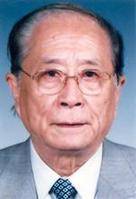 Shuhuan Wang (王澍寰)合作论文数Beijing Jishuitan Hospital3
 Wenjun Li (李文军)合作论文数Beijing Jishuitan Hospital3Lu-fei Dai合作论文数2Yaobin Yin合作论文数2Lingyu Wang合作论文数2Qin Hu (胡溱)合作论文数2WanShen Chang (常万绅)合作论文数2Guo Wen合作论文数2
 Yongwei Pan (潘勇卫)合作论文数Tsinghua University Affiliated Beijing Tsinghua Changgung Hospital2
 Shufeng Wang (王树锋)合作论文数Beijing Jishuitan Hospital2
 Yang Guo (郭阳)合作论文数Beijing Jishuitan Hospital2
 Wei Zhu (朱伟)合作论文数Beijing Jishuitan Hospital2
 Chang Liu合作论文数Department of Pathology & Immunology, Washington University School of Medicine in St. Louis2Wei Chen (陈玮)合作论文数1Jiuzheng Deng合作论文数1Lu Liu合作论文数1Cheng Shen合作论文数1Shen Jie合作论文数1Zhe Yi合作论文数1shuang wang (王爽)合作论文数1lei xu (徐雷)合作论文数1
 Rebecca QR Lim合作论文数Telethon Kids Inst, Univ Western Australia1




