Estrogen is very important to the differentiation of B lymphocytes; B lymphopoiesis induced by OVX was supposedly involved in osteoporosis. But the effects of B lymphocytes on the osteogenic differentiation of bone mesenchymal stem cells (BMSCs) are not clear. In this study, we detected bone quality and bone loss in a trabecular bone by electronic universal material testing machine and microcomputed tomography (micro-CT) in OVX and splenectomized-ovariectomy (SPX-OVX) rats. Additionally, changes in lymphocytes (B lymphocyte, CD4 + and CD8 + T lymphocytes, and macrophages) in the bone marrow were analyzed by flow cytometry. The osteogenesis of BMSCs cocultured with normal and LPS-pretreated B lymphocytes was detected by BCIP/NBT and Alizarin red S staining. Measurement of the Notch2, Notch4, Hey1, Hey2, Hes1, and runt-related transcription factor 2 (Runx2) expression in BMSCs cocultured with B lymphocytes was done using real-time PCR. The effects of dexamethasone and DAPT (inhibitor of Notch signaling) on osteogenesis of BMSCs were detected by BCIP/NBT, Alizarin red S staining, and real-time PCR. Osteoporosis happened in OVX rats, more serious in SPX-OVX rats, B lymphocytes increased in OVX rats, and sharply higher in SPX-OVX rats. Osteoporosis did not happen in SPX rats which is still companied with a high increase of B lymphocytes. LPS-pretreated B lymphocytes suppressed the osteogenesis of BMSCs, but the normal B lymphocytes could not. The LPS-pretreated B lymphocytes upregulated the expression of Notch4, Hes1, and Hey2 and downregulated the expression of Runx2 in BMSCs. Dexamethasone and DAPT could downregulate the high expression of Notch4, Hes1, Hey2 and upregulate the low expression of Runx2 in BMSCs which cocultured with LPS treated B lymphocytes, the inhibited ALP and Alizarin red staining in BMSCs which cocultured with LPS treated B lymphocytes also partly restored.
BACKGROUND:While it is known that irradiation can induce local and systemic bone loss over time, how focal irradiation induces systemic bone complications remains unclear. Immune cells are thought to be crucial to bone homeostasis, and abnormal immune cells lead to serious disruption of bone homeostasis, such as in acute lymphoblastic leukaemia. This disruption primarily occurs due to inhibition of the osteogenic differentiation of bone mesenchymal stem cells (BMSCs).METHODS:In this study, we detected local and systemic bone loss in trabecular bone by micro-computed tomography (micro-CT) and measurement of peroxisome proliferator-activated receptor gamma (PPARγ) and runt-related transcription factor 2 (RUNX2) expression in BMSCs using real-time polymerase chain reaction and western blotting. Additionally, changes in lymphocytes (B cells and CD4+ and CD8+ T cells) in the peripheral blood and bone marrow were analysed by flow cytometry. BMSC-derived osteoblasts and adipocytes, cultured in osteogenic or adipogenic media or co-cultured with lymphocytes, were detected by BCIP/NBT, Alizarin Red S and Oil Red O staining.RESULTS:Focal irradiation induced local and systemic bone loss in trabecular bone. Increased PPARγ expression and decreased RUNX2 expression were observed, accompanied by upregulated adipogenesis and downregulated osteogenesis of BMSCs. B cells and CD8+ T lymphocytes were increased in the blood and bone marrow after irradiation, while CD4+ T lymphocytes were decreased in the blood. Inhibition of RUNX2 expression and reduction of alkaline phosphatase activity and mineralization deposits were observed in lymphocyte-co-cultured BMSCs, accompanied by an increase in PPARγ expression and in the number of lipid droplets.CONCLUSIONS:Focal irradiation induced local and systemic bone loss in trabecular bone. Increased B cells and CD8+ T lymphocytes led to systemic bone loss by decreasing BMSC osteogenesis.
BACKGROUND:Bone-related complications are commonly reported in cancer patients receiving radiotherapy and are collectively referred to as the abscopal effect of irradiation, the mechanism of which remains poorly understood. When patients receive targeted radiotherapy to a tumor, the local skeleton is exposed to radiation, particularly within the bone marrow. We therefore investigated the hypothesis that single bone irradiation can induce deterioration of the skeleton outside the radiation field and is mediated by the bone marrow.METHODS:Using 4-month-old male Sprague-Dawley rats, the effects of irradiation (20 Gy, right distal femur and proximal tibia) on bone quality, microarchitecture and bone marrow, were evaluated prospectively by microcomputed tomography, histomorphometry, real-time polymerase chain reaction, and Western blot analysis.RESULTS:At 12 weeks post-irradiation, bone loss of the non-irradiated bone was induced and marrow adiposity was increased. Expression of runt-related transcription factor-2 by bone mesenchymal stem cells (BMSCs) decreased after irradiation by 88.0 % (P < 0.01) at the contralateral and 82.3 % (P < 0.01) at the irradiation site 2 weeks post-irradiation and decreased by 94.5 % (P < 0.001) at the contralateral and 44.1 % (P < 0.05) at the irradiation site 12 weeks post-irradiation. Interestingly, peroxisome proliferator-activated receptor gamma expression decreased by 61.8 % (P < 0.05) at the contralateral and by 48.3 % (P < 0.05) at the irradiation site 2 weeks post-irradiation but increased by 9-fold at the contralateral (P < 0.001) and by 13-fold (P < 0.001) at the irradiation site 12 weeks post-irradiation.CONCLUSIONS:These data highlight that radiation-induced bone complications are partly BMSC-mediated, with important implications for bone health maintenance in patients receiving radiotherapy.
Objective To study the influence of fluorine on signaling pathway of osteoprotegerin(OPG)/ receptor activator of NF-κB ligand(RANKL) in cultured rat osteoblasts.Methods Osteoblasts were isolated from skull of neonatal rats(< 24 hours) by enzyme digestion,and fluorine of different concentrations [0 (control),1 × 10-3,1 × 10-4,1 × 10-5,1 × l0-6 and 1 × 10⁻⁷ mol/L] were added into the culture medium of second generation of osteoblasts.The expressions of OPG and RANKL mRNA were determined using real-time PCR 24 and 48 hours after culturing.The expressions of OPG and RANKL protein were measured by Western blotting.Results ① After exposed to fluorine for 24 hours,the differences of RANKL and OPG mRNA expression had statistical significance between groups(F =30.95,22.62,all P < 0.01),the expression of RANKL mRNA(5.99 ± 0.39) in the 1 × 10⁻⁵ mol/L group and the expressions of OPG mRNA(3.52 ± 0.09,4.81 ± 0.15,3.68 ± 0.04) in the 1 × 10-4,1 × 10-5 and 1 × 10⁻⁶ mol/L groups were higher than those of the control group(3.20 ± 0.19,3.09 ± 0.58,all P < 0.05),but in the 1 × 10⁻³ mol/L group,RANKL mRNA(2.29 ± 0.18) was lower than that of the control group(P < 0.05).After exposed to fluorine for 48 hours,the differences of RANKL and OPG mRNA expression had statistical significance between groups(F =26.62,5.72,all P < 0.01),the expressions of RANKL and OPG mRNA(6.67 ± 0.49 and 5.05 ± 0.51) in the 1 × 10⁻⁵ mol/L group were higher than those of the control group(4.29 ± 0.07 and 4.34 ± 0.12,all P < 0.05),and in the 1 × 10⁻³ mol/L group the expression of OPG mRNA(3.63 ± 0.49) was lower than that of the control group(P < 0.05).② The expression of RANKL protein was not statistically significant between 24 hours and 48 hours groups (F =0.07,0.49,all P > 0.05) ; the differences of OPG protein expression had statistical significance between groups(F =3.26,P < 0.05),the expression of OPG protein in the 1 × 10⁻⁵ mol/L group(1.45 ± 0.10) was higher than that of the control group(1.05 ± 0.06,P < 0.05) at the 24 hours.After 48 hours,the expression of OPG protein was not statistically significant(F =0.44,P > 0.05).Conclusions At lower fluorine concentrations,bone formation is the main activity.But when fluorine concentration increased and time prolonged,the osteoclast differentiation and maturation are promoted,and the bone resorption is the main thing.
探索辐照后骨髓淋巴细胞对成骨细胞增殖分化的影响.采用酶消化法分离培养新生SD大鼠头盖骨成骨细胞,密度梯度离心法获取大鼠骨髓淋巴系细胞,137Csγ-射线辐照淋巴细胞和成骨细胞,剂量6 Gy,分组为0 Gy成骨细胞+0 Gy淋巴细胞(0OB0L),0 Gy成骨细胞+6 Gy淋巴细胞(0OB6L),6 Gy成骨细胞+6Gy淋巴细胞(6OB6L),共培养6h后,取各组淋巴细胞与正常成骨细胞共培养,MTT和PNPP法观察其对成骨细胞增殖和分化的影响,Real time PCR方法观察成骨细胞ALP、OCN、RANKL和OPG的基因表达变化.结果显示,辐照骨髓淋巴细胞抑制成骨细胞分化,成骨细胞ALP和OCN的表达显著降低,RANKL的表达增高,RANKL/OPG的比值则显著升高.辐照后的骨髓淋巴细胞抑制成骨细胞分化并且可能通过改变RANKL/OPG的比值进而影响破骨细胞的功能.
SD大鼠胫骨部位30 Gy照射2周和12周后处死,取血离心得血清;酶消化法分离培养新生SD大鼠头盖骨成骨细胞.采用MTT和PNPP法观察不同血清对成骨细胞增殖和分化的影响;用流式细胞仪观察成骨细胞周期变化;用Realtime PCR方法观察成骨细胞CyclinD、CyclinE、p21和p53的表达.结果显示:辐照2周和12周的大鼠血清抑制成骨细胞增殖,对成骨细胞ALP的表达无明显影响;辐照2周和12周的大鼠血清增加G1期成骨细胞比例,减少S期和G2期细胞比例;辐照2周大鼠血清抑制成骨细胞CyclinD和CyclinE的表达;12周大鼠血清促进成骨细胞p21和p53的表达,抑制成骨细胞CyclinE的表达.局部辐照大鼠血清通过抑制成骨细胞CyclinD和CyclinE表达,上调p21和p53的表达持续阻滞细胞于G1期抑制成骨细胞增殖.
Objective To explore the influence of fluorine on mRNA and protein expression of the insulin-like growth factor-1 (IGF-1) and its receptor of rat osteoblasts.Methods Osteoblasts were isolated from rat bone by enzyme digestion.Different fluorine concentration[0 (con-
Objective To investigate the roles of fibroblast growth factor 23 (FGF23) in osteoblast by RNA interference (RNAi) and its influence on cell differentiation by parathyroid hormone (PTH) in vitro. Methods The primary rat calvarial osteoblasts were cultured in MEM medium containing 10% charcoal stripped fetal bovine serum(CSFBS) and treated with rhPTH1-34 for 3 days.The changes of proliferation and alkaline phosphates (ALP) activity were measured by MTT and PNPP methods respectively,and the ALP and OCN mRNA levels were determined by real-time RT-PCR.Further FGF23 was transiently silenced by shRNA method in osteoblasts and the expression levels of FGF23,ALP and OCN were determined in transcriptional levels after rhPTH1-34 treatment. Results rhPTH1-34 prompted osteoblasts proliferation while less effect on its differentiation.The cellsproliferations were increased 31.6%~50.5% (P0.05) by rhPTH1-34 in range of 1×10⁻¹⁰-1×10⁻⁸ mol/L,while the cell ALP activities (calculated by D405/D570) were slightly but no significantly changed.Meanwhile,the mRNA levels of ALP and OCN were slightly increased 35% (P0.05) and 16% (P0.05) by 1×10-9 mol/L rhPTH1-34 in 2 hours treatment.The FGF23 expression was up-regulated (about 4 folds) in osteoblasts by 1×10⁻⁹ mol/L rhPTH1-34 in transcriptional level and which was turned down by the transfection of shRNA of FGF23.Further,the marked stimulating effects of rhPTH1-34 on ALP and OCN expression (about 1.8 and 5.8 folds respectively) were showed in the transfected osteoblasts. Conclusions The expression of FGF23 might involve in osteogenesis regulation by PTH.The up-regulation of FGF23 expression by PTH could suppress its stimulating effect on osteoblasts differentiation,and the underlying mechanisms remain to be clarified.
Objective To determine the impact of fluorine and aluminum,and both action combined on the number of rat osteoclasts and bone resorption cultured in vitro and to explore its mechanisms.Methods The osteoclasts and bone marrow stromal cells (BMSCs) isolated from long bone of new born rats were cultured,respectively,in TC199 medium (containing 10% fetal bovine serum) with fluoride,aluminum and fluoride combined with aluminum.The osteoclasts were inoculated in 96-well culture plate and ivory slice,BMSCs in 6-well culture plate,and culture medium was changed after 2 hours incubation.The cells were divided into control group,fluoride group,aluminum group and fluoride combined with aluminum group; the doses of sodium fluoride were 0,1.0 × 10-4,0,1.0 × 10-4 mol/L and the doses of aluminum chloride were 0,0,1.0 × 10-5,1.0 × 10-5 mol/L,respectively.Tartrate-resistant acid phosphatase (TRAP) staining positive cells were counted under light microscope after TRAP staining on the 5th day and the pit formed in ivory slices were measured by histomorphometry after staining with toludine blue.The expression of osteoprotegerin(OPG) and receptor activator of nuclear factor kappa-B ligand (RANKL) was detected by real-time fluorescence quantitative PCR in BMSCs after 8 h treatment.Results ① Fluoride,aluminum and the interactive effects of fluoride and aluminum all had impact on the numbers of osteoclasts (F =7.15,6.56 and 7.98,respectively,all P < 0.05).The numbers of osteoclasts in fluoride group,aluminum group and fluoride combined with aluminum group[(136.9 ± 22.99),(135.4 ± 23.5),(163.0 ± 24.4) per well] were higher than that in the control group[(92.5 ± 22.1) per well,all P < 0.05].② Fluoride,aluminum and the interactive effects of fluoride and aluminum all had impact on the resorption pit area on ivory slices(F =10.47,12.64,14.29,respectively,all P < 0.05).The resorption pit area on ivory slices in fluoride group,aluminum group and fluoride combined with aluminum group[(0.242 ± 0.031),(0.293 ± 0.026),(0.333 ± 0.016)mm2 per slice] was higher than that in the control group [(0.088 ± 0.030)mm2 per slice,all P < 0.05].③Fluoride,aluminum and the interactive effects of fluoride and aluminum all had impact on the expression ratios of RANKL/OPG in BMSCs (F =8.15,15.38,23.59,respectively,all P < 0.05).The expression ratios of RANKL/OPG in BMSCs in fluoride group,aluminum group and fluoride combined with aluminum group [(193.98 ± 137.93)%,(326.11 ± 176.78)%,(599.84 ± 275.82)%] were higher than that in the control group[(100.00 ± 56.02)%,all P < 0.05].Conclusions Both fluoride and aluminum can cause increase in the number of osteoclasts in vitro and promote cell differentiation and bone resorption activity,which may be related to increased expression ratio of RANKL/OPG mRNA in BMSCs.The stimulating effects of fluoride on osteoclasts differentiation and bone resorption is enhanced by aluminum.
目的 "99Tc-MDP"结合"骨康灵"联合治疗骨质疏松模型的分析。方法制作兔骨质疏松动物模型并进行实验分析。购置实验兔49只,分为7组:A组(正常对照组)、B组(骨质疏松对照组)、C组("99 Tc-MDP"治疗组)、D组("阿仑膦酸钠"治疗组)、E组("骨康灵"治疗组)、F组("99 Tc-MDP"联合"骨康灵"治疗组)、G组("阿仑膦酸钠"联合"骨康灵"治疗组)。采用肌肉注射"地塞米松磷酸钠注射液"(DX)制作兔骨质疏松模型,2次/每周,连续6周,以后骨质疏松模型继续维持"DX"肌肉注射,但剂量改为一次/周,直至试验结束。分别在骨质疏松的基础上进行治疗,疗程为16周。疗效评判指标:病理细胞学、骨形态计量分析、骨密度检测、生物力学试验、X线摄片、CT摄片、核素骨骼显像ROI的比值、血清BALP、BGP检测。各治疗组试验数据统计结果与B组相比较,依次评判疗效为显效、有效和无效。统计方法:SPSS13.0软件分析数据,所得数据采用x珋±s表示,采用方差分析和组间比较t检验。结果治疗16周后的下一周对各组受试动物进行相关指标检测,结果显示:A组与B组的骨形态计量(121.595±33.445和65.280±21.907)、骨密度(股骨头、L4分别为0.309±0.015、0.298±0.017和0.238±0.011、0.233±0.015)、骨生物力学(股骨头、L4分别为404.433±43.655、698.380±77.520和269.437±40.595、349.350±57.288)、核素骨骼显像(4.126±0.643和6.734±0.458)、BALP(9.000±2.828和42.833±12.714)、BGP(0.105±0.0147和0.176±0.0263),检测结果差异明显(P值均〈0.01),病理细胞学显示B组实验兔骨小梁排列稀疏,存在较明显的骨小梁断裂现象,而A组骨小梁排列规则、正常。但X线摄片和CT摄片比较差异不显著。证明本次实验制作兔骨质疏松动物模型成立。各治疗组与B组比较也存在不同程度的差异,方差分析示:形态计
目的以卵巢摘除法建立大鼠雌激素缺乏型骨质疏松模型,研究补肾中药复方和尼尔雌醇对卵巢摘除大鼠骨丢失和维生素D代谢的影响,探讨血清1,25(OH)2D水平与骨量丢失的相关性。方法手术摘除雌性大鼠双侧卵巢,以子宫重、子宫重/体重、子宫内膜厚度和血E2判断卵巢摘除是否完整;以骨干重、灰重、骨密度等骨量指标和骨组织形态计量、骨代谢生化标志物等鉴定骨质疏松模型建立。将实验大鼠随机分为假手术组(SHAM)、卵巢摘除组(OVX)、卵巢摘除尼尔雌醇干预组(OVX+CEE3)和卵巢摘除补肾中药干预组(OVX+TCM),每组10只。手术后35天开始灌服药物,给药13周后处死,检测上述指标和血25(OH)D、1,25(OH)2D、Ca、P等指标,观察尼尔雌醇和补肾中药对切卵大鼠骨量和骨结构的改善作用。结果 OVX大鼠双侧卵巢摘除完整,雌激素缺乏诱发骨质疏松指标明显;OVX组血清钙较SHAM组明显降低(P<0.01),给予尼尔雌醇干预后血清钙明显恢复(P<0.01),而补肾中药组血清钙仍在低水平。各组血25(OH)D水平并无明显改变,而OVX组血清1,25(OH)2D水平明显升高,较SHAM组[(38.45±10.99)nmol/L]增加64.1%(P<0.01);OVX+CEE3组血清1,25(OH)2D水平明显回落,较OVX组降低60.8%(P<0.01);OVX+TCM组血清1,25(OH)2D仍处于OVX组水平,明显高于SHAM组(P<0.01)。结论与尼尔雌醇比较,补肾中药复方改善卵巢摘除大鼠骨量和骨结构的作用不明显,可能与其未有效改善血清低钙和代偿性高1,25(OH)2D水平有关。
Objective To investigate the value of 99Tc-MDP combined with GUKANGLING for the treatment of osteoporosis model.Methods Rabbit osteoporosis model was established and analyzed.Forty-nine rabbits were bought and divided into 7 groups,including Group A(normal control),Group B(osteoporosis control),Group C(99Tc-MDP treatment group),Group D(lendronate sodium treatment group),Group E(GUKANGLING treatment group),Group F(99Tc-MDP combined with GUKANGLING treatment),and Group G(alendronate sodium combined with GUKANGLING treatment).Rabbit osteoporosis model was made by intramuscular injection of dexamethasone sodium phosphate,twice a week for 6 weeks continuously.Dexamethasone was then injected once a week until the experiment ended.The treatment was conducted for 16 weeks.Pathocytology,BMD,biomechanics,bone morphology,X-ray,CT,radionuclide bone scintigraphy,and serum BALP and BGP were used to compare the treatment results.SPSS 13.0 was used to analyze the data.A variance analysis and t-test were chosen as the statistics method.Results Relative indexes were examined on the next week after the 16-week treatment.Results showed that bone morphology(121.595±33.445 and 65.280±21.907),BMD(femoral head,L4 0.309±0.015,0.298±0.017 and 0.238±0.011,0.233±0.015),bone biomechanics(femoral head,L4 404.433±43.655,698.380±77.520 and 269.437±40.595,349.350±57.288),radionuclide bone scintighraphy(4.126±0.643 and 6.734±0.458),and serum BALP(9.000±2.828 and 42.833±12.714),BGP(0.105±0.015 and 0.176±0.026) were significantly different between Group A and Group B(P<0.01).The pathocytology of Group B showed diffused trabecula lining and more apparent trabecular broken.But in Group A,the trabecular was formulated in a normal state.This result proved the success in the osteoporosis animal model establishing.The treatment groups showed difference compared to Group B.The variance analysis showed bone morphology F=12.371,BMD F=8.832 in the femoral head and F=7.174 in L4,bone biomechanics F=10.548 in the femoral head and F=22.737 in L4,radionuclide bone scintighraphy F=24.830,BALP F=37.356,and BGP F=12.031,with statistical differences(P<0.01).Group F improved most apparently,and then Group C,G,and D,according to efficacy data.Group E did not showed apparent improvement in BMD and biomechanics,but a little improvement showed in bone morphology,radionuclide bone scintigraphy,and serum BALP and BGP.Conclusion The combining treatment of 99Tc-MDP and GUKANGLING show a better result.Combing with Chinese traditional medicine,99Tc-MDP may have activity of protecting SOD,suppressing the differentiation of mononuclear osteoclast precursor,decreasing the calcium lose.It may relate to the effect of benefiting qi for activating blood circulation and reinforcing liver and kidney to strengthen bones of KUKANGLING.A combining treatment of Chinese medicine and western medicine may enhance the clinical effect.
Objective To explore the effects of daidzein(DA) on the expressions of estrogen receptors(ER) and peroxisome proliferator-activated recepor γ(PPARγ) in osteoblasts and the influence of estrogen on these effects. Methods A mouse osteoblastic cell line MC3T3-E1 cultured in α-MEM containing 2% FBS was treated by 0.1 and 10 μmol/L DA.ER antagonist ICI182780 and PPARγ antagonist GW9662 in 0.1 μmol/L was added as required,and an equivalent amount of phosphate buffer solution(PBS) was used as control.For the study on estrogen effect,the cells were treated by DA in the serum-free medium with or without 10 nmol/L 17β-estradiol(E2).The expressions of ERα,ERβ and PPARγ were determined by real-time RT-PCR and Western blot analysis,respectively. Results DA inhibited ER,expression but stimulated PPARγ expression in the cells at the concentration of 0.1 and 10 μmol/L.The down-regulation of ERα by DA could be blocked by ICI182780,whereas the up-regulation of PPARγ could be repressed by GW9662 in transcription levels.Furthermore,the inhibitory effect of DA on ERβ expression was markedly enhanced,while its stimulatory effect on PPARγ expression was almost lost in serum-free medium with 10 nmol/L 17β-estradiol as determined by real-time RT-PCR. Conclusions Besides its direct roles in ERs and PPARγ mediated gene transcriptions,DA could exert indirect effect on cellular pharmacological responses by altering ER and PPARγ expressions.The predominant influence on receptors expression probably involved in the time-related biphasic effects of DA on osteogenesis,which was supposedly influenced by estrogen level.
骨髓基质干细胞(BMSCs)存在肾上腺素能受体,交感神经可能通过它们调节骨髓基质细胞的增殖和分化,本研究旨在观察肾上腺素受体对骨髓基质干细胞骨相关基因转录的调节作用.
Objective To compare the density of acetylcholine receptor (AchR) in orbicular muscle of mouth and gastrocnemius muscle and their affmity with rocuronium, trying to elucidate the mechanism for the difference in the sensitivity of the muscles innervated by facial and peripheral nerve respectively to muscle relaxant.Methods Eight pathogen-free adult male SD rats weighing 180-220 g were used in this study. Muscle strips were isolated from orbicular muscle of mouth and gastrocnemius muscle. Each muscle strip was further divided into 6 smaller and slender strips of same size using dissection microscope. One strip was stained with acetylcholinesterase to measure end-plate surface area (ESA). The other 5 strips were exposed to different concentrations of rocuronium (0, 2.5, 5.0, 7.5, 10.0μg/ml). The mean density of AchR at end-plate was obtained by AchR0/ESA. (AchR0 was defined as the number of AchR per end-plate without being exposed to rocuronium. AchRE was defined as the number of free AchR per end-plate after being exposed to different concentrations of rocuronium. ) The degree of saturation of AchR with different concentrations of rocuronium at each neuromuscular junction was calculated by (AchR0 - AchRE)/AchR0 which reflects the affinity of AchR with the rocuronium in orbicular muscle of mouth and gastrocnemius muscle. Results The density of AchR was significantly lower while the affinity with rocuronium was higher in gastrocnemius muscle than in orbicular muscle of mouth ( P < 0.05). Conclusion The density of AchR is lower and the affinity of AchR at end-plate with rocuronium is significantly greater in gastrocnemius muscle innervated by sciatic nerve than in orbicular muscle of mouth innervated by facial nerve. This may explain the mechanism for different sensitivity of the muscles innervated by facial and peripheral nerves to rocuronium.
Objective To determine the effects of fluoride on osteoclasts's quantity and bone resorption function in vitro and its mechanisms. Methods The osteoclasts and bone marrow stromal cells(BMSCs) isolated from long bone of new born rats were cultured respectively in TC199 medium (containing 10% fetal bovine serum) with fluoride. The osteoclasts were inoculated in 96-well culture plate and ivory slice, BMSCs were inoculated in 6- well culture plate, respectively, medium were changed after 2 hours incubation. They were divided into control group, low-dose fluoride, medium-dose fluoride and high-dose fluoride groups, the doses of sodium fluoride were 0,2.5 × 10-5,5.0 × 10-5,10.0 × 10-5 mol/L, respectively. Tartrate-resistant acid phosphatase(TRAP) staining positive cells were counted under light microscope after TRAP staining on the 2nd and the 5th day and the pit formed in ivory slices were measured by histomorphometry after staining with toludine blue. The expression of receptor activator of NK-κβ ligand(RANKL) and osteoprotegerin(OPC) was detected by real-time fluorescence quantitative (337.5 ± 70.5), (447.5 ± 43.4), (472.9 ± 34.8), (475.3 ± 24.3)/well in the control group, the low-dose, mediumdose and high-dose fluoride groups, respectively. The differences were statistically significant between these groups and the control group (all P < 0.05). After in vitro culture for 5 days, the numbers of osteoclasts were (92.5 ± 22.1), (123.0 ± 26.4), (135.5 ± 22.2), (136.9 ± 23.0) per well in the control group, the low-dose, medium-dose and high-dose fluoride groups, respectively. The differences were statistically significant between these groups and the (0.088 ± 0.030), (0.100 ± 0.018), (0.152 ± 0.015), (0.242 ± 0.031 )mm2 per piece in the control group, the lowdose, medium-dose and high-dose fluoride groups, respectively. The values of medium-dose and high-dose fluoride BMSCs in the control group, the low-dose, medium-dose and high-dose fluoride groups were 100.00 ± 56.02, 144.95 ± 97.21,223.25 ± 184.48,193.98 ± 137.93, respectively. The values of medium-dose and high-dose fluoride groups were significantly higher than that of control group (all P < 0.05). Conclusions Fluoride can cause increase in the number of osteoclasts in vitro and promote their cell differentiation and bone resorption activity, which may be related to increased expression ratio of RANKL/OPG mRNA in BMSCs.
Objective To observe the cellular and molecular mechanism of the better effect of the combination therapy using Gukangling and technetium methylene diphosphonate(99Tc-MDP) for glucocorticoid-induced osteoporosis than that of signal therapy.Methods SD Rats were randomly divided into DEX group,DEX-MDP group(2 mg/kg MDP),DEX-Z group(2 mg/kg Gukangling),and DEX-MDP-Z group.At the end of drug administration,blood from carotids of all rats was collected to produce drug containing serum.The effects on the proliferation and differentiation of osteoblasts were detected using MTT and PNPP methods.The effects on the number and function of osteoclasts were detected using the TRAP methods and observation of lacuna on bone slips.Rat bone marrow cells were collected from the femur.The expressions of RUNX2 and PPARγ of the bone marrow cells were detected using real-time PCR method.Results Compared with the control group and the single therapy group,the combination therapy slightly promoted osteoblast differentiation,and strongly inhibited osteoclast number and osteoclastic bone absorption.The combination therapy also enhanced the ratio of RUNX2 and PPARγ.Conclusion The combination therapy using Gukangling and 99Tc-MDP can effectively inhibit osteoclast number and osteoclast function,and can promote differentiation of bone marrow mesenchymal cells into osteoblast precursors.This may be one of the reasons for the superior of combination therapy over single therapy.
OBJECTIVETo investigate the roles of daidzein in the expressions of steroid receptor coactivator-1 (SRC-1) and nuclear receptor corepressor (NcoR) in MC3T3-E1 osteoblastic cells.METHODSMC3T3-E1 cells were cultured in α-minimal essential medium (α-MEM) containing 2% fetal bovine serum and treated with various concentrations of daidzein (10(-9), 10(-7) and 10(-5) mol/L) or 17β-estradiol at 10(-8) mol/L for 3 d. The protein levels of SRC-1 and NcoR in MC3T3-E1 cells were determined by Western blotting. Estrogen receptor (ER) antagonist ICI182780 at 10(-7) mol/L or specific ERα antagonist methyl-piperidino-pyrazole (MPP) at 10(-6) mol/L were used to block the corresponding receptors, and then MC3T3-E1 cells were treated with daidzein at 10(-7) mol/L or 10(-5) mol/L for 3 d. SRC-1 and NcoR protein levels were detected by Western blotting.RESULTSThe protein levels of SRC-1 increased by 2.5 fold (P<0.05) and 2 fold (P<0.05) by 10(-7) and 10(-5) mol/L of daidzein respectively, while the NcoR levels were not significantly altered. 17β-Estradiol at dose of 10(-8) mol/L did not affect the expression of SRC-1 but decreased NcoR protein expression by 35% (P<0.05). Compared with the control, daidzein at 10(-7) and 10(-5) mol/L did not increase SRC-1 expression when ERs were blocked by antagonist ICI182780. Daidzein at 10(-7) and 10(-5) mol/L up-regulated SRC-1 by 1.8 fold (P<0.05) and 2.4 fold (P<0.05) respectively while ERα was blocked by MPP.CONCLUSIONDaidzein increases protein level of SRC-1 and the ratio of SRC-1/NcoR. ERβ, instead of ERα, participates in the action of daidzein in regulating SRC-1 expression. Up-regulation of SRC-1 and increase of SRC-1/NcoR are part of the mechanism of the estrogenic effect of daidzein in improving osteogenesis.
 HongFu Wang (王洪复)合作论文数62
 Jianjun Gao (高建军)合作论文数Institute of Radiation Medicine, Fudan University62XiaoYa Xu (徐小雅)合作论文数23Shuzhu Gu (顾淑珠)合作论文数23Yi Zhou (周轶)合作论文数17Zhou Yi合作论文数9Qiaoling Ding合作论文数7ShengMei Feng (丰盛梅)合作论文数6Dao-lin Wei (魏道林)合作论文数5HuangYuan Huang (黄煌渊)合作论文数5Xia Xinlei合作论文数5Gangyong Huang (黄钢勇)合作论文数5Qiong ZOU (邹琼)合作论文数4Hao FANG (方浩)合作论文数4Jianmin Zhu (朱建民)合作论文数4MaoJuan Yu (喻茂娟)合作论文数4Jianyuan Jiang合作论文数4
 Dajin Li (李大金)合作论文数上海红房子医院妇科4
 Mingxiang Yu (于明香)合作论文数4Xingang Chen (陈新刚)合作论文数4
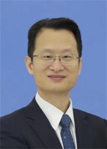 Xin Ma (马昕)合作论文数College for Elite Engineers, Fudan University;Huashan Hospital, Fudan University4
 Yanhong Gao (高艳虹)合作论文数Xinhua Hospital Affiliated to Shanghai Jiao Tong University School of Medicine4WANG Jin-feng合作论文数3L Wang合作论文数3Pei-kang XU (徐培康)合作论文数3
 Yingying Xu (徐莺莺)合作论文数School of Basic Medical Sciences, Fudan University;Shanghai Medical College of Fudan University3Jin-feng WANG (王金凤)合作论文数3HuaHua Pu (蒲画华)合作论文数3KeJia Gao (高克加)合作论文数3MingXiang Yu (于明香)合作论文数3ShaoFen Zhang (张绍芬)合作论文数3RuiXia Li (李瑞霞)合作论文数3WenJun Wang (王文君)合作论文数3
 Guoqiang Hua (华国强)合作论文数复旦大学放射医学研究所3
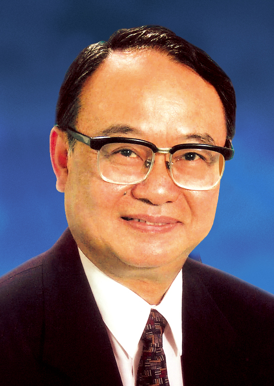 Kerong Dai (戴尅戎)合作论文数Shanghai Ninth People's Hospital, Shanghai Jiao Tong University School of Medicine3
 Zhifeng Yu (于志锋)合作论文数Shanghai Ninth People's Hospital, Shanghai Jiao Tong University School of Medicine3
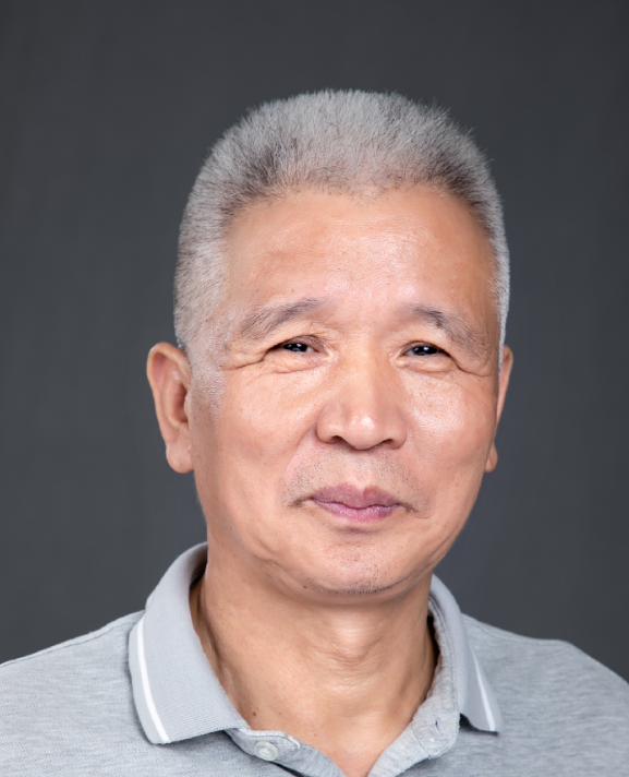 Wenjiang Zhou (周文江)合作论文数School of Pharmaceutical Sciences, Fudan University3
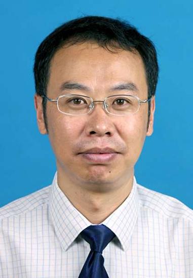 Tingting Tang (汤亭亭)合作论文数Shanghai Ninth Peoplo's Hospital, School of Medicine, Shanghai Jiao Tong University3Weiqiao Jin (金为翘)合作论文数2peifang fan (范佩芳)合作论文数2zhigen weng (翁志根)合作论文数2Jian-Jun Cao合作论文数2shijie chen (陈识杰)合作论文数2yonghe li (李永和)合作论文数2min le (乐敏)合作论文数2Wenjing Tang合作论文数2Guang DU (杜光)合作论文数2Li-hua WANG (王丽华)合作论文数2Ai-hong XU (徐爱红)合作论文数2




