Background The early progression of burn wounds poses a significant clinical challenge. Although inflammatory cell death is thought to be involved, the role of the newly discovered integrated cell death pathway PANoptosis in burn injuries has not yet been studied. Baicalin is a natural compound with anti-inflammatory properties and has therapeutic potential, but its impact on burn progression and PANoptosis is unknown. Methods A rat comb burn model was established. PANoptosis activation was assessed in the zone of stasis via Western blot, immunohistochemistry, and immunofluorescence for key markers (ZBP1, pMLKL, cleaved GSDMD, cleaved caspase-3, and ASC). In the intervention study, baicalin (100 mg/kg) or vehicle was administered for 7 days. PANoptosis markers, inflammatory cytokines (interleukin 1β, interleukin 6, interleukin 18, tumor necrosis factor α), myeloperoxidase activity, and histopathologic wound progression were evaluated. Results Burn injury induced time-dependent PANoptosis activation, characterized by coordinated upregulation of ZBP1, pMLKL, cleaved GSDMD, and cleaved caspase-3. Immunofluorescence confirmed PANoptosome assembly via ASC/cleaved caspase-3 colocalization. Baicalin treatment significantly suppressed all 3 PANoptosis executers, disrupted complex formation, reduced proinflammatory cytokines, and myeloperoxidase activity. Importantly, baicalin treatment significantly reduced the progression of burn wounds at day 7, as evidenced by diminished tissue necrosis and improved tissue architecture. Conclusion Our study identifies PANoptosis as a novel pathogenic mechanism contributing to burn wound progression. We further demonstrate that baicalin is an effective therapeutic agent that alleviates tissue damage possibly by inhibiting the PANoptosis pathway and its associated inflammatory cascade. These findings provide a new mechanistic understanding and highlight the therapeutic potential of baicalin for preventing burn wound deepening.
Background Radiotherapy has shown clinical effectiveness in scar carcinoma, but the underlying mechanisms—particularly its effects on the tumor immune microenvironment—remain unclear. This research intends to support a new hypothesis: that radiotherapy alters the tumor microenvironment in scar carcinoma by stimulating the “p53-Smad7-TGF-β" signaling pathway, which subsequently inhibits critical pathogenic mechanisms and alleviates immunosuppression. This exploratory translational study combined public transcriptomic data with experimental validation to investigate radiotherapy response in burn scar carcinoma. Methods The analysis combined public single-cell and bulk transcriptomic datasets with in vitro functional validation experiments. Additionally, two laboratory cell experiments were designed for functional validation. Bioinformatics analyses employed Seurat, CellChat, Monocle2, limma, and various machine learning techniques. The cell experiments utilized a human burn scar carcinoma cell line, with dose-response validation groups and mechanism recovery validation groups established. Parameters such as cell proliferation, key gene expression, mitochondrial membrane potential, and TGF-β secretion were assessed using the CCK-8 assay, q-PCR, TMRE detection, and ELISA techniques. Results Augur analysis unequivocally identified myeloid-derived suppressor cells (MDSCs) as the primary cell type exhibiting the most significant functional dysregulation following radiotherapy. Consistent results from multi-omics data indicate that the p53-Smad7 signaling pathway is activated post-radiotherapy, while the TGF-β/Smad pathway is suppressed. In vitro experiments demonstrated that post-radiotherapy, p53 and Smad7 mRNA expression increases dose-dependently, accompanied by reduced cellular proliferative activity; downregulation of p53 expression partially counteracts the radiation-induced elevation of Smad7 levels and suppression of TGF-β secretion, whereas introduction of a Smad7 activator restores these effects. Conclusion This study provides multi-omics and experimental evidence supporting the involvement of the p53-Smad7-TGF- axis in radiotherapy-induced immune remodeling in burn scar carcinoma. The activation of this axis constitutes the fundamental mechanism underlying the dual effects of radiotherapy—namely, direct suppression of tumor cell activity and enhancement of antitumor immunity.
Objective Hemorrhagic shock (HS) is a critical clinical condition in which cardiac dysfunction and failure are leading causes of mortality. Mitochondrial dysfunction is central to the pathogenesis of cardiac dysfunction in HS. Irisin has been shown to improve mitochondrial function and protect against ischemia-reperfusion injury (IRI), but its specific effects on myocardial injury in HS are unknown. This study investigates irisin's therapeutic potential in a rat model of HS.Methods For in vivo studies, a rat HS model was established via controlled blood withdrawal and Animals were allocated to four groups: Sham, HS, HS + Vehicle (HS + Veh), and HS + Irisin. Physiological responses were evaluated through temporal sampling at 1, 3, and 6 h post-HS. For in vitro studies, H9c2 cardiomyocytes were exposed to oxygen-glucose deprivation to establish a hypoxic model. Cells were categorized into six groups: normoxia (N), normoxia + AMPK inhibitor compound C (N + Cc), hypoxia (H), hypoxia + Cc (H + Cc), hypoxia + irisin (H + Irisin), and hypoxia + Cc + irisin (H + Cc + Irisin). Cellular functional outcomes were analyzed following 3-h hypoxia exposure.Results HS significantly reduced serum irisin levels. Exogenous irisin administration enhanced survival rates, stabilized mean arterial pressure (MAP), lowered lactate (LAC) levels, improved cardiac structure and function, and reduced myocardial injury biomarkers in HS rats. Mechanistically, irisin activated AMP-activated protein kinase (AMPK) and Sirtuin 1(SIRT1), to suppress the expression of dynamin-related protein 1 (Drp1) and fission protein 1 (Fis1), while upregulating mitofusin 1 (Mfn1). This modulation of mitochondrial dynamics preserved cardiomyocyte mitochondrial membrane potential (MMP), ATP production, and structural integrity. Hypoxic H9c2 cardiomyocytes exhibited consistent results. To confirm AMPK/Drp1-dependent mechanisms, Cc was administered to inhibit irisin-induced AMPK activation. Cc abolished irisin's suppression of Drp1/Fis1 and its Mfn1 upregulation. Furthermore, Cc eliminated irisin-mediated protection in both H9c2 cardiomyocytes and mitochondria.Conclusion Our study demonstrates that irisin ameliorates cardiac function and enhances early prognosis in HS. These cardioprotective effects are achieved through attenuation of myocardial damage and SIRT1/AMPK/Drp1 pathway-dependent restoration of mitochondrial homeostasis.
Hemorrhagic shock (HS) is one of the leading causes of death among young adults worldwide. Multiple organ dysfunction in HS is caused by an imbalance between tissue oxygen supply and demand, which is closely related to the poor prognosis of patient. Mitochondrial dysfunction is one of the key mechanisms contributing to multiple organ dysfunction in HS, while mitochondrial quality control regulates mitochondrial function through a series of processes, including mitochondrial biogenesis, mitochondrial dynamics, mitophagy, mitochondrial-derived vesicles, and mitochondrial protein homeostasis. Modulating mitochondrial quality control can improve organ dysfunction. This review aims to summarize the effects of mitochondrial dysfunction on organ function in HS and discuss the potential mechanisms of mitochondrial quality control, providing insights into the injury mechanisms underlying HS and guiding clinical management.
医疗救护飞机远航程空运救护4例特殊烧伤伤员,将伤员空运后送至北京进行后续治疗.4例烧伤伤员均为男性,平均年龄23.5岁,烧伤面积25%~62%体表总面积(total body surface area,TBSA),均伴有吸入性损伤,双眼灼伤,其中2例伴有皮肤裂伤.在空运后送过程中对烧伤伤员进行持续心电监测观察各项生命体征,进行静脉输液、换药、吸痰和吸氧等治疗,对部分心理不稳定伤员进行心理疏导.4例烧伤伤员在转运过程中生命体征平稳,被安全转运到目的地.
目的 观察生物胶体分散剂治疗植皮供区伤口的临床效果.方法 自2017年7月—2019年7月入选空军特色医学中心烧伤整形科的自体皮肤移植患者60例作为研究对象,随机分为治疗组和对照组,每组30例.治疗组的皮肤供区用生物胶体分散剂治疗,而对照组则用传统的油纱布治疗.观察患者皮肤供体区域的24 h出血量,换药时的疼痛评分,伤口愈合时间和疤痕增生程度.结果 治疗组和对照组植皮供区伤口术后24 h浸透纱布层数比较,差异统计学意义(P=0.002).治疗组和对照组供皮区于术后1、3、6、9 d换药时疼痛数值比较,其中术后1、9 d差异有统计学意义,术后3、6 d差异无统计学意义.治疗组创面愈合后3、6、9个月瘢痕评分较对照组显著减轻,差异均有统计学意义(P<0.05).结论 采用生物胶体分散剂治疗植皮供区伤口,各种组分协同作用,可促进止血、防止伤口感染、保护创面、减轻换药时疼痛、促进伤口愈合、改善疤痕增生.
目的 研究重组人表皮生长因子结合银离子敷料在负压封闭引流(vacuum sealing drainage,VSD)环境下对糖尿病足溃疡创面的治疗效果.方法 选取2型糖尿病足感染患者129例,随机分为3组,每组各43例,A组单纯使用银离子敷料治疗,B组在银离子敷料治疗基础上添加重组人表皮生长因子(rhEGF)治疗,C组在A和B组基础上增加VSD负压引流,观察治疗28 d后三组患者创面治疗效果.结果 三组治疗总有效率分别为:A组88.4%(38/43)、B组93.0%(40/43)、C组97.7%(42/43),差异有统计学意义(P<0.05),治疗后疼痛效果评分(VAS):C组(银离子敷料+rhEGF+VSD)的疼痛分值明显低于B组(银离子敷料+rhEGF)和A组(银离子敷料),差异有统计学意义(P<0.05);从创面修复情况、换药次数和抗生素使用情况对比发现,C组明显优于A组和B组.结论 重组人表皮生长因子(rhEGF)结合银离子敷料在负压封闭引流下(VSD)治疗糖尿病足,创面愈合时间短、肉芽生长速度快、疗效显著,值得临床推广应用.
The aim of this study was to investigate the correlation between cluster of differentiation 86 (CD86) gene rs1129055 and rs2715267 single nucleotide polymorphisms and sepsis susceptibility. One hundred twenty-five sepsis patients and 120 healthy controls were enrolled in this case-control study. CD86 polymorphisms rs1129055 and rs2715267 were genotyped through polymerase chain reaction-restriction fragment length polymorphism approach. Chi-square test was used to analyze differences in genotype and allele frequencies of the 2 polymorphisms between case and control groups. Odds ratios (ORs) and 95% confidence intervals (CIs) were used to present the association strength of the polymorphisms with sepsis susceptibility. AA genotype and A allele frequencies of CD86 rs1129055 were significantly lower in sepsis patients than in healthy controls (P<.05), revealing their significant associations with decreased disease susceptibility (OR= 0.351, 95% CI= 0.169-0.728; OR= 0.593, 95% CI= 0.415-0.847). Nevertheless, rs2715267 had no significant association with sepsis susceptibility (P>.05). AA genotype and A allele of CD86 polymorphism rs1129055 might be correlated with decreased sepsis susceptibility in Chinese Han population, but not rs2715267. Further study should be performed to verify our findings.
目的 观察可吸收修复胶原联合银离子敷料治疗供皮区创面的临床效果.方法 选取2016-07至2018-12医院收治的100例自体皮移植患者,随机分为治疗组和对照组,每组50例,治疗组采用可吸收修复胶原联合银离子敷料治疗供皮区创面,对照组采用传统凡士林纱布治疗.观察供皮区创面换药时疼痛程度、创面感染率、愈合时间,以及创面愈合后瘢痕指标评定.结果 治疗组供皮区创面换药时疼痛分数在术后3、6、9 d分别为6.24±2.23,4.13±2.37,1.49±1.31,显著低于对照组的7.73±2.14,5.24±1.59,2.43±1.66(P<0.05);治疗组供皮区创面感染率为2%,较对照组14%显著降低(P<0.05);治疗组创面愈合时间明显短于对照组(P<0.05);治疗组创面愈合后3、6、9个月温哥华瘢痕量表评分较对照组显著降低,差异均有统计学意义(P<0.05).结论 可吸收修复胶原联合银离子敷料治疗供皮区创面相比于传统凡士林纱布治疗是一种更优越的方式,适合临床推广使用.
The aim of this study was to investigate the correlation between cluster of differentiation 86 (CD86) gene rs1129055 and rs2715267 single nucleotide polymorphisms and sepsis susceptibility. One hundred twenty-five sepsis patients and 120 healthy controls were enrolled in this case-control study. CD86 polymorphisms rs1129055 and rs2715267 were genotyped through polymerase chain reaction-restriction fragment length polymorphism approach. Chi-square test was used to analyze differences in genotype and allele frequencies of the 2 polymorphisms between case and control groups. Odds ratios (ORs) and 95% confidence intervals (CIs) were used to present the association strength of the polymorphisms with sepsis susceptibility. AA genotype and A allele frequencies of CD86 rs1129055 were significantly lower in sepsis patients than in healthy controls (P< .05), revealing their significant associations with decreased disease susceptibility (OR=0.351, 95% CI=0.169–0.728; OR= 0.593, 95% CI=0.415–0.847). Nevertheless, rs2715267 had no significant association with sepsis susceptibility (P> .05). AA genotype and A allele of CD86 polymorphism rs1129055 might be correlated with decreased sepsis susceptibility in Chinese Han population, but not rs2715267. Further study should be performed to verify our findings. Abbreviations: CD86 = cluster of differentiation 86, CIs = confidence intervals, HWE = Hardy–Weinberg equilibrium, IL = interleukin, ORs = odds ratios, PCR = polymerase chain reaction, SNPs = single nucleotide polymorphisms.
Objective To compare a modified abdominal tube with traditional abdominal tube in the curative effect of repairing finger degloving injury.Methods Thirty patients with single finger degloving injuries admitted to Air Force General Hospital,PLA from Janua~ 2011 to December 2013 were divided into the experimental group and the control group according to the random number table,each contained 15 cases.Patients in experimental group received operation of modified abdominal tube,whereas patients in control group received operation of traditional abdominal tube.The postoperative swelling in the skin flap,appearance,infection,blood supply,sensory function recovery were compared between two groups.SPSS 18.0 was used for statistical analysis.The comparison for measurement data was performed by t test.Results The flap in two groups both survived well.One edge of flap in control group appeared black,with good recovery after dressing change.The experimental group did not appear this kind of situation.The postoperative flap in two groups both appeared some degree of swelling and purple,which was more obvious in the control group,accompanied with cyanosis.The wounds in two groups both had a small amount of effusion,through regular dressing treatment,both survived well with no infection.The capillary filling time in flap of experimental group was significantly shortened as compared to that of control group [(1.03 ±0.25) s,(0.91±0.21) svs.(1.24 ±0.33) s,(1.18±0.39) s],at 7 and 14 days post operation,respectively,the differences were statistically significant (t =-1.995,-2.361,P =0.0495,0.0254).The sensory function recovery in flap of experimental group was markedly improved as compared to that of control group[(2.66 ±0.38)s,(3.67±0.55)s vs.(1.97±0.36)s,(3.14 ±0.43)s],at 6 and 12 months post operation,respectively,the differences were statistically significant (t =5.105,2.940,with P values below 0.05).Conclusion Using this modified abdominal tube to repair single finger degloving injury can significantly increase blood supply of flaps,reduce the swelling,improve the external appearance of the skin flap,and have better feeling functional recovery.
Objective To study the effect of negative pressure wound therapy combined with local oxygen therapy on the treatment of pressure ulcers.Methods From January 2015 to December 2016,60 patients with pressure ulcers over Ⅲ stage admitted in Department of Burns and Plastic Surgery,Air Force General Hospital,People's Liberation Army,were divided into 3 groups according to the random number table method:conventional dressing change group,negative pressure wound therapy group and negative pressure wound therapy combined with local oxygen therapy group,20 cases in each group.Conventional dressing change group and negative pressure wound therapy group were respectively given conventional dressing method,negative pressure wound treatment.Negative pressure wound therapy combined with local oxygen therapy group was given negative pressure to attract wounds with local oxygen therapy.Immediately after admission,at the 1st,2nd,3rd,4th,5th,6th,7th and 8th week after treatment,the maximum length,width,depth,exudation,tissue type and wound shrinkage were observed.Date were processed with analysis of variance and LSD test.Results The length,width and depth of pressure ulcers in the negative pressure wound therapy group were significantly decreased as compared to conventional dressing change group,while the decrease in negative pressure wound therapy combined with local oxygen therapy group was more significant,the differences were statistically significant (with P values below 0.05).The length,width and depth in conventional dressing group at the 8th week after treatment were (3.04 ± 0.43),(3.63 ± 0.88),and (1.55 ±0.77) cm,while in the negative pressure wound therapy group were (2.14 ±0.71),(2.65 ± 1.27),and (1.05 ± 0.62) cm,and in negative pressure wound therapy combined with local oxygen therapy group,there were (1.17 ± 0.28),(1.39 ± 2.37),and (0.58 ± 0.45) cm.In the negative pressure wound therapy group,the exudation was reduced,the wound granulation tissue was increased,and the wound size was more reduced.All the above effect in the negative pressure wound therapy combined with local oxygen therapy group were obviously superior to negative pressure wound therapy group,the differences were statistically significant (with P values below 0.05).Conclusion Negative pressure wound therapy combined with local oxygen therapy can significantly promote wound healing,reduce exudation and promote the growth of granulation of pressure ulcers,it is better than the only use of negative pressure treatment,which can be applied as a new effective treatment method in the future.
目的 探讨一种改良的V-Y推进皮瓣修复指端软组织缺损的方法并观察其疗效.方法 对2011年以来收治的34例指端缺损患者采用改良V-Y推进皮瓣覆盖创面,观察皮瓣成活率、创面愈合情况、皮瓣外观、色泽和手指功能恢复情况.结果 术后皮瓣全部存活,创面I期愈合.随访时间为6~12个月.皮瓣质地柔软,外形圆润程度与正常指端接近(指甲缺损除外),无臃肿、棱角或色素沉着等异常.末节指腹指纹重新建立,指间关节活动正常,无明显钩甲畸形,指腹两点分辨觉为(5 .43 ± 0 .65)mm .根据中华医学会手外科学会上肢部分功能评定试用标准评定,患指主动活动为优22例,良12例,优良率100%.结论 改良V-Y推进皮瓣手术操作简单,修复缺损面积大,是一种修复指端缺损较为理想的方法.
Objective Observe the clinical effect of rhGM-CSF on treatment of wound dissolution of deep second de-gree burn. Method Devide 70 deep second degree burn patients into observation group(35 cases)and control group (35 cases)according to the random number table method. Two groups were both performed the routine debridement, observation group applied rhGM-CSF and control group applied gel matrix. Observe the wound healing,granulation growth,complete wound healing time,scar hyperplasia and adverse reaction incidence. Result:The observation group dissolution rate after 2 days,6 days,10 days and 14 days were higher than the control group,the difference was sta-tistically significant(P<0. 05). The average dissolution time of the observation group was shorter than the control group,the difference was statistically significant(P <0. 05). The average wound healing time of the observation group was shorter than the control group,the difference was statistically significant(P<0. 05). The effective rate of observation group after 14 days was higher than the control group,but the difference was not statistically significant (P>0. 05). The effective rate of observation group after 18 days was higher than the control group,but the difference was not statistically significant(P>0. 05). The effective rate were both 100% after 28 days. Both groups have no ad-verse reaction. Conclusion rhGM-CSF has a significant promotion effect on dissolution and healing of deep second degree burn wounds,and it is worth promoting in clinical practice.
目的:探讨小腿挤压伤伴撕脱伤患者的整体治疗方法,并分析其临床应用价值.方法:回顾性分析我院近5年来收治的23例小腿挤压伤伴撕脱伤患者的临床资料,分别采用行自体皮肤反削回植、异种皮覆盖或封闭负压吸引治疗+二期植皮、知名血管皮瓣转移、单纯清创缝合.结果:23例中,18例Ⅰ期愈合;5例局部皮肤坏死,经换药后Ⅱ期愈合2例,残余创面行植皮后Ⅱ期愈合l例;骨外露者经皮瓣转移修复后Ⅱ期愈合2例.随访3-16月,临床效果满意.结论:对于小腿挤压伤伴撕脱伤,依具体情况采用自体皮肤反削回植、异种皮覆盖或封闭负压吸引治疗+二期植皮、知名血管皮瓣转移、单纯清创缝合等方法修复创面对患者肢体功能恢复有较大的作用,临床效果较好,利于患者康复,具有一定的推广应用价值.
目的:探讨负压封闭引流技术应用于躯干部皮肤恶性肿瘤切除术的临床效果,并分析其临床应用价值.方法:回顾性分析2012年5月~2015年3月我院收治的11例躯干部皮肤恶性肿瘤患者的临床资料,均予行肿物切除术并同时行自体皮游离移植术,并外用持续负压封闭引流术于植皮术区.结果:所有患者均接受肿物切除+植皮+封闭负压引流技术治疗.9例患者于4~9天后拆除负压材料,皮片全部成活.1例患者治疗后24小时内吸引管堵塞,经术后于24小时内行管路冲洗疏通后再次行负压吸引,于术后5天首次拆除负压材料,见皮片部分成活,部分皮片仍有浮动,血运未明显建立,予以扩大皮片引流孔后再次行负压封闭引流术,术后9天再次拆除负压材料,见皮片成活较好.1例患者治疗4天后出现新鲜渗血经引流管引出,予拆除负压材料,所植皮片下有积血块,有新鲜渗血,予以清创后再次行负压封闭引流术,术后9天再次拆除负压材料,见皮片成活较好.随访半年到3年,所植皮片无破溃,肿瘤无复发.结论:躯干部皮肤恶性肿瘤切除术中,植皮联合封闭式负压引流技术可使所植皮片固定确实,充分引流渗液积血,利于皮片成活,对无法有效包扎固定肢体特殊部位(肩部、臀部、躯干部)等术区植皮提供了有效方法,具有一定的推广应用价值.
目的:观察重组人酸性成纤维细胞生长因子( rh-aFGF)治疗深Ⅱ度烧伤的临床效果。方法将65例深Ⅱ度烧伤患者按随机数字表法分为观察组30例和对照组35例,两组均行削痂术,观察组削痂术后应用rh-aFGF,对照组削痂术后不使用rh-aFGF,观察创面愈合情况、肉芽生长情况、创面完全愈合时间、瘢痕增生情况、不良反应发生情况。结果观察组的平均愈合时间短于对照组(P<0.05),观察组第10、15、19天愈合率均高于对照组(P<0.05),观察组肉芽生长情况优于对照组(P<0.05);术后1年观察组瘢痕评分优于对照组,平均瘢痕高度低于对照组,平均瘢痕面积小于对照组,差异有显著性(P<0.05);观察组水肿评分、渗出评分、脓液评分优于对照组(P<0.05),观察组细菌培养阳性率低于对照组,差异无显著性(P>0.05);无不良反应发生。结论削痂术后应用rh-aFGF能够促进烧伤创面肉芽生长,缩短愈合时间,加快烧伤创面的愈合速度,减少瘢痕形成。
目的 观察蜡疗联合矫形器治疗在手植皮手术后康复治疗中的疗效.方法 选取2009-06至2013-06,在空军总医院烧伤整形科收治的手部植皮患者63例,按康复方法分为综合康复组(32例)和常规康复组(31例).常规康复组给予压力治疗、日常生活动作训练、主动训练和药物治疗.综合康复组还加用蜡疗和矫形器治疗.两组患者在康复治疗6个月后对患者手部疼痛、瘢痕增生情况和手功能情况进行评价.结果 康复开始时综合康复组疼痛评分和常规康复组差异无统计学意义(P=0.086).经康复治疗后,综合康复组疼痛评分为(1.56±0.27)分明显低于常规康复组(2.92±0.36)分,差异有统计学意义(P =0.012).综合康复组瘢痕增生在治疗后评分(4.36±0.78)分明显低于常规治疗组(5.92±0.91)分,差异有统计学意义(P<0.05).综合康复组手功能TAM和Barthel评分分别为(238.5±12.7)分和(87.2±6.76)分,均明显高于常规康复组的(58.2±5.9)分和(70.2±4.2)分,差异有统计学意义(P<0.05).结论 蜡疗联合矫形器治疗能促进植皮手术后手功能的恢复.
目的:比较邮票植皮、meek植皮以及自体微粒皮移植用于大面积烧伤患者创面修复的临床效果和可行性.方法:回顾性分析我院烧伤科收治的120例大面积烧伤患者的临床资料,根据不同的手术植皮方法分为微粒皮植皮组、Meek植皮组和邮票皮组,每组40例.三组患者入院后均给予常规基础治疗,再根据选择植皮方式的不同实施微粒皮植皮、Meek植皮和邮票皮植皮.对比三组患者的植皮成活率、创面一期愈合率、愈合时间、死亡率、1%烧伤面积(1% TBSA)治疗费用(元)以及康复效果(康复优良率).结果:邮票皮组植皮成活率明显高于Meek植皮组(P<0.05),Meek植皮组植皮成活率明显高于微粒皮植皮组(P<0.05).Meek植皮组和邮票皮组创面愈合时间均明显短于微粒皮植皮组(P<0.05).邮票皮组1% TBSA治疗费用明显低于Meek植皮组(P<0.05),Meek植皮组1% TBSA治疗费用明显低于微粒皮植皮组(P<0.05).Meek植皮组和邮票皮组的创面一期愈合率均明显高于微粒皮植皮组口<0.05);Meek植皮组和邮票皮组的死亡率均明显低于微粒皮植皮组(P<0.05);meek植皮组的瘢痕最轻,关节功能恢复最好;微粒皮植皮组瘢痕最重,关节功能恢复最差.结论:不同植皮方式用于大面积烧伤创面修复的临床效果各不相同.邮票皮成活率高、抗感染能力强,但扩张比例低,创面愈合效果差;meek皮扩张比例高、康复效果好,但抗感染能力差;微粒皮扩张比例最高,但成活率低,治疗成本高.临床上,应该根据患者的自身情况选择不同的植皮方式.
目的:探讨转化生长因子β1(TGF-β1)基因-509C/T位点的多态性与瘢痕疙瘩之间的相关性。方法选取瘢痕疙瘩患者159例作为病例组,其中轻度瘢痕疙瘩67例,中度瘢痕疙瘩54例,重度瘢痕疙瘩38例。另选年龄及性别与病例组匹配的本院同期外科住院无病理性瘢痕者164例作为对照组。采用聚合酶链反应-限制性片段长度多态性(PCR-RFLP)方法检测两组人群TGF-β1基因-509C/T位点的单核苷酸多态性。结果瘢痕疙瘩患者-509C/T位点的C等位基因频率及CC基因型频率均明显高于正常对照组(χ2=4.392,P=0.036;χ2=5.078,P=0.024),而其CT、TT基因型频率与对照组比较,差异无显著性(χ2=0.783,P=0.376;χ2=1.201,P=0.273)。说明携带TGF-β1基因-509C/T位点的C等位基因及CC基因型者患瘢痕疙瘩的风险增加(分别OR=1.394,95% CI为1.021~1.903;OR=1.818,95% CI为1.077~3.070)。同时重度瘢痕疙瘩组-509C/T位点的C等位基因频率及CC基因型频率均明显高于对照组(P<0.05)。结论瘢痕疙瘩患者存在TGF-β1基因-509C/T位点的多态性,-509位点的C等位基因及CC基因型可能影响瘢痕疙瘩的发生发展, TGF-β1基因-509C/T位点的多态性可能是导致瘢痕疙瘩的一个危险因子。
 JingNing Cai (蔡景宁)合作论文数14XiaoFan Xie (谢晓繁)合作论文数12WeiHong Cao (曹卫红)合作论文数12ShiJian Wu (吴世建)合作论文数11
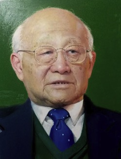 Zhiyong Sheng (盛志勇)合作论文数Chinese PLA General Hospital11Sen Hu (胡森)合作论文数Chinese Pla General Hospital11Bin Li (李斌)合作论文数10BaoLong Li (李宝龙)合作论文数8Mengjing Xiao合作论文数7
 Jiake Chai (柴家科)合作论文数Chinese People's Liberation Arm General Hospital7
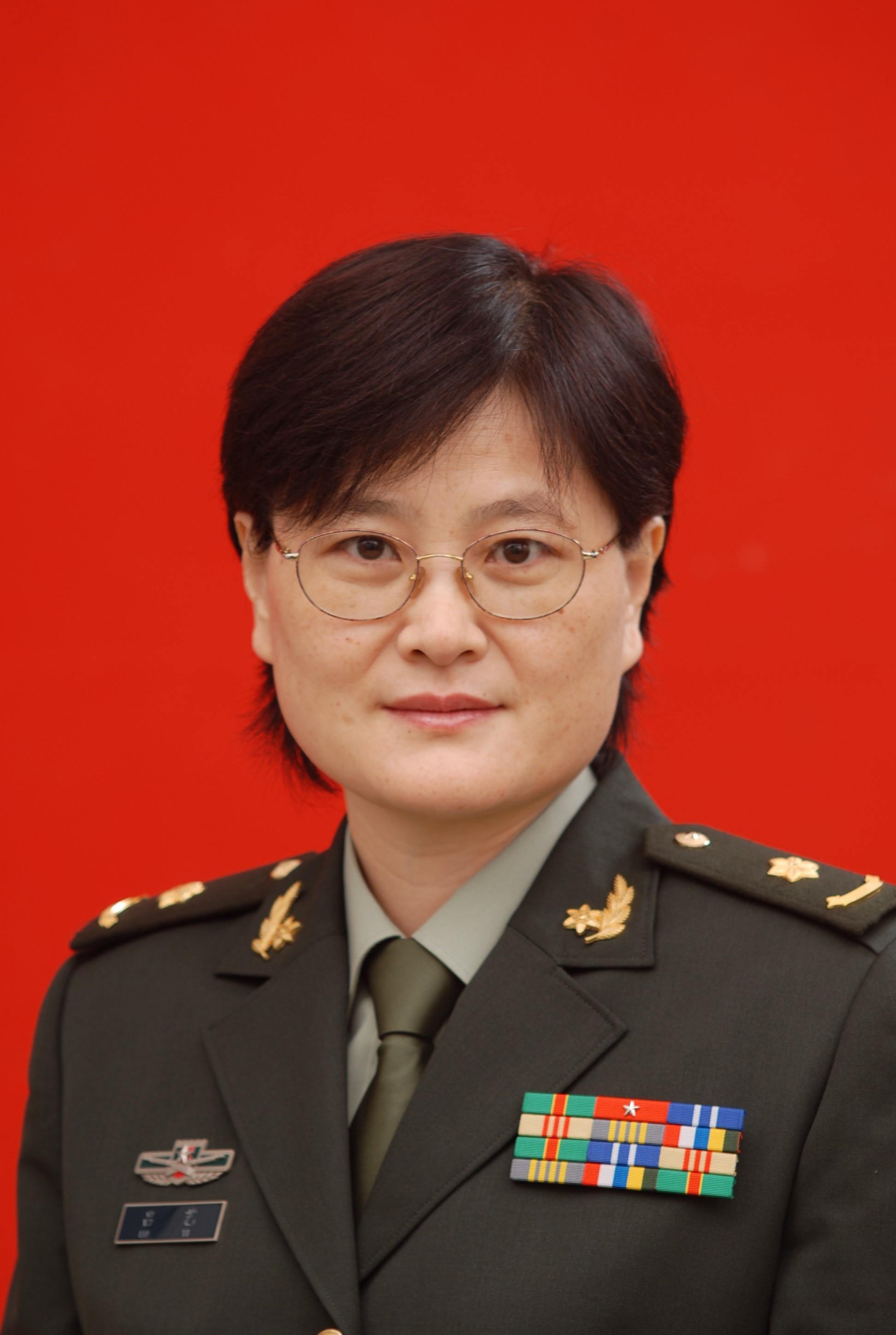 Yi Lv (吕艺)合作论文数Fourth Medical Center PLA General Hospital5Tianjun Sun合作论文数4Bin LI (李斌)合作论文数3Yunfei Chi合作论文数3YeQiong Li (李晔琼)合作论文数3QingGang Hu (胡清刚)合作论文数3GuoYong Zhou (周国勇)合作论文数3Wei Liu (刘伟)合作论文数3
 Chuanan Shen (申传安)合作论文数Fourth Medical Center of PLA General Hospital3Weiping Song (宋卫平)合作论文数3
 Hongming Yang (杨红明)合作论文数Chinese People's Liberation Army General Hospital3
 Chiyu Jia (贾赤宇)合作论文数School of Medicine, Xiamen University;Xiang‘an Hospital of Xiamen University3Hui-Nan Yin合作论文数3
 Daifeng Hao (郝岱峰)合作论文数中国人民解放军总医院第一附属(304)医院烧伤整形外科3jian li (李健)合作论文数2Shuang Liu (刘爽)合作论文数2Jianhua Cai合作论文数2Rui Feng合作论文数2Qi Song合作论文数2
 Li Ma合作论文数2
 Yujie Jiang (蒋玉洁)合作论文数2YuFei Chen (陈宇飞)合作论文数2Xibo Zhang合作论文数2
 Jiangyang Lu (陆江阳)合作论文数Fourth Medical Center of PLA General Hospital2
 Hongjie Duan (段红杰)合作论文数Fourth Medical Center of PLA General Hospital2Lan HE (何兰)合作论文数2Shan Xu (徐珊)合作论文数2Huping Deng (邓虎平)合作论文数2Shaofang Han合作论文数1Hailiang Bai合作论文数1jianbing xiao (肖建兵)合作论文数1Fangfang BI (毕芳芳)合作论文数1Fei XIE (谢飞)合作论文数1Chenlong JIA (贾辰龙)合作论文数1Z. Sheng合作论文数1Hong-guang Wang (王红光)合作论文数1Na SUN (孙娜)合作论文数1




