Lung cancer is one of the most common malignant tumors worldwide, seriously threatening human health. PAB (pseudolaric acid B), an extract of pseudolarix amabilis, has exhibited notable anticancer properties. Nevertheless, the molecular mechanisms underlying PAB-induced anticancer activities remain controversial in lung cancer especially. Herein we aim to investigate the role of IAPs in PAB-induced anticancer effects. PAB selectively inhibits the viability of lung cancer cells and downregulates the expression of IAPs. Transcriptomic analysis suggests that PAB induces lung cancer cells ferroptosis. PAB indeed promotes ferroptotic cell death since PAB enhances lipid oxidation and Fe2+ content. Interestingly, PAB markedly enhances Survivin expression. Suppression of Survivin abolishes PAB-induced ferroptosis. PAB significantly strengthens JNK and ERK kinase activities. We further demonstrate suppression of JNK/ERK reversed PAB-mediated-cytotoxicity, meanwhile restores the expression of Survivin and ferroptosis-related proteins. Consistently, xenograft tumor model results also support that PAB induces ferroptosis through Survivin upregulation. Collectively, we demonstrate PAB induces ferroptosis in lung cancer cells in vivo and in vitro depending on JNK and ERK-mediated Survivin upregulation, providing novel insight for clinical administration of PAB in lung cancer.
BACKGROUND:The role of solute carrier family 6 member 17 (SLC6A17) in lung adenocarcinoma (LUAD) is unclear. OBJECTIVES:To address this gap in knowledge, we employed bioinformatics analysis and experimental validation. METHODS:This research aimed to scrutinize the expression patterns of the SLC6A17 gene across a spectrum of cancers and specifically within LUAD, utilizing data extracted from The Cancer Genome Atlas (TCGA). The correlation between SLC6A17 expression and LUAD prognosis was investigated to assess its diagnostic relevance. The study delved into the possible regulatory mechanisms of SLC6A17, focusing on its links to immune cell infiltration and drug response in LUAD. The examination of SLC6A17 expression was extended to single-cell sequencing data in LUAD, alongside an evaluation of the gene's genomic alterations and clinical implications within this disease context. Validation of SLC6A17 expression levels was conducted using datasets from GSE87340 and various cell lines, employing quantitative real-time polymerase chain reaction (qRTPCR) techniques. RESULTS:SLC6A17 exhibited aberrant expression in both pan-cancer and LUAD. Increased expression of SLC6A17 in LUAD patients was significantly associated with poorer overall survival (p = 0.008), progress-free survival (p = 0.019), and disease specific survival (p = 0.030). In LUAD patients, the levels of SLC6A17 expression were found to be a significant standalone indicator of prognosis, with a p-value of 0.031. SLC6A17 exhibited associations with various pathways, including focal adhesion, ECM receptor interaction, cell cycle, linoleic acid metabolism, pathways in cancer, and more. SLC6A17 expression demonstrated correlations with immune infiltration in LUAD. SLC6A17 expression revealed a notably inverse relationship with several substances, including AR-42, T0901317, tubastatin A, SB52334, and amuvatinib, within the context of LUAD. SLC6A17 was found to be significantly positively regulated in LUAD cell lines. CONCLUSION:These findings suggest that SLC6A17 indicates the potential of a potential prognostic biomarker and immunotherapeutic target for patients with LUAD.
OBJECTIVE:Esophageal cancer is one of the most common digestive cancers in the world. Because of the limitation and resistence of the traditional chemotherapy drugs, it is important to explore new therapeutic targets and strategies for this refractory cancer. Recently, targeting deubiquitinases has emerged as a promising avenue for the development of anti-tumor drugs. However, the role and underlying mechanism of NSC632839, a broad-spectrum deubiquitinases inhibitor, in esophageal squamous cell carcinoma in vitro remain elusive. METHODS:Cell Counting Kit-8 assay, colony formation assay, EdU proliferation experiment and cell morphology observation were used to detect the effect of NSC632839 on cell growth. Flow cytometry was employed to detect cell apoptosis and cell cycle arrest. Immunoblot and immunofluorescence was used to evaluate the expression level of cell cycle-, apoptosis-, and autophagy-related proteins. RESULTS:NSC632839 inhibited the proliferation of Kyse30 and Kyse450 cells. Mechanistically, NSC632839 induced the formation of multipolar spindles, and its concomitant spindle assembly checkpoint-dependent mitotic arrest, followed by CREB-Noxa-mediated apoptosis. Reversine, a classical MPS1 kinase inhibitor known for its ability to inhibit the spindle assembly checkpoint, could rescue NSC632839-induced cell cycle arrest and apoptosis. Additionally, NSC632839 could trigger pro-survival autophagy. Combination of autophagy inhibitor, CQ and BafA1, with NSC632839 could induce stronger cell proliferation inhibition and apoptosis than NSC632839 alone. CONCLUSIONS:These findings provided a novel anti-cancer mechanism of NSC632839 and highlighted it as a potential anti-tumor agent for the treatment of esophageal cancer.
The induction of immunogenic cell death (ICD) impedes tumor progression via both tumor cell-intrinsic and -extrinsic mechanisms, representing a robust therapeutic strategy. However, ICD-targeted therapy remains to be explored and optimized. Through kinome-wide CRISPR-Cas9 screen, NUAK family SNF1-like kinase 1 (NUAK1) is identified as a potential target. The ICD-provoking effect of NUAK1 inhibition depends on the production of reactive oxygen species (ROS), consequent to the downregulation of nuclear factor erythroid 2-related factor 2 (NRF2)-mediated antioxidant gene expression. Moreover, the mevalonate pathway/cholesterol biosynthesis, activated by spliced form of X-box binding protein 1 (XBP1s) downstream of ICD-induced endoplasmic reticulum (ER) stress, functions as a negative feedback mechanism. Targeting the mevalonate pathway with CRISPR knockout or the 3-hydroxy-3-methylglutaryl-coenzyme A reductase (HMGCR) inhibitor simvastatin amplifies NUAK1 inhibition-mediated ICD and antitumor activity, while cholesterol dampens ROS and ICD, and therefore also dampens tumor suppression. The combination of NUAK1 inhibitor and statin enhances the efficacy of anti-PD-1 therapy. Collectively, our study unveils the promise of blocking the mevalonate-cholesterol pathway in conjunction with ICD-targeted immunotherapy.
Casein Sodium coated curcumin nanocapsules (Cas@Cur) were fabricated by a pH shift method, which improved the water solubility of curcumin (Cur). Hydrogen bonds and hydrophobic interactions were the main forces for the formation of Cas@Cur. Chitosan films (CS) reinforced with pomegranate cellulose nanocrystals (PCNCs), Cas@Cur, PCNC/Cas@Cur, and PCNC/Cur were developed and named CP, CS-Cas@Cur, CP-Cas@Cur and CP-Cur, respectively. The addition of Cas@Cur decreased the moisture content, crystallinity and water contact angle of chitosan film, and increased its water solubility and light barrier property. The CP-Cur film presented the roughest cross-sectional SEM image owing to the hydrophobicity of Cur. CP-Cas@Cur film exhibited the excellent cumulative release of Cur, and was 1.60 and 3.70 times of that of CP-Cur in the semi-fatty and fatty food simulation systems at 2 h, respectively, owing to the controlled-release function of PCNCs and great water solubility of Cas@Cur. Furthermore, the CP-Cas@Cur film displayed excellent antioxidant property, antibacterial activity and sensitive color responsiveness to pH and NH3. Interestingly, the CP-Cas@Cur films exhibited a visible color change at pH 3-7. The application of CP-Cas@Cur film in the preservation of milk and shrimp indicated its potential for the visual monitoring of food freshness.
Ubiquitin-specific protease 24 (USP24) is a deubiquitinase that regulates protein stability, localization, and activity, exhibiting pronounced tissue-specific functions. Depending on the tumor context, USP24 can act either as an oncogene or suppressor, reflecting its strong dependence on tissue-specific substrates and signaling environments. Its dual roles in DNA damage repair and autophagy further underscore its functional complexity. This review summarizes the structural features of USP24, its diverse cellular functions, and its involvement in cancer and other diseases, and highlights recent advances in USP24 inhibitors. Targeting USP24 represents a novel and clinically relevant strategy, capable of selectively modulating its tissue-dependent activities, thereby offering potential avenues for therapeutic intervention and clinical translation across multiple disease contexts.
Background: A direct comparison of programmed cell death-(ligand)1 (PD-(L)1) in combination with solvent-based paclitaxel (sb-paclitaxel) or albumin-bound paclitaxel (nab-paclitaxel) in advanced squamous non-small cell lung cancer (NSCLC) is currently lacking. Therefore, we conducted this network meta-analysis (NMA) to compare these two combination regimens. Methods: We systematically searched articles from PubMed, Embase, Cochrane Library, and Web of Science, as well as abstracts from ESMO, ASCO, and WCLC, to identify phase III randomized controlled trials (RCTs) investigating first-line applications of PD-(L)1 inhibitors combined with sb-paclitaxel or nab-paclitaxel in the treatment of advanced squamous NSCLC. Results: The present study consisted of 10 RCTs involving a total of 4352 patients. We compared ICIs+sb-paclitaxel to ICIs+nab-paclitaxel, PD-1+chemotherapy (chem) to PD-L1+chem, and different ICIs combination chemotherapy regimens. Our results revealed that compared to ICIs+nab-paclitaxel, ICIs+sb-paclitaxel exhibited a slightly better OS (hazard ratio (HR): 0.79, 95%CI: 0.58-1.08) and PFS (HR: 0.84, 95%CI: 0.65-1.07), although the differences were not statistically significant. In contrast to PD-L1+chem, PD-1+chem markedly prolonged OS (HR: 0.75, 95%CI: 0.60-0.93) and PFS (HR: 0.79, 95%CI: 0.66-0.96). In terms of the safety profile, there were no differences in the incidence of grade ≥3 TRAEs between ICIs+nab-paclitaxel and ICIs+sb-paclitaxel (RR: 1.05, 95%CI: 0.53-1.99), PD-1+chem and PD-L1+chem (RR: 0.93, 95% CI :0.68-1.28) as well as comparisons among various ICIs+chem regimens. Conclusion: The efficacy and safety of ICIs+sb-paclitaxel is comparable to that of ICIs+nab-paclitaxel in advanced squamous NSCLC. However, ICIs+sb-paclitaxel exhibits a slightly improved PFS, OS, and similar safety profile, making it a potential preferred choice for first-line treatment.
Context Forsythoside A (FSA) was extracted from Forsythia suspensa, a traditional Chinese medicine, which has been demonstrated to exert anti-inflammatory, antibacterial, and other pharmacological effects. However, the anticancer effect of FSA in esophageal squamous cell carcinoma (ESCC) has not been documented.Objective The present study aimed to elucidate the mechanism of FSA against ESCC.Materials and methods Network pharmacology and molecular docking were employed to predict the mechanism. FSA was utilized to treat ESCC cell lines KYSE450 and KYSE30, followed by CCK-8 assay, cell cloning formation assay, flow cytometry, Western blot, RNA-seq analysis, and subsequent in vivo experiments.Results Network pharmacology and molecular docking predicted that the therapeutic effect of FSA in ESCC is mediated through proteins such as BCL2 and BAX, influencing KEGG pathways associated with apoptosis. In vitro experiments showed that FSA inhibited cell proliferation and plate clone formation, promoted cell apoptosis and impacted the cell cycle distribution of G2/M phase by regulating BCL2, BAX, and p21. Further RNA-seq in KYSE450 cells showed that FSA regulated the expression of 223 genes, specifically affecting the biological process of epidermal development. In vivo experiments showed that gastric administration of FSA resulted in notable reductions in both tumor volume and weight by regulating BCL2, BAX, and p21. 16S rRNA sequencing showed that FSA led to significant changes of beta diversity. Abundance of 11 specific bacterial taxa were considerably changed following administration of FSA.Conclusions This study presents a novel candidate drug against ESCC and establishes a foundation for future clinical application.
In this work, we utilized the molecular hybridization strategy to design and synthesize novel 1,2,3-triazole benzothiazole derivatives K1-26. The antiproliferative activities against MGC-803, Kyse30 and HCT-116 cells were explored, and their structure-activity relationship were preliminarily conducted and summarized. Among them, compound K18, exhibited the strongest proliferation inhibitory activity, with esophageal cancer cells Kyse30 and EC-109 being the most sensitive to its effects (IC50 values were 0.042 and 0.038 μM, respectively). Compound K18 effectively inhibited tubulin polymerization (IC50 = 0.446 μM), thereby hindering tubulin polymerize into filamentous microtubules in Kyse30 and EC-109 cells. Additionally, compound K18 induced the degradation of oncogenic protein YAP via the UPS pathway. Based on these dual molecular-level effects, compound K18 could induce G2/M phase arrest and cell apoptosis in Kyse30 and EC-109 cells, as well as regulate the expression levels of cell cycle and apoptosis-related proteins. In summary, our findings highlight a novel 1,2,3-triazole benzothiazole derivative K18, which possesses significant potential for treating esophageal cancers.
AQP3 (aquaporin 3 (Gill blood group)), a member of the AQP family, is an aquaglyceroporin which transports water, glycerol and small solutes across the plasma membrane. Beyond its role in fluid transport, AQP3 plays a significant role in regulating various aspects of tumor cell behavior, including cell proliferation, migration, and invasion. Nevertheless, the underlying regulatory mechanism of AQP3 in tumors remains unclear. Here, for the first time, we report that AQP3 is a direct target for ubiquitination by the SCFFBXW5 complex. In addition, we revealed that downregulation of FBXW5 significantly induced AQP3 expression to prompt macroautophagic/autophagic cell death in hepatocellular carcinoma (HCC) cells. Mechanistically, AQP3 accumulation induced by FBXW5 knockdown led to the degradation of PDPK1/PDK1 in a lysosomal-dependent manner, thus inactivating the AKT-MTOR pathway and inducing autophagic death in HCC. Taken together, our findings revealed a previously undiscovered regulatory mechanism through which FBXW5 degraded AQP3 to suppress autophagic cell death via the PDPK1-AKT-MTOR axis in HCC cells.
Low glucose is a common microenvironment for rapidly growing solid tumors, which has developed multiple approaches to survive under glucose deprivation. However, the specific regulatory mechanism remains largely elusive. In this study, we demonstrate that glucose deprivation, while not amino acid or serum starvation, transactivates the expression of DCAF1. This enhances the K48-linked polyubiquitination and proteasome-dependent degradation of Rheb, inhibits mTORC1 activity, induces autophagy, and facilitates cancer cell survival under glucose deprivation conditions. This study identified DCAF1 as a new cellular glucose sensor and uncovered new insights into mechanism of DCAF1-mediated inactivation of Rheb-mTORC1 pathway for promoting cancer cell survival in response to glucose deprivation.
G-protein-coupled receptors (GPRs) are critical regulators of various biological behaviors, and their role in gastric cancer (GC) progression is gaining increasing attention. Among them, the immune regulatory mechanisms mediated by chemokine receptor 4 (CXCR4) remain insufficiently understood. This study aims to explore the immune regulatory functions of CXCR4 and the heterogeneity of the tumor microenvironment (TME) by examining GPR-related gene expression in GC. Through multi-omics approaches, including spatial transcriptomics and single-cell RNA sequencing, we investigated the oncogenic mechanisms of CXCR4, particularly its role in T cell immune exhaustion. In vitro experiments, including ELISA, PCR, CCK8 assays, cell scratch assays, and colony formation assays, were used to validate the role of CXCR4 in the migration and invasion of AGS and SNU-1 cell lines. CXCR4 silencing using siRNA further demonstrated its regulatory effects on these cellular processes. Our results revealed a strong correlation between elevated CXCR4 expression and increased exhaustion of regulatory T cells (Tregs) in the TME. Furthermore, heightened CXCR4 expression was linked to increased TME heterogeneity, driven by oxidative stress and activation of the NF-κB pathway, promoting immune evasion and tumor progression. Silencing CXCR4 significantly inhibited the invasive and proliferative abilities of AGS and SNU-1 cells, while also reducing the expression of pro-inflammatory cytokines IL-1β and interleukin-6, thus alleviating chronic inflammation and improving TME conditions. In conclusion, our comprehensive investigation highlights CXCR4 as a key mediator of TME dynamics and immune modulation in GC. Targeting CXCR4 presents a promising therapeutic strategy to slow tumor progression by reducing Tregs-mediated immune exhaustion and TME heterogeneity, positioning it as a novel therapeutic target in GC treatment.
As the continuation of our work on the development of tubulin inhibitors with potential anticancer activities, novel bis-substituted aromatic amide dithiocarbamate derivatives were designed by contacting bis-substituted aryl scaffolds (potential anti-tubulin fragments) with N-containing heterocycles (potential anti-tubulin fragments) in one hybrid using the anticancer dithioformate unit as the linker. The antiproliferative activity against three digestive tract tumor cells was evaluated and preliminary structure activity relationships were summarized. Among these compounds, compound 20q exhibited most potent antiproliferative activity against MGC-803, HCT-116, Kyse30 and Kyse450 cells with IC50 values of 0.084, 0.227, 0.069 and 0.078 μM, respectively. In further studies, compound 20q was identified as a novel tubulin inhibitor targeting the colchicine binding site. Compound 20q could inhibit the microtubule assembly and disrupt cytoskeleton in Kyse30 and Kyse450 cells. The results of molecular docking suggested that compound 20q could tightly bind into the colchicine binding site of tubulin by hydrogen bonds and hydrophobic interactions. Compound 20q dose-dependently inhibited the cell growth and colony formation, effectively arrested cells at the G2/M phase and induce mitochondrial apoptosis in Kyse30 and Kyse450 cells. In addition, Compound 20q could regulate the expression of G2/M phase and mitochondrial apoptosis related proteins. Collectively, compound 20q was here reported as a novel tubulin inhibitor with potential anticancer activities.
Increasing evidence suggests that targeting ubiquitin-specific peptidase 8 (USP8) serves as an attractive anti-cancer strategy. However, the role of USP8 inhibitor, DUB-IN-1, in esophageal squamous cell carcinoma (ESCC) cells still needs to be explored. Here, immunohistochemistry was employed to examine the expression of USP8 in ESCC tissues. Cell Counting Kit-8 (CCK-8) was used to evaluate cell proliferation ability, and propidium iodide (PI) was selected to test the effect of DUB-IN-1 on cell cycle. AnnexinV-FITC/PI staining and the activity of caspase 3 were detedcted to evaluate apoptosis. Transmission electron microscope, microtubule-associated protein 1 light-chain 3 (LC3) expression, and acridine orange (AO) staining were selected to check if there was autophagy. Comet assay and.-H2AX immunofluorescence was used to monitor DNA damage. Rescue experiment was used to determine the key role of of p53 in cell cycle, apoptosis, and autophagy. Results revealed that the leve of USP8 was higher in ESCC tissues than that in tissues adjacent to carcinoma. DUB-IN-1, an USP8 inhibitor, caused DNA damage, led to G2/M phase block by p53-p21 axis, and triggered apoptosis by regulating the p53 target proteins including Bax, Noxa, and Puma. Besides, DUB-IN-1 could stimulate autophagy through p53-dependent adenosine 5'-monophosphate (AMP)-activated protein kinase (AMPK) activation. Taken together, this study revealed the cytotoxic effects and the mechanism of DUB-IN-1, which indicated that DUB-IN-1 may be a novel inhibitor targeting USP8 that can kill ESCC cells.
Esophageal squamous cell carcinoma (ESCC) is a common digestive cancer with high mortality rate due to late diagnosis and drug resistance. It is important to identify new molecular target and develop new anticancer strategy. ML323 is a novel USP1 inhibitor and exhibits anticancer activity against several cancers. Herein, we investigated whether ML323 has some cytotoxity effect on ESCC cells and explored the underlying mechanisms. Results revealed that ML323 impeded esophageal cancer cell viability and colony formation. Meanwhile, ML323 blocked cells at G0/G1 phase concomitant with the reduced protein level of c-Myc, cyclin D1, CDK4 and CDK6. ML323 treatment also triggered DNA damage and active p53. Then, ML323 induced apoptosis by p53-Noxa. Additionally, it stimulated protective autophagy. Co-treatment with CQ or BafA1, two classical autophagy inhibitors, enhanced the cytotoxity of ML323. These findings suggested that USP1 inhibitor (ML323) could be used as a viable anti-ESCC approach.
Objective: The hyperactivated neddylation pathway plays an important role in tumorigenesis and is emerging as a promising anticancer target. We aimed to study whether NEDD8 (neural precursor cell expressed, developmentally down-regulated 8) might serve as a therapeutic target in esophageal squamous cell carcinoma (ESCC). Methods: The clinical relevance of NEDD8 expression was evaluated by using The Cancer Genome Atlas (TCGA) database and tissue arrays. NEDD8-knockdown ESCC cells generated with the CRISPR/Cas9 system were used to explore the anticancer effects and mechanisms. Quantitative proteomic analysis was used to examine the variations in NEDD8 knockdown-induced biological pathways. The cell cycle and apoptosis were assessed with fluorescence activated cell sorting. A subcutaneous-transplantation mouse tumor model was established to investigate the anticancer potential of NEDD8 silencing in vivo. Results: NEDD8 was upregulated at both the mRNA and protein expression levels in ESCC, and NEDD8 overexpression was associated with poorer overall patient survival (mRNA level: P = 0.028, protein level: P = 0.026, log-rank test). Downregulation of NEDD8 significantly suppressed tumor growth both in vitro and in vivo. Quantitative proteomic analysis revealed that downregulation of NEDD8 induced cell cycle arrest, DNA damage, and apoptosis in ESCC cells. Mechanistic studies demonstrated that NEDD8 knockdown led to the accumulation of cullin-RING E3 ubiquitin ligases (CRLs) substrates through inactivation of CRLs, thus suppressing the malignant phenotype by inducing cell cycle arrest and apoptosis in ESCC. Rescue experiments demonstrated that the induction of apoptosis after NEDD8 silencing was attenuated by DR5 knockdown. Conclusions: Our study elucidated the anti-ESCC effects and underlying mechanisms of NEDD8 knockdown, and validated NEDD8 as a potential target for ESCC therapy.
The deubiquitinating enzyme USP1 (ubiquitin-specific protease 1) plays a role in the progression of various tumors, emerging as a potential therapeutic target. This study aimed to determine the role of USP1 as a therapeutic target in hepatocellular carcinoma (HCC). We detected USP1 expression in the tumor and adjacent tissues of patients with HCC using immunohistochemical staining. We evaluated the effect of the USP1 inhibitor ML-323 on HCC cell proliferation and cell cycle using a CCK-8 cell-counting kit and plate cloning assays, and propidium iodide, respectively. Apoptosis was detected by annexin V-FITC/Propidium Iodide (PI) staining and caspase 3 (casp3) activity. Transmission electron microscopy and LC3B immunofluorescence were used to detect autophagy. Western blotting was used to detect the accumulation of ubiquitinated proteins, the expression of endoplasmic reticulum (ER) stress-related proteins, and the AMPK-ULK1/ATG13 signaling pathway. We demonstrated that ML-323 inhibits the growth of HCC cells and induces G1 phase cell cycle arrest by regulating cyclin expression. ML-323 treatment resulted in the accumulation of ubiquitinated proteins, induced ER stress, and triggered Noxa-dependent apoptosis, which was regulated by the Activating Transcription Factor 4(ATF4). Moreover, active ER stress induces protective autophagy by increasing AMPK phosphorylation; therefore, we inhibited ER stress using 4-Phenylbutyric acid (4-PBA), which resulted in ER stress reduction, apoptosis, and autophagy in ML-323-treated HCC cells. In addition, blocking autophagy using the AMPK inhibitor compound C (CC), chloroquine (CQ), or bafilomycin A1 (BafA1) enhanced the cytotoxic effect of ML-323. Our findings revealed that targeting USP1 may be a potential strategy for the treatment of HCC.
Human esophageal squamous cell carcinoma (ESCC) is one of the most lethal cancers in human digestive system. It is necessary to discover novel antitumor agents for the treatment of esophageal cancers because of its poor prognosis. Indoline has been reported as an efficient anticancer fragment to design novel anticancer agents. In this work, indoline derivatives were designed, synthesized and explored their anticancer activity. Compound 9d, which exhibited potent antiproliferative activity with IC50 values of 1.84 μM (MGC-803 cells), 6.82 μM (A549 cells), 1.61 μM (Kyse30 cells), 1.49 μM (Kyse450 cells), 2.08 μM (Kyse510 cells) and 2.24 μM (EC-109 cells), respectively. The most active compound 9d was identified as a tubulin inhibitor targeting colchicine binding site with an IC50 value of 3.4 µM. Compound 9d could strongly suppress the tubulin polymerization in Kyse450 cells. The results of molecular docking also suggested compound 9d could tightly bind into the colchicine binding site of tubulin. Besides, compound 9d inhibited the growth of KYSE450 cells in a time and dose-dependent manner. All the results suggest that the indoline derivatives may be a class of novel tubulin inhibitors with potential anticancer activity, and which is worthy of further study.
郑州大学在"双一流"建设的时代背景下,突出人才培养的核心地位,通过制定科学的教学标准体系、构建完善的质量保障系统、打造高水平的教师队伍,历经多年的探索与实践,逐步形成将"标准"的理念贯穿于学、教、管全过程的地方综合性大学本科教学质量保障体系,为培养创新型人才和创建一流本科教育提供了坚实的保障.
 Ziming Dong (董子明)合作论文数19Pei Li (李沛)合作论文数17Beibei Sha合作论文数16Miaomiao Li合作论文数11Tao Hu (胡涛)合作论文数Tsinghua university11Yaxin Sun合作论文数7Jingyang Zhang合作论文数7Longhao Wang合作论文数6
 Jianxiang Shi合作论文数Henan Key Laboratory of Tumor Molecular Biomarkers, Zhengzhou University6Xingge Liu合作论文数5JiMin Zhao (赵继敏)合作论文数5
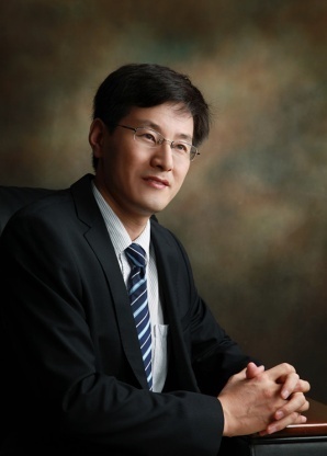 Lijun Jia (贾立军)合作论文数Longhua Hospital, Shanghai University of Traditional Chinese Medicine5
 Yi Zhang (张毅)合作论文数Biological Cell Therapy Center, The First Affiliated Hospital of Zhengzhou University5Qianyun Hao合作论文数4Wenjing Huang合作论文数4Shan Zhao合作论文数4Wenbo Cao合作论文数4Saijun Mo (莫赛军)合作论文数4
 Sai-Yang Zhang (张赛扬)合作论文数School of Basic Medical Sciences, Zhengzhou University4
 Robert Hoffman合作论文数AntiCancer, Inc.;Department of Surgery, Medical Center, University of California, San Diego3Xuan-Yu Hu合作论文数3Yangcheng Ma合作论文数3Zhenzhen Liu合作论文数3
 Yanan Jiang合作论文数3Junfen Ma合作论文数3Yanyu Jiang合作论文数3Yuan Zhang合作论文数Zhengzhou Univ, Affiliated Hosp 1, Dept Clin Lab, Zhengzhou 45005, Henan, Peoples R China3Jian Song合作论文数3Lili Cai合作论文数3HongKai Lian (连鸿凯)合作论文数3ChunYao Wang (王纯耀)合作论文数3
 Deyin Guo (郭德银)合作论文数School of Medicine, Sun Yat-sen University3
 Qiong Fu (扶琼)合作论文数上海交通大学医学院附属仁济医院3Liang Yupei合作论文数3Li Li-Hui合作论文数3
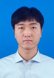 Xiaoyu Chen (陈小榆)合作论文数Coll Informat Sci & Engn, Yanshan Univ3Ningjing LEI (雷宁静)合作论文数2Yongqing Heng合作论文数2Zhaoyang Shang合作论文数2Ying Du合作论文数2yilin zhang合作论文数2Lifeng Jiang合作论文数2Shuaiheng Hou (侯帅恒)合作论文数2Shiwen Wang合作论文数2Zheng Li合作论文数2
 Shuo Li合作论文数2
 Peng Gao合作论文数Dept Neurosurg, Zhengzhou Univ2Yue Du合作论文数2




