目的 探讨血脂异常对不稳定型心绞痛(UAP)和急性心肌梗死(AMI)患者临床识别的价值和意义.方法 本研究纳入武汉大学人民医院于2016年1月至2018年12月收治的UAP患者122例、AMI患者185例.采用全自动生化分析仪检测所有患者血清中三酰甘油(TG)、总胆固醇(TC)、高密度脂蛋白胆固醇(HDL-C)、低密度脂蛋白胆固醇(LDL-C)、心肌肌钙蛋白I(cTnI)等.ROC曲线分析观察血脂指标联合或单独检测对UAP和AMI患者临床识别的敏感性和特异性.结果 Mann-Whitney U检验结果显示AMI患者血清中血糖值Glu、脑钠肽前体(proBNP)、cTnI显著高于UAP组患者,差异具有统计学意义(P<0.05);Unpaired studentt检验结果显示AMI患者血清中TC、LDL-C、纤维蛋白原(FIB)的水平显著高于UAP组患者,差异具有统计学意义(P<0.05).ROC曲线分析TC、LDL-C的曲线下面积分别为0.623和0.654,而TC联合LDL-C的曲线下面积仅占0.664.结论 血脂指标TC联合LDL-C对于临床识别UAP和AMI的诊断价值不大.因此,血脂指标异常仅只能作为UAP和AMI患者的辅助诊断之一,临床早期鉴别诊断两者尚需借助其他检查手段.
目的 探讨血清成纤维细胞生长因子(FGF21)在老年冠状动脉粥样硬化心脏病(冠心病)患者中的表达水平,及其与肥胖、糖脂代谢和冠状动脉(冠脉)狭窄程度之间的关系.方法 纳入老年冠心病患者38例(试验组),选取同期冠脉造影正常的老年患者42例(对照组).收集两组患者的一般临床资料;检测空腹血糖、脂代谢指标[总胆固醇(TC)、三酰甘油(TG)、低密度脂蛋白胆固醇(LDL-C)和高密度脂蛋白胆固醇(HDL-C)];酶联免疫吸附法检测所有患者血清FGF21水平;运用Gensini评分量化试验组患者的冠脉狭窄程度;采用Pearson分析探讨老年冠心病患者血清FGF21水平与肥胖、糖脂代谢指标、冠脉狭窄程度之间的相关性.结果 两组患者在年龄、性别方面差异无统计学意义(P>0.05).试验组患者的体质指数(BMI)、腰臀比、空腹血糖、TC、TG和LDL-C显著高于对照组,差异具有统计学意义(P<0.05).与对照组相比较,试验组血清FGF21水平显著升高,差异具有统计学意义(P<0.05).相关性结果显示,老年冠心病患者血清FGF21水平与BMI、腰臀比、空腹血糖、TC、TG、LDL-C、Gensini评分呈正相关,相关系数r分别为0.573、0.495、0.614、0.426、0.271、0.380和0.689,差异具有统计学意义(P<0.05).结论 血清FGF21在老年冠心病患者中的表达显著升高,且与肥胖、高血糖、高血脂和冠脉狭窄程度密切相关.
The ubiquitin-proteasome system (UPS) degrades a protein molecule via 2 main steps: ubiquitination and proteasomal degradation. Extraproteasomal ubiquitin receptors are thought to couple the 2 steps, but this proposition has not been tested in vivo with vertebrates. More importantly, impaired UPS performance plays a major role in cardiac pathogenesis, including myocardial ischemia-reperfusion injury (IRI), but the molecular basis of UPS impairment remains poorly understood. Ubiquilin1 is a bona fide extraproteasomal ubiquitin receptor. Here, we report that mice with a cardiomyocyte-restricted knockout of Ubiquilin1 (Ubqln1-CKO mice) accumulated a surrogate UPS substrate (GFPdgn) and increased myocardial ubiquitinated proteins without altering proteasome activities, resulting in late-onset cardiomyopathy and a markedly shortened life span. When subject to regional myocardial ischemia-reperfusion, young Ubqln1-CKO mice showed substantially exacerbated cardiac malfunction and enlarged infarct size, and conversely, mice with transgenic Ubqln1 overexpression displayed attenuated IRI. Furthermore, Ubqln1 overexpression facilitated proteasomal degradation of oxidized proteins and the degradation of a UPS surrogate substrate in cultured cardiomyocytes without increasing autophagic flux. These findings demonstrate that Ubiquilin1 is essential to cardiac ubiquitination-proteasome coupling and that an inadequacy in the coupling represents a major pathogenic factor for myocardial IRI; therefore, strategies to strengthen coupling have the potential to reduce IRI.
目的 对一个扩张型心肌病(dilated cardiomyopathy,DCM)家系行候选致病基因全外显子高通量测序,以寻找该家系的致病基因,并分析其基因型和表型的关系.方法 收集武汉大学人民医院就诊的1例DCM患者及其家系成员的临床资料及血液标本.与先证者及其家属签订知情同意书,绘制家谱图,由我院临床分子诊断中心对先证者进行候选致病基因全外显子高通量测序,获得可疑突变后,用Sanger测序对家系其他成员进行验证,寻找致病基因.结果 家系先证者6号染色体外显子上存在受磷蛋白(phospholamban,PLN)基因的精氨酸缺失突变c.36_38delAAG(p.Arg13del),为该家系的可疑致病基因.先证者目前心脏扩大,心功能显著下降,且超声心动图提示左心室附壁血栓形成,心电图提示肢导低电压以及胸导联R波极度减低.先证者母亲及其大姐因心脏病死亡,二姐目前患有扩张型心肌病,其子女未检测到致病基因.受磷蛋白作为肌质网钙离子循环中的调节蛋白,它的基因表达、分布、功能与心室的收缩功能密切相关.结论 DCM家系中存在PLN基因缺失突变:PLN c.36_38delAAG(p.Arg13del),是家族性扩张型心肌病的重要致病基因,此突变在汉族人群中尚属首次报道.
Objective To screen the candidate pathogenic gene among family members of a dilated cardiomy-opathy (DCM), and find the relationship between the genotype and the phenotype. Methods The proband and family members came from Daye City, Hubei Province. The proband was diagnosed with dilated cardiomyopathy at Renmin Hospital of Wuhan University in April 2017, and some of his family member were dead then. The inheritance atlas was drawn, and analysis of genetic characteristics and clinical phenotype was performed. The candidated gene exon of the proband was sequenced by high-throughput sequencing, and ultimately the target area of the exon and mutations of can-didate genes were screened. Then bidirectional sequencing of Sanger was used to sequence other family members which were matching with gender and age to testify whether there are the above mutations. Results In this family, the proband and his sister carry one missense mutations, with TTN c.100126A>G (p.Thr33376Ala). The heart function of proband was declined and accompanied by malignant arrhythmia. But his sister has no obvious clinical symptoms. Conclusion The patient of this family carries the genetic mutation of TTN. TTN truncating mutations are a common cause of dilated cardiomyopathy, which maybe improve our understanding of the pathophysiology of dilated cardiomy-opathy.
BACKGROUND AND OBJECTIVE:Angiotensin-converting enzyme (ACE) inhibitors have been reported to decrease myocardial remodeling and faciliate cardiac function improvement in the setting myocardial infarction by affecting bradykinin. The purpose of this study was to evaluate the combination effects of perindopril and bradykinin (BK) in rats with myocardial infarction.METHODS:Wistar Rats underwent to left anterior descending (LAD) coronary artery ligation were allocated into MI group (n=6); Perindopril group (n=7); Perindopril+BK group (n=7). An additional sham operation group (Sham group, n=6) were also established. After 4 weeks, the left ventricle function, myocardial tissue morphology, myocardial collagen volume faction, infracted ventricular wall thickness, myocardial infarction area and neovascular formation were evaluated.RESULTS:Combination treatment with perindopril and BK were showed significant improvement on LVEDV, LVEF and LVFS than MI group. Moreover, a significant improvement on LVEF was found in Perindopril+BK group than Perindopril group but not on LVEDV and LVFS between these two groups. Furthermore, neo-vessel density was significantly increased in Perindopril+BK group than other groups while no significant improvement on vessel density was found after the treatment of perindopril. In addition, myocardial infarction thickness improvement was found in Perindopril and group than MI group while combination treatment with perindopril and BK can significant improve the myocardial infarction thickness than perindopril only.CONCLUSIONS:Combination treatment with ACE inhibitor perindopril and BK can significantly improve the ventricle function in the rat model of myocardial infarction. Our data suggest BK can serve as adjuvant treatment in myocardial infarction treatment.
Objective:To observe the effects of Notch signaling pathway on the differentiation of embryonic stem cells to myocardial cells.Methods:The experiments were divided into two parts:regulating the switch of Notch signaling pathway to study the effects on differentiation rates of embryonic stem cells to cardiomyocytes,and determining the expression of myocardium differentiation related proteins.Mouse embryonic stem cells were labeled with DAPI fluorescence(200 μg/ml).Then Jagged protein(5 mg/L),an activator of notch signal,or MW167(100 μmol/L),an inhibitor of notch signal,was added to culture embryonic stem cells for 14 days.The appearance of spontaneously contractive cells was observed under phase contrast microscope.The expression of myocardial specific contractive protein was identified by cell immunofluorescence assay,and the differentiation ratio of embryonic stem cells to cardiomyocytes was calculated.The expression of myocardial differentiation related proteins were identified by Western blot.Results:At the 14th day,there were about 15% embryonic stem cells differentiated into spontaneously contractive cardiomyocytes,and the differentiation ratio was significantly reduced with Jagged treatment,whereas dramatically increased with MW167 treatment.Western blot results suggested that the expression of early myocardial differentiation related proteins were significantly reduced when cells were treated with Jagged,whereas dramatically increased when the cells were treated with MW167.Conclusion:Jagged protein inhibits the differentiation of embryonic stem cells to cardiomyocytes through activating notch signaling pathway,however,MW167 can promote the differentiation of embryonic stem cells to cardiomyocytes through inhibiting notch signaling pathway.
Background/Aims : To investigate the beneficial effects of atorvastatin added to the cell therapy with bone marrow-derived mesenchymal stromal cells (BMSCs) in a rabbit model of acute myocardial infarction (AMI). Methods: Rabbits were randomly divided into control group (n=10), bone marrow stem cells transplantation group (n=10), and BMSCs + atorvastatin group (n=10). AMI was established by ligating the left descending coronary artery. The left ventricular (LV) function was evaluated by echocardiography. H&E staining and Masson's Trichrome staining were performed to evaluate inflammatory cell infiltration and cardiac fibrosis. Immunohistochemistry and TUNEL were conducted to assess survival, differentiation, and apoptosis of transplanted cells and cardiomyocytes. Results: BMSCs decreased LV systolic and diastolic diameters and increased LV ejection fractions, LV fractional shortening, LV systolic pressure and LV end-diastolic pressure. Atorvastatin synergistically enhanced the BMSCs-induced improvements of ischemic cardiac dysfunction. Atorvastatin reduced inflammatory cell infiltration, cardiac fibrosis, and derangement of myocardial morphology/structure. Atorvastatin added a protective effect to cardiomyocytes against apoptotic cell death in infarct and peri-infarct areas, and also increased the survival rate of implanted BMSCs in acute myocardial ischemia. Atorvastatin also promoted cardiac differentiation of implanted BMSCs in infarct myocardium. Conclusion: Atorvastatin acts to improve the microenvironment both by synergistically enhancing the existing effects of BMSCs and by adding new therapeutic effects to BMSCs transplantation, and this combinational therapy is a superior cell/pharmacological therapeutic approach that merits future preclinical and clinical studies.
Objective:To explore the role of plasma Pentraxin 3(PTX3) in the development of acute coronary syndrome(ACS),and to assess the prognostic value of PTX3 in patients with ACS, through detecting the plasma levels of PTX3 in ACS patients and normal controls.Methods; Eighty consecutive patients with ACS,including 32 cases of ST-segment elevation myocardial infarction (STEMI) patients,23 cases of non-ST-segment elevation myocardial infarction(NSTEMI) patients,and 25 cases of unstable angina pectoris(UAP) patients,were included in this study.Another 20 patients who were excluded coronary artery disease by coronary angiography were set as the control group on the corresponding period.All hospitalized patients were detected plasma PTX3 by ELISA,and followed up for 3 months after discharge to observe the situation about adverse cardiovascular events.Results:Plasma PTX3 levels in ACS group were significant- ly higher than in control group(P<0.001).In patients with ACS,plasma PTX3 levels significant correlated with white blood cell count(WBC) 0=0.359,P = 0.001),hs-CRP(r=0.405, P<0.000 1) and NT-ProBNP 0=0.883,P<0.000 1).Compared with patients with PTX3<5. 51μg/L,patients with higher plasma PTX3 concentration had a higher 3-month combined cardiovascular event rate(39.5%vs 17.5%,P<0.05).Multiple logistic regression analysis showed that only PTX3(Or=4.11,95%CI:1.37-12.38,P = 0.012) and NT-ProBNP(0=2.56,95% CI:1.12-4.53,P = 0.021),but not hs-CRP,were independent predictors for the 3-month cardiovascular end point events in patients with ACS.Conclusion:PTX3 may contribute to the pathogenesis of atherosclerosis.PTX3 and NT-proBNP,but not hs-CRP,were powerful independent prognostic indicators for the 3-month cardiovascular end point events in patients with ACS.Combined detection of PTX3 and NT-proBNP may substantially contribute to the early risk stratification in patients with ACS.
Human papillomavirus (HPV) 16 infection and RASSF1A expression play important roles in tumor development and progression. However, the precise mechanisms underlying their concerted function in the development of reproductive system tumors still remain to be elucidated. In the present study, we showed that HPV16-E6 selectively upregulates RASSF1A expression via degradation of p53, which interacts with the RASSF1A promoter and regulates apoptosis. Overexpression of p53 triggered a decrease in endogenous RASSF1A in SiHa cells, accompanied by apoptosis. Similarly, knockdown of endogenous HPV16-E6 in SiHa cells with RNA interference (RNAi) led to downregulation of RASSF1A mediated by p53 and the subsequent induction of apoptosis. These findings collectively suggest that HPV16 infection regulates p53-mediated RASSF1A expression and suppresses apoptosis. Moreover, RASSF1A may form an element of the negative autoregulatory feedback loops that act on the HPV16 response and are involved in p53-dependent apoptosis. Our results provide novel insights into the cellular mechanism of tumor development, and present a starting point for the development of novel strategies in cancer treatment and effective diagnosis.
Objective To investigate the effects of trimetazidine(TMZ) on the survival of mesenchymal stem cells(MSCs) in an ex-vitro model of hypoxia and subsequent activities of transplanted MSCs in rat hearts with acute myocardial infarction(AMI).Methods MSCs were cultured in serum-free medium and exposed to hypoxia for 12h with or without TMZ.The viability and apoptosis of MSCs at passage 3 were examined by transmission electron microscope.Thirty wistar rats were divided randomly into 3 groups,including AMI control group,MSCs transplantation group,and TMZ+MSCs group.MSCs were injected into peri-infarct myocardium(MSCs and TMZ+MSCs groups) thirty minutes after coronary artery ligation.The rats in TMZ+MSCs group were additionally fed TMZ from 3 days before AMI to 28 days after AMI.Cardiac structure and function were assessed by echocardiography 28 days after transplantation.The survival and differentiation of transplanted cells were detected by immunofluorescent staining.The cellular apoptosis in the peri-infarct region was detected with TUNEL assay.Blood samples were collected before the start of TMZ therapy and 24h,48h after AMI,and inflammatory cytokines(CRP,TNF-α) were measured.Results In hypoxic culture,the TMZ-treated MSCs displayed a two-fold decrease in apoptosis under serum-free medium and hypoxia environment.In vivo,cardiac infarct size was significantly smaller,cardiac function significantly improved in the MSCs and TMZ+MSCs groups than those in the control group.Combined treatment with TMZ and MSCs implantation demonstrated a further decrease in MSCs apoptosis,a further increase in MSCs viability,a further decrease in infarct size,and a further improvement in cardiac function,compared with those of MSCs group.The baseline levels of inflammatory cytokines(CRP,TNF-α) were not significantly different among the groups.However,all parameters at 24h were lower in TMZ+MSCs group than those in MSCs group.Conclusion Implantation of MSCs combined with TMZ treatment is superior to MSCs monotherapy for MSCs viability and cardiac function recovery.The inhibition of inflammatory cytokines expression may be the potential mechanism.
Bone marrow mesenchymal stem cells (MSCs) have shown potential for cardiac repair following myocardial injury, but this approach is limited by their poor viability after transplantation. The present study was to investigate whether trimetazidine (TMZ) could improve survival of MSCs in an ex vitro model of hypoxia, as well as survival, differentiation, and subsequent activities of transplanted MSCs in rat hearts with acute myocardial infarction (AMI). MSCs at passage 3 were examined for their viability and apoptosis under a transmission electron microscope, and by using flow cytometry following culture in serum-free medium and exposure to hypoxia (5% CO2, 95% N2) for 12 h with or without TMZ. Thirty Wistar rats were divided into 3 groups (n=10 each group), including group I (AMI control), group II (MSCs transplantation alone), and group III (TMZ+MSCs). Rat MSCs (4×10⁷) were injected into peri-infarct myocardium (MSCs group and TMZ+MSCs group) 30 min after coronary artery ligation. The rats in TMZ+MSCs group were additionally fed on TMZ (2.08 mg·kg−1·day−1) from day 3 before AMI to day 28 after AMI. Cardiac structure and function were assessed by echocardiography at 28th day after transplantation. Blood samples were collected before the start of TMZ therapy (baseline), and 24 and 48 h after AMI, and inflammatory cytokines (CRP, TNF-α) were measured. Then the survival and differentiation of transplanted cells in vivo were detected by immunofluorescent staining. The cellular apoptosis in the peri-infarct region was detected by using TUNEL assay. Furthermore, apoptosis-related proteins (Bcl-2, Bax) within the post-infarcted myocardium were detected by using Western blotting. In hypoxic culture, the TMZ-treated MSCs displayed a two-fold decrease in apoptosis under serum-free medium and hypoxia environment. In vivo, cardiac infarct size was significantly reduced, and cardiac function significantly improved in MSCs and TMZ+MSCs groups as compared with those in the AMI control group. Combined treatment of TMZ with MSCs implantation demonstrated further decreased MSCs apoptosis, further increased MSCs viability, further decreased infarct size, and further improved cardiac function as compared with MSCs alone. The baseline levels of inflammatory cytokines (CRP, TNF-α) had no significant difference among the groups. In contrast, all parameters at 24 h were lower in TMZ+MSCs group than those in MSCs group. Furthermore, Western blotting indicated that the expression of anti-apoptotic protein Bcl-2 was up-regulated, while the pro-apoptotic protein Bax was down-regulated in the TMZ+MSCs group, compared with that in the MSCs group. It is suggested that implantation of MSCs combined with TMZ treatment is superior to MSCs monotherapy for MSCs viability and cardiac function recovery.
Objective:To observe the effects of Jagged protein on the differentiation of embryonic stem cells to myocardial cells.Methods:Mouse embryonic stem cells were labelled by fluorescent dye DAPI(200 mg/L).Then Jagged protein(5 mg/L),the activator of notch signal,was added to embryonic stem cells in vitro for 14 days.The spontaneously contractive cells were observed under phase contrast microscope,ultra microstructure of spontaneously contractive cells were observed under transmission electron microscope.The expression of myocardial specific contactive protein was identified by cell immunofluorescent assay,and the differentiation ratio of embryonic stem cells to cardiomyocytes was calculatd.Results:The spontaneous differentiation of embryonic stem cells is strong.On the 14th day,there were about 15% embryonic stem cells differentiated into spontaneously contractive cardiomyocytes.When Jagged protein was added,the differentiation ratio was significantly reduced.Conclusion:Jagged protein added to embryonic stem cells activates the notch signal,which would inhibit the differentiation of embryonic stem cells to cardiomyocytes.
Objective: To investigate the control state, impact factors, and cardiovascular comorbidities in clinic patients with hypertension. Methods: One hundred clinic hypertension patients were retrospectively analyzed, and age, gender, blood pressure, family medical history, body mass index, smoking history, blood biochemical parameters, related target organ damage, comorbidities, etc, were recorded. Results: The treatment rate of all patients was 100% and the blood pressure control rate was 32%. Calcium antagonists, and angiotensin converting enzyme inhibitors, β-blockers, and angiotensin II receptor antagonists were commonly used. The proportion of patients with complications of coronary heart disease, arrhythmia, diabetes and stroke were 6%, 22%, 11%, and 8% respectively. 84% patients had one or more risk factors such as smoking, obesity, blood lipid abnormality, diabetes and family hypertension history. The control rate of smoking was 32.1%, and the control rate of TC, TG, and HDL-C respectively in the patients with hyperlipidemia history was 37.8%, 25.8% and 69.2% respectively, glucose control rate in patients with diabetes history was 45.4%. Conclusion: The treatment rate of the clinic hypertension patients was high but the control rate was low, the combined risk factors were more and were not controlled satisfactory, the damage to target organ was common and serious. The comprehensive control of hypertension should include controlling both the risk factors and comorbidities.
AIM: To investigate whether Trimetazidine(TMZ) can improve the survival of Bone marrow mesenchymal stem cells(MSCs) in an ex-vitro model of hypoxia,as well as survival,differentiation,and subsequent activities of transplanted MSCs in rat hearts with acute myocardial infarction(AMI),and discuss the mechanism. METHODS: MSCs at passage 3 were examined for their viability and apoptosis by transmission electron microscope,and flow cytometry following cultured in serum-free medium and exposed to hypoxia(5%CO2,95%N2) for 12 h with or without TMZ.Thirty wistar rats were divided into 3 groups(n=10 in every group),including group I(AMI control),group II(MSCs transplantation alone),and group III(TMZ+MSCs).Rat MSCs(4×10⁷cells) were injected into peri-infarct myocardium(MSCs and TMZ+MSCs groups) thirty minutes after coronary artery ligation.The rats in TMZ+MSCs group were additionally fed TMZ with 2.08 mg·kg-1·d-1 from 3 days before AMI to 28 days after AMI.Cardiac structure and function was assessed by echocardiography 28 days after transplantation.The survival and differentiation of transplanted cells were detected by immunofluorescent staining.The cellular apoptosis in the peri-infarct region was detected with TUNEL assay.Furthermore,apoptosis-related proteins(Bcl-2,Bax) within the post-infarcted myocardium were detected with Western blotting.RESULTS:In hypoxic culture,the TMZ-treated MSCs displayed a two-fold decrease in apoptosis under serum-free medium and hypoxia environment.In vivo,cardiac infarct size was significantly smaller,cardiac function significantly improved in the MSCs and TMZ+MSCs groups than those in the control group.Combined treatment with TMZ and MSCs implantation demonstrated a further decrease in MSCs apoptosis,a further increase in MSCs viability,a further decrease in infarct size,and a further improvement in cardiac function as compared with MSCs alone.Western blotting indicated that the expression of anti-apoptotic protein Bcl-2 was upregulated,while the pro-apoptotic protein Bax was downregulated in the TMZ+MSCs group,compared with that in the MSCs group.CONCLUSION: Implantation of MSCs combined with TMZ treatment is superior to MSCs monotherapy for MSCs viability and cardiac function recovery.The up-regulating of Bcl-2 maybe the potential mechanism.
Objective:To investigate the improvement of trimetazidine(TMZ) on viability of bone marrow mesenchymal stem cells(MSCs) in an in vitro model of hypoxia culture and in vivo model of acute myocardial infarction(AMI) in rats. Methods:In vitro:Rats' MSCs were cultured in serum-free medium and exposed to hypoxia condition,and then examined at the third passage for their viability and apoptosis by transmission electron microscopy and flow cytometry.In vivo:AMI model was created by coronary artery ligation.30 Wista rats were randomly divided into 3 groups,n=10 in each group.AMI control group,the serum-free medium was injected;MSCs alone group,MSCs(4×10⁷cells) were injected into the borderline of infarcted myocar-dium and MSCs+TMZ group,the rats were fed with TMZ 2.08mg/(kg·day) from 3 days before AMI to 28 days after AMI.Cardiac structure and function were assessed by echocardiography 28 days after MSCs implantation.The viability and apoptosis of implanted MSCs were detected at the borderline of infarcted myocardium. Results:In vitro:TMZ-treated MSCs presented half amount lower apoptosis rate than those without TMZ-treated MSCs under hypoxia culture.In vivo:Compared with AMI control group,the myocardial infarction sizes were significantly smaller in MSCs alone group and in MSCs+TMZ group,the cardiac function was significantly improved.Compared with MSCs alone group,MSCs had the higher viability and lower apoptosis,had the smaller size of infarction and the better cardiac function in MSCs + TMZ group. Conclusion:The combination of MSCs with TMZ implantation is superior to MSCs alone for higher MSCs viability and better cardiac function recovery in AMI model in rats.
Backgrounds: Experimental and clinical studies have suggested that cell implantation could improve cardiac function after myocardial infarction (MI). However, this technique was limited by decreased engraftment and survival of transplanted cells within the ischemic tissue. The present study was performed to investigate whether implantation of bone marrow-derived mononuclear cells (BMMNCs) encapsulated in hydrogel could increase cell engraftment and help to restore cardiac function of MI rabbits. Methods: MI was induced in rabbits by coronary artery ligation. One week later, cell culture medium, Dex-PCL-HEMA/PNIPAAm hydrogel, BMMNCs in medium or BMMNCs in hydrogel were injected into the infarcted area of the left ventricle (LV). Results: Increased cell engraftment was observed 48 h after injection when cells were encapsulated in hydrogel; 30 days after treatment, echocardiographic studies showed that injection of BMMNCs in hydrogel preserved LV ejection fraction and attenuated LV dilatation compared with other groups. Histological analysis indicated that injection of BMMNCs in hydrogel enhanced neovascular formation and prevented scar expansion compared with the other groups. Conclusion: Injection of hydrogel-encapsulated BMMNCs increased cell engraftment and improved LV function; this technique may serve as an effective approach to restore infarcted myocardium.
Objective To observe the rat cardiac size and cardiac function changes before and after trimetazidine administration plus bone-marrow stem cells transplanting through echocardiography.Methods Forty wistar rats were divided into the following 4 groups randomly:control group (T),myocardial infarction group (Ⅱ),bone marrow stem calls transplantation group (Ⅲ),and bone marrow stem cells transplantation plus trimetazidine administration group(Ⅳ).The rats' left anterior coronary artery in group Ⅱ,Ⅲ and Ⅳwas ligated to produce myocardial infarction model,then bone-marrow stem cells were injected around the infarcted area into the later two groups.Furthermore,rats in group Ⅳ were administrated with trimetazidine.The size and systolic function of the hearts were measured 4 weeks after transplantation.The left ventricular systolic pressure(LVSP) and the end-diastolic pressure(LVEDP) were also measured at the end of experiment.Results The left ventricular diameter of rats in group Ⅲ and Ⅳ was smaller than that in group Ⅱ,and the ventricular systolic function increased,LVSP increased and LVEDP decreased statistically in group Ⅲ and Ⅳ.the amelioration of cardiac size and function was significantly notable in group Ⅳ than that in group Ⅲ.Conclusions Bone marrow stem cells transplantation can release the enlargement of left ventricle and improve cardiac function after myocardial infarction.The therapeutic efficacy can be further elevated if administrated with trimetazidine simultaneously.
Short-term administration of losartan reduced the aortic surface lesion area and mean intimal thickness. The mechanisms for reducing atherosclerotic progression by losartan may be related to decreased macrophage proliferation and accumulation in the arterial wall, decreased activation of nuclear factor-kappa B and expression of its target gene ICAM-1. Objective — Recent studies suggest that angiotensin II (Ang II) may contribute to the vascular inflammatory response and atherosclerosis. Losartan is a specific AT| receptor antagonist which can effectively inhibit the effects of Ang II. However, the effects of losartan on atherogenesis have been rarely demonstrated. We designed this study to investigate the effects of short-term administration of losartan on atherosclerotic lesions in the aorta from rabbits fed a cholesterol-enriched diet and the possible mechanisms of its anti-atherogenic effects. Methods and results — The rabbits were randomly divided into three groups: (a) cholesterol group;(b) losartan-treated group;(c) normal control group. We observed that mean serum lipid levels in the cholesterol group were significantly higher than those in normal control rabbits, while blood pressure between two groups did not change significantly.Treatment with losartan did not affect serum lipid levels or systolic blood pressure but did reduce the aortic surface lesion area and mean intimal thickness.The number of macrophages markedly decreased after administration of losartan. Losartan also attenuated the activation of nuclear factor-kappa B and the expression of its target gene ICAM-1. Conclusions — In summary, losartan inhibited atherosclerotic progression by decreasing macrophage proliferation and accumulation in the arterial wall.The mechanisms for reducing atherosclerotic progression by losartan may be related to decreased activation of nuclear factor-kappa B.
Objective To observe effects of bone marrow stem cell implantation on ventricular arrhythmia after acute myocardial infarction in rat. Methods Thirty rats were randomly divided into two groups. Cell implantation group was treated with bone marrow stem cell injection, and control group was treated with the same quantity of serum-free medium injection. Then Holter was used to observe arrhythmia on the first and twenty-eighth day after injection. Results On the first day after injection, there wasn′t difference in the incidence rate of ventricular arrhythmia between the two groups. While on the twenty-eighth day after injection, the incidence rate of ventricular arrhythmia was decreased significantly in cell implantation group(P0.05). Conclusions Bone marrow stem cell implantation can effectively reduce the occurrence of ventricular arrhythmia after acute myocardial infarction.
 Gengshan Li (李庚山)合作论文数Renmin Hospital of Wuhan University26
 Jing Wang (王晶)合作论文数13
 Yihao Tian (田毅浩)合作论文数School of Basic Medical Sciences, Wuhan University13
 Jianjun Li (李建军)合作论文数Fuwai Hospital, Chinese Academy of Medical Sciences10
 Xuejun Jiang (蒋学俊)合作论文数Renmin Hospital of Wuhan University9
 Jingjun Lv (吕菁君)合作论文数Emergency Department, Renmin Hospital, Wuhan University8
 Qing Zhou (周青)合作论文数Renmin Hospital, Wuhan University7
 NingXu Li (李凝旭)合作论文数Liyuan Hospital, Tongji Medical College, Huazhong University of Science and Technology5Jun PENG (彭俊)合作论文数4YongNan Lv (吕永楠)合作论文数4Wen Jiang (蒋雯)合作论文数4
 Gangyan Zhu (朱刚艳)合作论文数Renmin Hospital of Wuhan University4Wenwei Huang合作论文数4
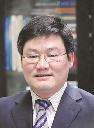 Hong Jiang (江洪)合作论文数Cardiovascular Department, Renmin Hospital, Wuhan University4ShiQi Tang (唐世琪)合作论文数3SongLin Wu (邬松林)合作论文数3HongMei Ma (马红梅)合作论文数3FuLiang Wang (汪福良)合作论文数3GuoSheng Lin (林国生)合作论文数3Zhe Qu (曲哲)合作论文数3YuPing Zhang (张玉平)合作论文数3Ying Liu (刘颖)合作论文数3Ling Xia (夏泠)合作论文数Zhongnan Hospital of Wuhan University;The Second Clinical College of Wuhan University3
 Ruiqiang Guo (郭瑞强)合作论文数Renmin Hospital of Wuhan University;Wuhan University First Clinical College3Jingwei Wang合作论文数2LIN Gui-zhen (林桂珍)合作论文数2Xia Li (李夏)合作论文数2
 Xiaoyan Li (李晓艳)合作论文数武汉大学第一临床学院2Jiayun Hu (胡家芸)合作论文数2
 Congxin Huang (黄从新)合作论文数Department of Cardiology, Renmin Hospital of Wuhan University2
 Chengjun Hu (胡成俊)合作论文数School of Basic Medical Sciences, Wuhan University2Penglong Wu合作论文数1jianjun li (李建军)合作论文数1Chang Liu合作论文数1Guocao LI (李国草)合作论文数1Zongwei Zhang合作论文数1Jing Zhang合作论文数1wang jing (王晶)合作论文数1Tao Lin合作论文数1Bo Pan (潘博)合作论文数1Jun Peng合作论文数1Mingxia Fan合作论文数1min dai (戴敏)合作论文数1Faqian Li合作论文数1ChunHong Fang (方春红)合作论文数1LaYuan Xiong (熊腊元)合作论文数1




