Sulfurmustard (SM) is a typical representative of highly-toxic chemical warfare agent that is difficult to be prevented and treated. SM could produce various biomarkers with protein, glutathione and DNA, etc, besides oxidative and hydrolytic metabolism. The tripeptide-adduct hydroxyethylthioethyl-Cys-Pro-Pro (HETE-CPF) originating from Cys-34 on the human serum albumin (HSA) is a very important retrospective biomarker, which is usually used to identify SM exposure due to its long half-life and good stability. In this work, a novel synthetic route of HETE-CPF was developed with chloroethanol as the starting material. Compared with the original synthetic route using 2-chloroethyl mercaptoethanol as the starting material, the new synthetic method had less side reactions, higher yield and easier purification. Furthermore, a highly sensitive analysis method for HETE-CPF in whole blood samples was established, and the detection sensitivity was up to 2 ng/mL, which was enough to identify human plasma sample after exposure to SM. Meanwhile, it was also identified that after SM exposure via caudal vein in rats, the exposure biomarkers formed with plasma albumin were significantly different from those of human plasma, which was HETE-Cys-Pro-Tyr (HETE-CPY), not HETE-CPF. The fundamental reason might be due to their different primary sequence of albumin.
OBJECTIVE:Our goal was to analyze the demographic and pathologic characteristics as well as prognosis in nonsmoking and nondrinking (NSND) oral squamous cell carcinoma (SCC) patients compared with typical oral SCC patients.PATIENTS AND METHODS:A total of 353 patients were retrospectively enrolled and divided into two groups: the NSND group and the current smoking/current drinking (CSCD) group. Demographic, pathologic, and molecular data were compared between the two groups. The main research endpoints were locoregional control (LRC) and disease-specific survival (DSS).RESULTS:In the NSND group, 16.3%, 41.9%, and 53.5% of patients were aged no more than 40 years, were female, and had an educational background of high school or above compared to 3.7%, 6.0%, and 38.2% of patients in the CSCD group, respectively. A total of 15.1% of the NSND patients had SCC of the lower gingiva and floor of the mouth, which was lower than the 35.6% of patients in the CSCD group. CSCD patients were likely to have an advanced disease stage (48.7% vs 32.5%, p=0.042) and poorly differentiated cancer (26.6% vs 16.3%, p=0.042). The NSND patients had a mean Ki-67 index of 24.5%, which was lower than the mean of 35.7% in the CSCD patients. The two groups had no HPV infection and similar p16 expression (4.7% vs 10.1%, p=0.132), but there was higher expression of p53 (38.6% vs 17.4%, p<0.001) and p63 (59.9% vs 29.1%, p<0.001) in the CSCD group. The 5-year LRC rates for NSND patients and CSCD patients were 48% and 38%, respectively, and the difference was significant (p=0.048). The 5-year DSS rates for NSND patients and CSCD patients were 56% and 39%, respectively, and the difference was significant (p=0.047). Further, a Cox model confirmed the independence of smoking and drinking status for affecting LRC and DSS.CONCLUSION:NSND oral SCC patients are a different entity. HPV infection has a limited role in carcinogenesis in NSND patients, and p16 expression is associated with worse locoregional control.
Sulfur mustard (SM) is a typically representative alkylating agent with high reactivity. SM can produce a variety of hydrolytic and oxidative metabolites including various proteins and nucleic acid adducts. Among them, divinyl sulfone (DVS) is an important oxidative metabolite that should be paid more attention, which has high reactivity and toxicity close to SM. In this study, the human serum albumin (HSA) and human plasma were exposed to DVS and its two-phase metabolite DVS-GSH, respectively. After digestion with proteinase K and purification by solid phase extraction (SPE), UPLC-Q-extractive orbitrap high rosolution mass spectrometry (HRMS) was used to identify some albumin adducts. According to the analysis results, three novel biomarkers, DVS-Cys-Pro-Phe, Phe-Pro-Cys-DVS-Cys-Pro-Phe and GSH-DVS-Cys-Pro-Phe, were successfully identified by UPLC-Q-exactive orbitrap/HRMS. The results showed that both DVS and DVS-GSH could react with Cys-34 of albumin due to their highly reactive alkene bonds. These novel damage biomarkers not only provided new clues for SM exposure and diagnosis, but also provided evidence for elucidating the damage mechanism of SM from a new perspective.
Objective: To determine the scavenging effect and the change of metabolism of paraquat (PQ) using hemoperfusion (HP) once and twice within 12 hours after intoxication and explore the better scheme of HP. Methods: 18 beagles were randomly divided into 3 groups. Single HP group, Double HP group and Control group. Peripheral veins blood was collected at different times within 48 hours after exposure in each group. Toxin concentration was measured, analyzed and compared among 3 groups. Results: 6 hours after exposure, Single HP group and Double HP group has finished the first HP treatment, and the concentration of PQ was lower than that of the control group, the difference was statistically significant (P<0.05) . 10 hours after exposure, there was no statistical difference of toxin concentration among 3 groups (P>0.05) . 12 hours after exposure, Double HP group has finished the second HP treatment, the concentration of PQ was significantly lower than that of Single HP group and Control group (P<0.05) . 24 hours and 48 hours after exposure, there was no statistical difference of toxin concentration among 3 groups (P>0.05) . Statistical difference were not observed in toxicokinetical parameters among 3 groups (P>0.05) . Conclusion: HP treatment once and twice within 12 hours after intoxication could effectively reduce the toxin concentration in the peripheral veins blood after HP for about 4 hours, then the toxin concentration would return to the same level as Control group quickly. It was suggested that at the beginning of poisoning, HP treatment once or twice could not significantly change the metabolism of paraquat.
OBJECTIVE:To study the adsorption effect of activated charcoal suspension on paraquat (PQ) in gastrointestinal tract of beagles exposed to PQ.METHODS:Twenty healthy male beagles were randomly divided into experimental group and control group, with 6 beagles in each group. 20% PQ solution (a dose of 30 mg/kg) was prescribed through stomach for beagles in both groups. After exposure to PQ for 30 minutes, the beagles in experimental group were given activated charcoal suspension (1.0 g/kg of type I activated charcoal powder mixed with 100 mL of normal saline) by gavage, while the control group was only given equal volume of normal saline. After exposure to PQ for 10 minutes, 30 minutes, and 1, 2, 4, 8, 12, 24, and 48 hours, blood was collected from hepatic portal veins and peripheral veins to detect the PQ concentration change in the plasma. The toxicokinetics software DAS 2.1.1 was applied to analyze PQ concentration and compare the change in toxicokinetics parameters between the both groups. The change in vital signs including heart rate (HR), respiratory rate (RR) and pulse oxygen saturation (SpO2) was dynamically monitored 10 minutes before exposure, 4 hours and each day from the 1st to the 7th day after exposure.RESULTS:After exposure to PQ, the poison concentration in the plasma of hepatic portal veins and peripheral veins in the control group rose quickly and reached peak 4 hours later. It fell quickly at first, and fell slowly 8 hours later. But in the experimental group, the increase rate to the peak was significantly slow. Besides, PQ peak fell more obviously than that in the control group and it was about 50% of the control group (μg/L: 123.50±11.67 vs. 255.18±12.29 in blood from hepatic portal veins, 122.35±11.72 vs. 250.86±11.15 in blood from peripheral veins). After 8 hours it fell much more quickly than that of the control group. After exposure to PQ for 48 hours, PQ concentration in the plasma was still lower than that of the control group (μg/L: 0.53±0.18 vs. 15.98±5.58 in blood from hepatic portal veins, 0.31±0.01 vs. 15.03±4.82 in blood from peripheral veins, both P < 0.01). With the toxicokinetics analysis, compared with the control group, the maximum concentration (Cmax) and area under the curve (AUC) of PQ in the plasma of hepatic portal veins and peripheral veins in the experimental group were significantly decreased [Cmax (μg/L): 125.07±9.49 vs. 255.18±12.29 in blood from hepatic portal veins, 123.38±9.52 vs. 250.86±11.15 in blood from peripheral veins; AUC (mg×L-1×h-1): 1.6±0.2 vs. 3.3±0.4 in blood from hepatic portal veins, 1.5±0.2 vs. 3.2±0.3 in blood from peripheral veins], time to the peak (Tmax) of PQ was slowed (hours: 5.3±1.9 vs. 4.0±0.0 in blood from hepatic portal veins, 4.7±1.5 vs. 4.0±0.0 in blood from peripheral veins), and PQ plasma half-life (t1/2) and mean retention time (MRT) were significantly shortened [t1/2 (hours): 3.8±1.2 vs. 15.4±3.7 in blood from hepatic portal veins, 3.5±1.0 vs. 15.5±2.7 in blood from peripheral veins; MRT (hours): 8.0±1.5 vs. 13.4±1.2 in blood from hepatic portal veins, 7.6±1.3 vs. 13.3±1.2 in blood from peripheral veins; all P < 0.01]. After exposure to PQ, HR and RR in both the experimental group and the control group increased and reached to the peak about the 4th day and then the increase rate began to slow down gradually; SpO2 slowed down gradually and reached to the valley about the 4th day and then it began to recover, but the change range of vital signs in the experimental group was smaller than that of the control group, and the parameters were significantly better than those of control group [4-day HR (bpm): 134.50±3.04 vs. 142.00±6.43, 4-day RR (times/min): 31.00±0.58 vs. 34.33±0.94, 4-day SpO2: 0.900±0.006 vs. 0.873±0.005, all P < 0.05].CONCLUSIONS:Activated charcoal administrated at 30 minutes after PQ poisoning can slow down the increase rate of PQ concentration in the plasma, decrease the peak concentration and has less influence on vital signs in beagles.
Paraquat (PQ) as a kind of sterile and herbicides, has effects of contact-kill and systemic action, which can be absorbed quickly by green plants and make them wither and die. Therefore, it is widely used in the agricultural production and occupies a large part in our country's pesticide market. PQ poisoning has become one of the most common pesticide poisoning in our country. PQ can be passivated when combining with soil, but it has great toxicity for human. There are still no specific antidotes for PQ poisoning at home and abroad, and the death rate of PQ oral poisoning is up to 95%. Clinically comprehensive treatment is adopted, including gastric lavage, intentional diarrhea, diuresis and blood perfusion. However, the therapeutic effect is not good and the case fatality rate keeps high. It has become one of the hot issues for emergency medicine study to search PQ's special efficiency measures. This paper briefly reviews PQ's poisoning mechanism and its clinical treatment progress to provide new exploration direction and treatment ideas for basic research and clinical treatment of PQ.
Objective To establish an animal model by placing one end of PICC in the hepatic portal vein of a beagle dog and leaving the other end out of its body.Methods Six Beagle dogs were given respiration anesthesia through orotracheal intubation.An incision was made through the right rectus abdominalis to locate the superior mesenteric vein (SMA) and the main hepatic portal vein.The left branch of SMA was separated and cut to put PICC into the main hepatic portal vein before being ligated and fixed.The other end of PICC was elicited through the right abdominal wall and passed beneath the skin to the back neck and fastened in case of movement.Results The anesthetic effect was good and all the operations were successful.The mean operation time was about an hour and the mean blood loss was about 15 ml.The incision healed 5-7 d after operation.Conclusion The establishment of the model can improve the effects of liver-targeting drugs,which can cut down the dosage,lower the cost of treatment and experiment and reduce the adverse effect of medicines.Through PICC,we can directly draw blood from the hepatic portal vein to measure the blood concentration before the first pass elimination.Then according to the concentration,we can calculate the absorption rate in the gastrointestinal tract,which can facilitate related experimental studies.
A disturbance of energy metabolism reduces cardiac function in acute severe hemorrhagic patients. Alternatively, adequate energy supply reduces heart failure and increases survival. However, the approach to regulating energy metabolism conductive to vital organs is limited, and the underlying molecular mechanism remains unknown. This study assesses the ability of histone deacetylase inhibitors (HDACIs) to preserve cardiac energy metabolism during lethal hemorrhagic injury. In the lethally hemorrhagic rat and hypoxic myocardial cells, energy metabolism and heart function were well maintained following HDACI treatment, as evident by continuous ATP production with normal cardiac contraction. Valproic acid (VPA) regulated the energy metabolism of hemorrhagic heart by reducing lactate synthesis and protecting the mitochondrial ultrastructure and respiration, which were attributable to the inhibition of lactate dehydrogenase A activity and the increased myeloid cell leukemia-1 (mcl-1) gene expression, ultimately facilitating ATP production and consumption. MCL-1, the key target of VPA, mediated this cardioprotective effect under acute severe hemorrhage conditions. Our results suggest that HDACIs promote cardioprotection by improving energy metabolism during hemorrhagic injury and could therefore be an effective strategy to counteract this process in the clinical setting.
BACKGROUND: Hemorrhage is a major cause of morbidity and mortality among trauma patients. The pathophysiologic changes following acute severe hemorrhage and tissue hypoxia lead to an imbalance of protein acetylation. Histone deacetylase inhibitors (HDACIs) were reported to restore the acetylation imbalance and serve as potential drugs for treating severe hemorrhage. However, the molecular mechanism of HDACI-mediated cytoprotection remains unclear. In this study, we examined the myocardial protective effects and respective mechanism of the HDACI valproic acid (VPA) administered during hemorrhagic and hypoxic stress in vivo and in vitro.METHODS: In vivo, the therapeutic effect of VPA was evaluated in acute severe hemorrhagic rats, and the expressions of BCL-2 signal pathway molecules were observed in rat heart tissues. To explore the molecular mechanism of VPA-mediated myocardial protection, a cobalt chloride (CoCl2)-induced hypoxia model of rat H9c2 cardiomyoblasts was applied to mimic hypoxic injury raised by acute hemorrhage.RESULTS: VPA administration significantly improved the 4-hour survival rate of hemorrhagic animals from 55% to 100% and protected H9c2 cells against CoCl2-induced hypoxic injury at a dose of between 12.5 mu M and 100 mu M. Increased expression of BCL-2 messenger RNA was observed following VPA treatment in the heart tissues of hemorrhagic rats (approximately 4.9-fold) and in H9c2 cells that survived CoCl2-induced hypoxia (approximately 4.9-fold). Western blot analysis showed a concomitant increase in BCL-2 protein expression and Akt phosphorylation following VPA treatment. The cytoprotective activity of VPA was diminished by triciribine-mediated inhibition of Akt activation and by silencing of BCL-2 gene expression.CONCLUSION: These findings suggest that VPA protects myocardial cells from hemorrhagic and hypoxic stress through the Akt/BCL-2 survival pathway, indicating a potential use of HDACIs for acute severe hemorrhage treatment. Copyright (C) 2016 Wolters Kluwer Health, Inc. All rights reserved.
Objective The goal of this study was to investigate the effect of hypertonic saline with 6% Dextran-70 (HSD) resuscitation on organ damage and the resuscitation efficiency of the combination of HSD and lactated ringers (LR) in a model of hemorrhage shock in dogs. Methods Beagles were bled to hold their mean arterial pressure (MAP) at 50±5 mmHg for 1 h. After hemorrhage, beagles were divided into three groups (n = 7) to receive pre-hospital resuscitation for 1 h (R1): HSD (4 ml/kg), LR (40 ml/kg), and HSD+LR (a combination of 4 ml/kg HSD and 40 ml/kg LR). Next, LR was transfused into all groups as in-hospital resuscitation (R2). After two hours of observation (R3), autologous blood was transfused. Hemodynamic responses and systemic oxygenation were measured at predetermined phases. Three days after resuscitation, the animals were sacrificed and tissues including kidney, lung, liver and intestinal were obtained for pathological analysis. Results Although the initial resuscitation with HSD was shown to be faster than LR with regard to an ascending MAP, the HSD group showed a similar hemodynamic performance compared to the LR group throughout the experiment. Compared with the LR group, the systemic oxygenation performance in the HSD group was similar but showed a lower venous-to-arterial CO2 gradient (Pv-aCO2) at R3 (p < 0.05). Additionally, the histology score of the kidneys, lungs and liver were significantly lower in the HSD group than in the LR group (p < 0.05). The HSD+LR group showed a superior hemodynamic response but higher extravascular lung water (EVLW) and lower arterial oxygen tension (PaO2) than the other groups (p < 0.05). The HSD+LR group showed a marginally improved systemic oxygenation performance and lower histology score than other groups. Conclusions Resuscitation after hemorrhagic shock with a bolus of HSD showed a similar hemodynamic response compared with LR at ten times the volume of HSD, but HSD showed superior efficacy in organ protection. Our findings suggest that resuscitation with the combination of HSD and LR in the pre-hospital setting is an effective treatment.
目的 探讨小鼠腹腔注射百草枯对其血清总抗氧化态的影响.方法 小鼠随机分为对照组和百草枯染毒组(PQ组),PQ组小鼠又分别按照50、70、100、150、200和300 mg/kg的百草枯染毒剂量分成6组,给予一次性腹腔注射百草枯;PQ组小鼠分别在染毒后1h和3h留取血样,与对照组比较血清ABTS自由基清除率;每个血样采集点均有5个样本.结果 不同染毒剂量组小鼠染毒后1h、3h的血清ABTS自由基清除率显著低于对照组小鼠(P<0.05);血清ABTS自由基清除率与染毒剂量和检测时间之间无显著相关性.结论 百草枯染毒后小鼠的血清ABTS自由基清除率下降,血清总抗氧化态降低.
目的 研究黑青稞籽皮提取物对心肌细胞缺氧/复氧(H/R)损伤的保护作用及其机制.方法 取大鼠胚胎心肌细胞(H9c2),随机分为5组:正常对照组(C组)、缺氧/复氧组(H/R组)、缺氧/复氧+低、中、高剂量药物组(H/R+L、M、H组).流式细胞术检测细胞凋亡、细胞线粒体跨膜电位(△Ψm)和细胞内活性氧(ROS)产生情况;Western blot方法检测B细胞淋巴瘤/白血病-2基因(Bcl-2),Bcl-2相关X蛋白(Bax),总丝氨酸苏氨酸蛋白激酶(tAkt)和磷酸化丝氨酸/苏氨酸蛋白激酶(pAkt)蛋白表达变化.结果 药物组[(H/R+L)组至(H/R+H组)]细胞凋亡比例为(26.7±2.9)%至(6.1±1.1)%,较H/R组[(57.3±10.4)%]明显降低,差异有统计学意义(P<0.05);与H/R组比较,药物组△Ψm升高,差异有高度统计学意义(P<0.01),ROS水平下降,差异有高度统计学意义(P<0.01),Bcl-2及pAkt表达升高,Bax及tAkt表达无明显变化.结论 黑青稞籽皮提取物对心肌细胞缺氧/复氧损伤具有保护作用,可抑制心肌细胞凋亡,其可能与Bcl-2/Bax调控和激活磷脂酰肌醇-3激酶/丝氨酸苏氨酸蛋白激酶(PI3K-Akt)信号通路有关.
Pancreatic cancer is a lethal human malignancy with an extremely poor prognosis and urgently requires new therapies. Histone deacetylase inhibitors (HDACIs) represent a new class of anticancer agents and have shown promising antitumor activities in preclinical models of pancreatic cancer. In this study, we sought to determine the antitumor effects of a novel HDACI, chidamide (CS055), in pancreatic cancer cells alone or in combination with gemcitabine. Treatments of BxPC-3 or PANC-1 pancreatic cancer cell lines with chidamide resulted in dose- and time-dependent growth arrest, accompanied by induction of p21 expression. When combined in a sequential schedule, chidamide synergistically enhanced gemcitabine-induced cell growth arrest and apoptosis, accompanied by cooperative downregulation of Mcl-1 and loss of mitochondrial membrane potential (ΔΨm). Chidamide enhanced gemcitabine-induced DNA double-strand breaks and S phase arrest, and abrogated the G2/M cell cycle checkpoint, potentially through suppression of CHK1 expression. Our results suggest that chidamide has a therapeutic potential for treating pancreatic cancer, especially in combination with gemcitabine.
Objective To evaluate the effect of frozen RBC washing machines of the dialysis type and centrifuge type in processing frozen red blood cells.Methods Frozen red blood cells were thawed and divided equally into two blood bags,processed by the two types of machines respectively and preserved at 4℃ after deglycerolization.The content and recovery of hemoglobin,the supernatant osmolarities,and the supernatant hemoglobin were detected to estimate the effect of the two machines.The supernatant hemoglobin and concentrations of Na+ and K+ were measured at d 0,1,3,5 and 7 during blood preservation at 4℃ to observe the time limits for preservation at 4℃ after deglycerolization and compare the effect of the two machines.Results For cells processed by the dialysis and centrifuge type of machines,the content of hemoglobin was(17.2±0.51) and(16.8±0.46)g,respectively;osmolarities representing residual glycerol were(315±8) and(322±8)mOsm.The free hemoglobin values were(0.65±0.08) and(0.72±0.09)g/L.The above indexes all met national standards and there was no significant difference between the two groups.The red blood cells processed by the two machines could be preserved at 4℃ for three days.Conclusion Under level 2-3 sea conditions,the dialysis and centrifuge types of frozen RBC washing machines could both be used on large-tonnage ships and the red blood cells processed meet national standards.
Objective To observe the effect of high dose of methylprednisolone on Balb /c mice with acute paraquat( PQ)poisoning. Methods Balb / c mice were randomly divided into poisoning group( 30 mice),treatment group( 30 mice),and medrol control group( 5 mice). The poisoning group and treatment group were randomly divided into 6 sub-groups according to different PQ doses( 50,70,100,150,200,and 300 mg /( kg·d),respectively) by i. p. injection; after recieving the PQ injection,the mice in treatment group were immediately received 200 mg / kg of methylprednisolone i. p. injection. The control mice were only received 200 mg / kg of methylprednisolone by i. p. injection. The LD50values between poisoning group and treatment group were compared each other. Results( 1) At 24,48 and 72 h after exposure to paraquat,the LD50values were216. 67 mg / kg,216. 67 mg / kg and 188. 24 mg / kg,respectively,in poisoning group,and 70. 42 mg / kg,70. 42 mg / kg and87. 66 mg / kg in treatment group,respectively.( 2) The LD50values in treatment group were smaller than that in poisoning group at either 24,48 or 72 h after exposure to paraquat. Conclusion A single intraperitoneal injection of high-dose methylprednisolone could accelerate the death of Balb / c mice with paraquat poisoning.
Acute coagulopathy induced by combat trauma is a main cause of death for severe hemorrhagic patients.Damage control resuscitation(DCR) which focused on coagulopathy prevention in the early stage was proposed by US Army Institute of Surgical Research in 2006.This resuscitation strategy could dramatically reduce the mortality of severe hemorrhagic patients.The impact of coagulopathy on combat trauma treatment,the mechanism of coagulopathy induced by hemorrhage and the application of DCR in combat trauma treatments are reviewed.The implication of DCR for military transfusion is also discussed.
突发事件医学应急力量的设备器材和药品的采购、贮存与管理,应遵照“统一规划、分级储备、确保急需、突出重点、品种齐全、动态储备”的原则。根据化学中毒应急救援队伍承担的任务,需要配备的装备器材主要包括医疗救治、洗消和检测的专业器材,药品包括抗毒药物和常用急救药物等,同时也需配备必要的防护和辅助器材。
皮肤是化学毒剂及恐怖性化学毒物损伤的主要染毒途径。化学毒剂污染皮肤后首要的医学处置措施即为染毒部位的洗消[1]。洗消作为化学染毒医学应急救援的首要措施,为人员的生命安全及维持作战能力提供决定性的保障。为此,美军《化生放核防御现代化计划》将防化洗消作为持续作战之核心能力之一[2]。
Objective To compare the effect of different decontamination methods of dichlorvos poisoning with skin contact.Methods On the establishment of skin exposure to dichlorvos model,the levels of blood acetylcholinesterase(AchE) were tested in different time point,and the effect of different decontaminate liquid(water,soap water,sodium bicarbonate solution) were analyzed combined with the start time of the decontamination.Results With skin exposed to dichlorvos after 1 h,the blood AchE had decreased to 30% or less,which was severe poisoning,the decontamination effect was not significant;the sooner was the decontamination,the smaller was the AchE inhibition,and lower level of intoxication;there were no statistically significant difference between different experiment groups in decontamination effect after 30 minutes.Conclusion In the case of dichlorvos poisoning,it is significant to begin decontaminating as soon as possible to reduce absorption,decrease the severity,and save time for post-hospital treatment,it is the best of less than 10 minutes;alkaline decontamination solution have lost the advantages when the poisoning time has lasted for more than 30 minutes,then decontamination should immediately begin with no consideration of decontamination solution.
应急准备是针对可能发生的事故,为迅速、有序地开展应急行动而预先进行的组织准备、行动方案和应急保障。为保证迅速、有序、有效地开展化学中毒事件的应急与救援行动,需要做好相应的准备工作,主要包括医疗卫生救援机构、救援队伍与人员、装备与药品、通讯与保障、培训与演练、保障与救援预案等。救援任务承担的主体主要包括地方卫生管理部门、卫生监督机构、疾病控制、“120”紧急救援中心、化学中毒救治基地或指定救治机构,以及卫生应急专业队伍。这里主要简述救援队伍的准备与建设。
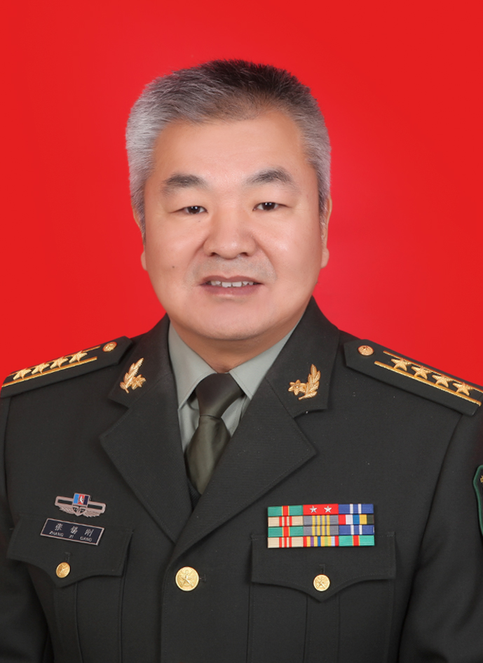 Xigang Zhang (张锡刚)合作论文数中国人民解放军总医院第五医学中心(原307医院)20
 YanBo Li (李彦博)合作论文数9WeiDong Luo (罗卫东)合作论文数8YunZhi Fa (法云智)合作论文数6HaoChun Wang (王浩春)合作论文数6
 QingYi Meng (孟庆义)合作论文数Chinese PLA General Hospital6
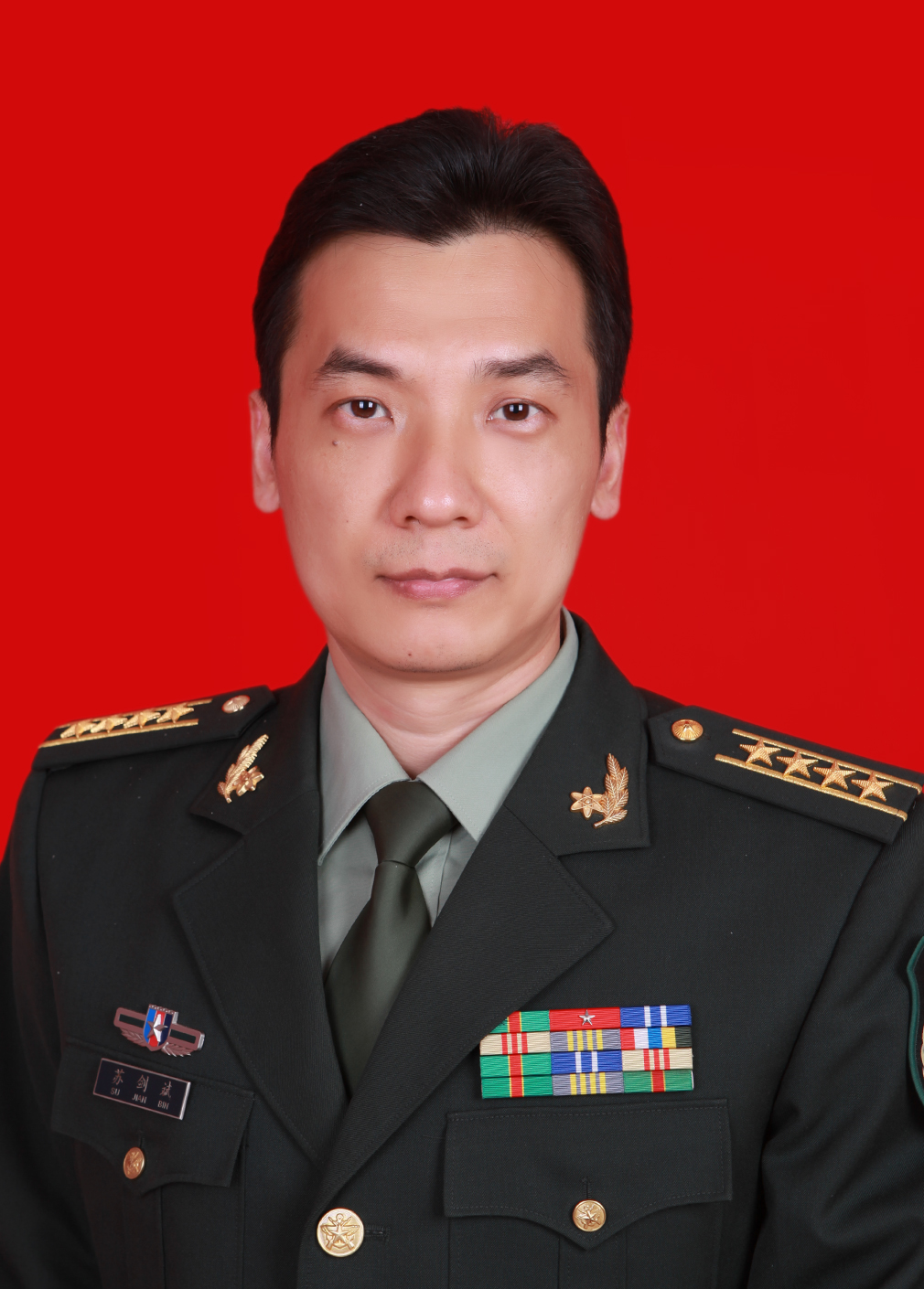 Jianbin Su (苏剑斌)合作论文数Chinese People's Liberation Army General Hospital6Xia Xia (夏侠)合作论文数5Peng Liu (刘鹏)合作论文数4
 Zhixin Qiao合作论文数4Sugang Liu (刘素刚)合作论文数中国人民解放军军事科学院军事医学研究院4Yang Zhan合作论文数3ShaoQing Huang (黄韶清)合作论文数3
 Rigao Ding (丁日高)合作论文数中国人民解放军军事医学科学院毒物药物研究所3He Min合作论文数3Xuanlin Wang合作论文数3
 Suping Ren (任素萍)合作论文数3Qiyuan Kuai合作论文数2Yanbing Wang合作论文数2Wei-yi GAO (高维谊)合作论文数2Xueping Tang合作论文数2Yuhao Pei (裴禹皓)合作论文数2Wu Jian-Feng合作论文数2Yu Wang合作论文数2Bai-sheng SUN (孙百胜)合作论文数2YueSu Zhou (周越塑)合作论文数2MingXue Zhu (朱明学)合作论文数2YaDong Xia (夏亚东)合作论文数2ChengWen Sun (孙成文)合作论文数2MaoXing Yue (岳茂兴)合作论文数2Jing He (赫兢)合作论文数2HuiYang Zhang (张慧洋)合作论文数2HaiYing An (安海英)合作论文数2RongGui Wei (魏荣贵)合作论文数2Guang Li (李光)合作论文数2
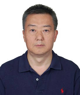 Jianwei Xie (谢剑炜)合作论文数Academy of Military Medical Sciences2
 Qun Yu (于群)合作论文数School of Chemistry and Chemical Engineering, Shandong University2Lihua Su (苏丽华)合作论文数2Chunyan Wang (王春燕)合作论文数2
 Xin Yuan (苑鑫)合作论文数解放军总医院第五医学中心呼吸与危重症医学科(防生科)2Jia Chen (陈佳)合作论文数Institute of Pharmacology and Toxicology, Academy of Military Medical Sciences2Ying Wang合作论文数2Huasong Ma (马华松)合作论文数战略支援部队特色医学中心脊柱外科2Haoyu PEI (裴皓玉)合作论文数1Yulong Liu合作论文数1
 Xu Zhang合作论文数1Chunyan WANG (王春燕)合作论文数1Gong-Qing ZHANG (张公庆)合作论文数1




