Accurate pre‑operative diagnosis of vocal cord lesions remains challenging. We developed a multimodal deep learning model that integrates laryngoscopic images, voice recordings, and biochemical markers to improve diagnostic accuracy. In this retrospective study (2020–2025), a total of 425 patients were enrolled. Of these, 374 patients treated between January 2020 and May 2025 formed the development cohort (contributing 1,947 aligned multimodal samples), and the remaining 51 consecutive cases treated between June 2025 and December 2025 were reserved as an independent temporal test set. Using an early‑fusion strategy and a Transformer encoder, we built a unified diagnostic framework. Model performance was evaluated by accuracy, average precision, AUC, and loss. During training, the model achieved a sample‑level training accuracy of 98.59
Background We developed an intelligent diagnostic model for vocal cord lesions by integrating multimodal clinical data—laryngoscopic images, voice recordings, and a biochemical indicator—within a deep learning framework. Methods In this retrospective study (2020–2024), data from 374 patients with vocal lesions were assembled into 1,973 aligned multimodal samples, each combining laryngoscopic images, voice recordings, and a key biochemical indicator. We applied a multi-scale feature fusion strategy with modal embedding and extracted high-level representations using a Transformer encoder. Model performance was assessed by accuracy, average precision (AP), area under the curve (AUC), and loss. Results The model achieved strong performance: training and validation accuracy were 99.83% and 95.38%, respectively. Both AP and AUC on the validation set exceeded 99%, and loss values were low (0.0059 training, 0.1 validation), indicating effective learning and good generalization without marked overfitting. Conclusions The proposed multimodal fusion model has substantial potential to improve detection of vocal cord lesions and reduce missed diagnoses. The findings support a move toward precision medicine in otolaryngology and provide a scalable framework for integrating heterogeneous clinical data in broader medical AI applications.
Background:Nuclear protein in testis (NUT) carcinoma is an extremely rare and highly aggressive epithelial malignancy driven by NUTM1 rearrangements. Sinonasal involvement is uncommon and often presents with non-specific clinical and radiologic features, leading to delayed diagnosis. Optimal management remains undefined, and outcomes are poor when complete resection is not feasible. Case presentation:A 31-year-old man developed progressive numbness and swelling of the left cheek after tooth extraction. Imaging revealed a soft-tissue mass involving the left maxillary sinus with adjacent maxillofacial soft-tissue extension. Endoscopic biopsy demonstrated a poorly differentiated carcinoma with diffuse punctate nuclear NUT expression, high proliferative index (Ki-67 ~50%), and PD-L1 expression in both tumor cells and immune cells. ^18F-FDG PET-CT showed no regional or distant metastases. Given unresectability, the patient received toripalimab (240 mg) combined with docetaxel and cisplatin every 3 weeks. MRI after three cycles showed early radiologic improvement, and further tumor regression was observed after six cycles, consistent with a partial response. The patient subsequently continued on toripalimab-based maintenance therapy with ongoing stable residual disease at the latest follow-up (approximately 5 months after therapy initiation and 6 months from diagnosis). Conclusion:To our knowledge, this is the first reported case of toripalimab-based chemoimmunotherapy demonstrating an early partial response and short-term disease control in unresectable maxillary sinus NUT carcinoma. It supports the potential role of PD-1 blockade integrated with platinum-taxane chemotherapy as a component of multimodal management for sinonasal NUT carcinomas.
[This corrects the article DOI: 10.3389/fimmu.2026.1763340.].
OBJECTIVE:To develop an intelligent diagnostic model based on a Local-Higher Order Graph Neural Network (LHGNN) to achieve noninvasive auxiliary multi-class pathological classification of vocal fold tumors by analyzing Mel-spectrograms of voice data. METHODS:Patients with pathologically confirmed vocal fold polyps (n = 1272), premalignant lesions (n = 180), and malignant tumors (n = 197) were retrospectively identified. Patients were divided into training and validation sets in a ratio of 8:2. The patient's vowel and continuous sentence audio were collected, and the audio was segmented and converted into Mel-spectrograms. The data was enhanced by random occlusion, adding noise and time shifting. We used a convolutional backbone network-based LHGNN architecture, whose core consists of multiple cascaded LHG modules. The model was evaluated using metrics including accuracy, recall, F1-score, and the area under the curve (AUC) value. RESULTS:The final dataset comprised 4745 audio samples after balanced sampling (1603 vocal fold polyps, 1598 premalignant lesions, 1544 vocal fold carcinomas). The validation set achieved optimal performance after 461 training epochs: overall accuracy of 92.94%, F1-score of 0.9289, recall of 0.9281, and AUC of 0.9877. The multi-class confusion matrix demonstrated identification accuracies of 91.35% for vocal fold polyps, 90.99% for premalignant lesions, and 94.05% for malignant tumors. CONCLUSION:This study successfully validated the high efficacy of the LHGNN model in the multi-class classification of vocal fold tumors. Its excellent performance highlights the significant potential of deep learning in vocal pathological analysis. The model provides a new tool for the noninvasive preliminary screening of vocal fold tumors. LEVEL OF EVIDENCE: 3:
BckgroundLaryngopharyngeal reflux (LPR) is a widespread global health issue. Its recurring symptoms and impact on quality of life create significant economic burdens for individuals and society. To examine the links between lifestyle, diet, and LPR symptoms (LPRS) in college students, and to build an LPRS screening model using a Genetic Algorithm (GA)-Stacking method.Patients and MethodsA cross-sectional study of 502 undergraduates from 21 universities in Jilin Province, China, using an electronic questionnaire. LPRS were assessed via the Reflux Symptom Index (RSI). Associations were analyzed with multiple methods, and a GA-Stacking screening model was developed.ResultsLPRS prevalence was 50.20% (252/502). Significant risk factors included frequent fried food consumption (OR: 1.89; 95% CI, 1.35-2.64), late-evening meals (OR: 2.15; 95% CI, 1.54-3.01), and low physical activity (OR: 1.72; 95% CI, 1.23-2.41). The GA-Stacking model performed well, with a recall of 0.909, accuracy of 0.927, and AUC of 0.96 (95% CI, 0.94-0.98).ConclusionsModifiable factors like fried food intake and meal timing are strongly linked to LPRS in students. The GA-Stacking model effectively identifies high-risk individuals for early intervention, highlighting the role of lifestyle changes and informing targeted health strategies.
ABSTRACT Extracellular vesicles (EVs) from immune cells represent a novel drug delivery system with inherent anti‐tumor properties. To remodel the immunosuppressive tumor microenvironment and mediate multimodal therapy, we produced neutrophil nanovesicles at high yield and loaded them with drug (doxorubicin, DOX), near‐infrared region II fluorescent dye (FD1080), and PD‐1 inhibitor (TFA) through a simple extrusion method and further modified them with DSPE‐PEG2000‐cRGD to enhance their tumor‐targeting ability. The RGD‐NNV@FD1080&DOX/TFA exhibited excellent photothermal effect and efficiently suppressed tumor progression in mouse xenograft tumor, PDX, and lung metastasis models. Upon laser irradiation, the RGD‐NNV@FD1080&DOX/TFA elicited the activation of anti‐tumor immunity with increased infiltration of mature dendritic cells (DCs), CD8 + T cells, and decreased infiltration of regulatory T cells (Tregs) in tumors. Single‐cell RNA sequencing revealed that the combination therapy increased the subtype of cytotoxic and memory T cells while decreasing that of exhausted T cells, and re‐polarized M2 TAMs and N2 TANs. The re‐challenge model further confirmed that RGD‐NNV@FD1080&DOX/TFA exerted effective long‐term immune memory in mouse models and displayed excellent biosafety in vivo. Overall, we designed a simple and potentially clinically applicable nanovesicle‐based nanomedicine delivery system that exhibits a highly efficient anti‐tumor effect by remodeling the tumor immune microenvironment and activating anti‐tumor immunity.
Exosomes mediate cellular communications and have a profound impact on cancer progression. N2 neutrophils, which are polarized by factors from cancers, extensively infiltrate into tumor tissues and promote cancer progression via distinct mechanisms. However, the role and underlying mechanism of exosomes derived from N2 neutrophils (N2-EXO) in cancer remain to be investigated. Herein, we reported that N2-EXO enhanced the proliferation and metastasis of gastric cancer (GC) cells by promoting their stemness. In addition, miR-223-3p and miR-425-5p, which were highly expressed in N2-EXO from GC patients, promoted cancer metastasis and reduced cancer sensitivity to oxaliplatin. The cancer-promoting effect of N2-EXO was abolished by the addition of miRNA inhibitor both in vitro and in vivo. Mechanically, miR-223-3p and miR-425-5p directly targeted FOXO3 and PTEN genes, respectively, which synergistically promoted GC progression by regulating PI3K/AKT signaling pathway. Taken together, our results reveal a novel mechanism by which N2-EXO promotes GC progression, providing new insights into the function of exosomes from N2 neutrophils in cancer.
Peroxiredoxin 2 (PRDX2) is an antioxidant enzyme that has been reported to be overexpressed in various cancers. However, the role of PRDX2 in gastric cancer progression and its underlying mechanism remains unclear. Herein, we revealed the function of PRDX2 in gastric cancer progression and explored its molecule mechanism. We identified that PRDX2 was upregulated and associated with poor prognosis in gastric cancer. The knockdown of PRDX2 inhibited the proliferation, migration and invasion of gastric cancer cells in vitro and suppressed tumor growth in vivo. Mechanistically, PRDX2 interacted with PKM2 (pyruvate kinase isozyme type M2) and protected PKM2 from ubiquitination and degradation, which enhanced glycolysis in gastric cancer cells. The interaction between PRDX2 and PKM2 also enhanced the binding affinity between PKM2 and importin α5, which induced PKM2 nuclear translocation and activated STAT3 signaling pathway. In addition, STAT3 (signal transducer and activator of transcription 3) was identified to bind to PRDX2 gene promoter and upregulate PRDX2 expression, which forms a positive regulatory feedback loop in gastric cancer cells. The present study unravels the biological role of PRDX2 in cancer progression and illustrates the underlying molecular mechanism, which may provide a potential therapeutic target for gastric cancer.
PURPOSE:In the context of rapid development of modern medical technology, the explosive growth of medical data has imposed a heavy diagnostic burden on professional physicians. Especially in the field of computer-assisted treatment research on laryngoscope imaging data, existing studies are still insufficient, which prompts this study to develop a new feature extraction and classification method. The purpose of this study is to improve the accuracy and efficiency of diagnosis for laryngopharyngeal reflux disease by using computer-assisted treatment technology and laryngoscope imaging data and drug treatment results. This not only has important significance in relieving the work pressure of physicians, but also has broad practical value and realistic significance. METHODS:This study utilized the laryngoscope images provided by the Department of Otolaryngology, Jilin University Second Hospital, and proposed an innovative image feature extraction method that integrates distribution features and texture features. The local binary pattern method was used to capture the texture information of the image, while the gray histogram method was used to extract the distribution characteristics of the image. This technology effectively achieved the fusion of features, and the performance of the five classic classification algorithms was compared and analyzed for the features obtained. CONCLUSIONS:The study results show that the feature extraction method proposed in this paper, when combined with the random forest discriminant algorithm, achieves an accuracy rate of 96.61% in laryngoscope image classification, demonstrating excellent performance. Furthermore, the algorithm has low requirements on the number of samples, further proving its high efficiency and practicality in actual applications.
PURPOSE:Invasive fungal infections are a serious threat to immunocompromised individuals, with laryngeal involvement being rare and often presenting with atypical symptoms that complicate early diagnosis. METHODS:The authors report a case of chronic invasive fungal laryngeal cartilage necrosis, initially manifesting as bilateral vocal cord fixation. RESULTS:Pathology suggested Mucorales infection. Postoperative recovery included resolution of both laryngeal and pulmonary issues after a total laryngectomy. There has been no recurrence at follow-up visits. CONCLUSION:Early diagnosis, aggressive surgical debridement, and full-course antifungal treatment are essential for improving outcomes in extrapulmonary fungal infections.
The pathogenesis of obstructive sleep apnea hypopnea syndrome (OSAHS) is multifactorial and has garnered increasing attention due to its significant societal impact and long-term health consequences. Investigation into the underlying mechanisms of OSAHS remains critical to advancing public health. The genioglossus (GG), recognized as a key muscle in maintaining upper airway patency, exhibits structural and functional changes that correlate with OSAHS severity. As such, therapeutic strategies targeting genioglossus dysfunction have become a focal point in OSAHS management. Human umbilical cord mesenchymal stem cells (hUCMSCs), characterized by their capacity for self-renewal and multipotent differentiation, demonstrate promising regenerative potential. These cells are readily obtainable, and can be efficiently isolated, cultured, expanded, and purified. Their low immunogenicity enhances their suitability for allogeneic transplantation. Given these properties, investigation into the potential protective effects of hUCMSCs on genioglossus injury in individuals with OSAHS holds considerable therapeutic promise.
Neutrophil-derived extracellular vesicles (NEVs) are critically involved in disease progression and are considered potential biomarkers. However, the tedious processes of NEV separation and detection restrain their use. Herein, we presented an integrated microfluidic chip for NEV (IMCN) analysis, which achieved immune-separation of CD66b+ NEVs and multiplexed detection of their contained miRNAs (termed NEV signatures) by using 10 μL serum samples. The optimized microchannel and flow rate of the IMCN chip enabled efficient capture of NEVs (>90%). After recognition of the captured NEVs by a specific CD63 aptamer, on-chip rolling circle amplification (RCA) reaction was triggered by the released aptamers and miRNAs from heat-lysed NEVs. Then, the RCA products bound to molecular beacons (MBs), initiating allosteric hairpin structures and amplified "turn on" fluorescence signals (RCA-MB assay). Clinical sample analysis showed that NEV signatures had a high area under curve (AUC) in distinguishing between healthy control (HC) and gastric cancer (GC) (0.891), benign gastric diseases (BGD) and GC (0.857). Notably, the AUC reached 0.912 with a combination of five biomarkers (NEV signatures, CEA, and CA199) to differentiate GC from HC, and the diagnostic accuracy was further increased by using a machine learning (ML)-based ensemble classification system. Therefore, the developed IMCN chip is a valuable platform for NEV analysis and may have potential use in GC diagnosis.
OBJECTIVE:To investigate the application value, technical advantages, clinical efficacy, educational impact, and challenges in promotion of the Da Vinci robotic surgical system (Transoral Robotic Surgery, TORS) in complex head and neck surgeries, providing a reference for the advancement of precise and intelligent surgery in this field. METHODS:The technical principles and evolution of the Da Vinci system were analyzed. Its clinical application data in laryngeal, oropharyngeal, obstructive sleep apnea (OSA), nasal cavity, and thyroid surgeries were reviewed. The supporting role of digital technologies (AI, 3D visualization, VR, 3D printing) was assessed. Challenges related to cost and training requirements were summarized. RESULTS:Leveraging advantages such as instrument flexibility, high-definition 3D visualization, tremor filtration, and precise manipulation, the Da Vinci system significantly enhanced outcomes in head and neck surgery: precise resection of laryngeal cancer reduced operative risks; efficient treatment of early-stage oropharyngeal squamous cell carcinoma (OPSCC) was achieved with fewer complications; favorable long-term survival rates were observed for OPSCC; OSA symptoms were effectively improved; and it demonstrated both minimally invasive benefits and therapeutic efficacy in recurrent nasal cavity cancers and thyroid surgeries. Digital technologies enhanced surgical precision and medical training efficiency. However, high unit costs and stringent training requirements limit its adoption in small and medium-sized hospitals. CONCLUSION:The Da Vinci system, integrated with digital technologies, significantly improves the safety, precision, and patient prognosis in head and neck surgery, while elevating medical education standards. High costs and intensive training needs are issues for why it is not widely used. Future tasks should emphasize cost-cutting to enhance patient care access and improve quality across the medical spectrum.
BACKGROUND:Circular RNAs (circRNAs) play important roles in cancer progression and metastasis. However, the expression profiles and biological roles of circRNAs in non-small cell lung cancer (NSCLC) remain unclear.METHODS:In this study, we identified a novel circRNA, hsa_circ_0006834 (termed circ6834), in NSCLC by RNA-seq and investigated the biological role of circ6834 in NSCLC progression in vitro and in vivo. Finally, the molecular mechanism of circ6834 was revealed by tagged RNA affinity purification (TRAP), western blot, RNA immunoprecipitation, dual luciferase reporter gene assays and rescue experiments.RESULTS:Our results showed that circ6834 was downregulated in NSCLC tumor tissues and cell lines. Circ6834 overexpression inhibited NSCLC cell growth and metastasis both in vitro and in vivo, while circ6834 knockdown had the opposite effect. We found that TGF-β treatment decreased circ6834 expression, which was associated with the QKI reduction in NSCLC cells and circ6834 antagonized TGF-β-induced EMT and metastasis in NSCLC cells. Mechanistically, circ6834 bound to AHNAK protein, a key regulator of TGF-β/Smad signaling, and inhibited its stability by enhancing TRIM25-mediated ubiquitination and degradation. In addition, circ6834 acted as a miRNA sponge for miR-873-5p and upregulated TXNIP gene expression, which together inactivated the TGF-β/Smad signaling pathway in NSCLC cells.CONCLUSION:In conclusion, circ6834 is a tumor-suppressive circRNA that inhibits NSCLC progression by forming a negative regulatory feedback loop with the TGF-β/Smad signaling pathway and represents a novel therapeutic target for NSCLC.
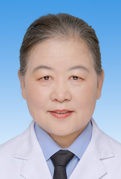 Chunshun Jin (金春顺)合作论文数The Second Norman Bethune Hospital of Jilin University30
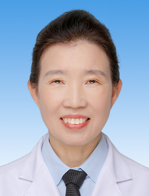 Lianji Wen (文连姬)合作论文数The Second Norman Bethune Hospital of Jilin University29
 Xue Zhao (赵雪)合作论文数The Second Norman Bethune Hospital of Jilin University23
 Yan Liu (刘岩)合作论文数The Second Norman Bethune Hospital of Jilin University23
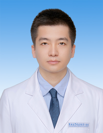 Jinzhang Cheng (程金章)合作论文数The Second Norman Bethune Hospital of Jilin University20
 Maoye Wang合作论文数15Ying Li (李赢)合作论文数School of Chemical and Material Engineering, Jiangnan University14Zhao Xue合作论文数13
 Chengbi Xu (许承弼)合作论文数The Second Norman Bethune Hospital of Jilin University13
 Jiahui Zhang合作论文数12
 Di Wu (吴翟)合作论文数School of Physics, Zhengzhou University12Jianmei Gu合作论文数12
 Kai Xue (薛凯)合作论文数The Second Norman Bethune Hospital of Jilin University11Liu Xueshi-Bojie合作论文数10
 Yin Zhao (赵胤)合作论文数The Second Norman Bethune Hospital of Jilin University10Zhi-Ru Li (李志儒)合作论文数10Huihui ZHANG (张慧慧)合作论文数9
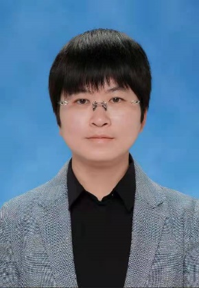 Xiaoxin Zhang (张小昕)合作论文数School of Medicine, Jiangsu University9Tingting Gu (谷婷婷)合作论文数8GuangHong Han (韩光红)合作论文数8Qi Xinmeng合作论文数8Yang Jingpu合作论文数8Runbi Ji合作论文数8bo jie liu shi xue (刘学识博杰)合作论文数6Haixian Guo合作论文数6Yanshi Li合作论文数6Si-Yi Li合作论文数6Min Fu合作论文数6
 Min Pan合作论文数Dept Otorhinolaryngol, Chongqing Med Univ6Ji Lian (连姬)合作论文数6Di Wang合作论文数6He Hui-Min合作论文数6
 Guohua Hu (胡国华)合作论文数The First Affiliated Hospital of Chongqing Medical University6Xu Zhang合作论文数6
 Sun Wei-Ming合作论文数5Zhigang YAN (闫志刚)合作论文数5Jiayuan Liu合作论文数5
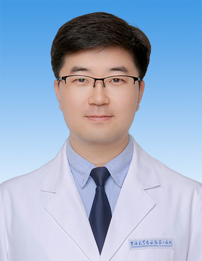 Chengxun Jin (金成勋)合作论文数The Second Norman Bethune Hospital of Jilin University5
 Ding Xin (辛丁)合作论文数The Second Hospital of Jilin University5Zhihai Wang合作论文数5
 Jinqiu Li (李金秋)合作论文数The Second Hospital of Jilin University5Hui Yang合作论文数4Yixin Li合作论文数4Xiaomin BIAN (边晓敏)合作论文数4LI Shaonan (李绍楠)合作论文数4Shujun Gao合作论文数4




