Following the publication of this paper, it was drawn to the Editor's attention by a concerned reader that, regarding the cell invasion assay data shown in Fig. 4A, the 'Control' and 'pU‑siNC' data panels appeared to share an overlapping section of data, such that data which were intended to show the results of differently performed experiments had apparently been derived from the same original source. The authors were contacted by the Editorial Office to offer an explanation for this apparent anomaly in the presentation of the data in this paper; however, up to this time, no response from them has been forthcoming. Owing to the fact that the Editorial Office has been made aware of potential issues surrounding the scientific integrity of this paper, we are issuing an Expression of Concern to notify readers of this potential problem while the Editorial Office continues to investigate this matter further. [Molecular Medicine Reports 3: 903‑907, 2010; DOI: 10.3892/mmr.2010.355].
This study aimed to explore the bidirectional causal relationship between inflammatory bowel disease (IBD) and Hashimoto thyroiditis (HT) using Mendelian randomization (MR) analysis. While both conditions are characterized by persistent inflammation and immune dysregulation, the direction of causality remains unclear. We performed a 2-sample bidirectional MR analysis using summary statistics from genome-wide association studies for IBD and HT. Genetic variants strongly associated with IBD and HT were selected as instrumental variables for forward and reverse MR analyses, respectively. Various MR methods, including inverse-variance weighted (IVW), MR-Egger regression, weighted median, and weighted mode, were employed to assess causal relationships. Sensitivity analyses were conducted to evaluate the reliability of results, including tests for pleiotropy and heterogeneity. Data were sourced from individuals of European descent to minimize population stratification bias. In the forward MR analysis, no strong evidence of a causal effect of IBD on HT was found, with the IVW method yielding an odds ratio (OR) of 0.9617 (95% CI: 0.7549-1.2251; P = .7519) Similarly, in the reverse MR analysis, no significant causal effect of HT on IBD was observed, with the IVW method showing an OR of 0.9991 (95% CI: 0.9693-1.0298, P = .9527). Sensitivity analyses confirmed the absence of heterogeneity or pleiotropic effects that could influence the results. The MR-PRESSO analysis did not detect any outlier SNPs. This bidirectional MR analysis provides no evidence for a causal relationship between IBD and HT in either direction.
Hepatocellular carcinoma (HCC) is one of the deadliest malignancies worldwide featured with the poor prognosis and high mortality in affected patients. Given its insensitivity to conventional systemic chemotherapy, the development of novel modalities for HCC management is highly urgent. Sonodynamic therapy (SDT) has gained considerable momentum in cancer therapy. Especially, through synergistic SDT/chemotherapy, SDT would enhance the chemotherapeutic process on inhibiting tumor growth, which holds great potential on combating HCC. In this work, we report on the design/fabrication of targeted biodegradable nanosonosensitizers based on hollow mesoporous organosilica nanoparticles (HMONs), followed by pore-engineering including covalent anchoring of protoporphyrin (PpIX, HMONs-PpIX) and conjugation of arginine-glycine-aspartic acid in order to specifically targeting HCC cells. Such nanosonosensitizers provide efficient loading and controllable stimuli-responsive release of chemotherapeutic agents for HCC-targeting chemotherapy, thus promoting an enhancing chemotherapeutic process via the unique sonotoxicity under ultrasound irradiation. The HMONs matrix with biologically active organic groups in the framework (disulfide bond) are endowed with intrinsic tumor microenvironment-responsive biodegradability and improved biocompatibility/biosafety. In particular, a synergistic inhibition effect of drug-loaded HMONs-PpIX-arginine-glycine-aspartic acid on HCC growth has been systematically demonstrated both in vitro and in vivo (84.7% inhibition rate), which brings insights and meets the versatile therapeutic requirements for HCC management.
Detailed description of Supplementary Figures such as genes expression in cell lines/tumor tissues, high magnification images and so on.
PDF file - 85K, Table S1 (Real-time PCR primer sequence) and supplementary experimental procedures
PDF file, 983K, Supplemental Figure S1 p-AMPK/AMPK ratios of 19 HCC specimens. Supplemental Figure S2 Western blot analysis of p-AMPK(Thr172), PARP, CyclinD1. Supplemental Figure S3 AICAR inhibits human HCC cells growth. Supplemental Figure S4 Transfection of siRNA target AMPKalpha1 and AMPKalpha2 effectively knockdown expression of AMPK in SMMC7721 cells. Supplemental Figure S5 Metformin inhibits HCC cells growth via activation of AMPK. Supplemental Figure S6 Metformin causes cycle arrest and induces apoptosis in HCC cells. Supplemental Figure S7 Therapeutic metformin/AMPK activation sensitizes HCC cells towards chemotherapy. Supplemental Figure S8 Therapeutic metformin/AMPK activation inhibited IL-6/STAT3 signaling activity Supplemental Figure S9 Correlation between expression of p-AMPK (Thr172) and P65 nuclear staining in human HCC specimens. Supplemental Figure S10 AICAR treatment repressed NF-kappaB reporter gene activity. Supplemental Figure S11 SMMC7721 or HepG2 cells were transfected with vector, IkappaBalpha superrepressor (IkappaBalphaSR) or P65. Supplemental Figure S12 IkappaBalphaSR prevented metformin-induced upregulation of PTEN expression in SMMC7721 cells Supplemental Figure S13 Metformin did not significantly affect body weight, average blood glucose level and liver function of the mice. Supplemental Figure S14 Western blot analysis on the expressions of p-AMPK, p-PTEN, PTEN, p-STAT3 and STAT3 from respective cell homogenate Supplemental Table S1 Primer Sequences Used for RT-PCR.
Supplementary Data from COOH-Terminal Truncated HBV X Protein Plays Key Role in Hepatocarcinogenesis
Methods not listed in the main manuscript including patient inclusion criteria, ChIP, ISH, IF and so on.
Supplementary Table 1-9. Supplementary Table 1. Clinicopathologic features of 245 HCC patients with PVTT in training cohort. Supplementary Table 2. Clinicopathologic features of 372 HCC Patients in validation cohort. Supplementary Table 3. Primers and siRNA Sequences. Supplementary Table 4. Antibodies used in the study. Supplementary Table 5. lncRNAs expressed differentially both in PVTT Compared with PT and in CSQT-2 Compared with Hep3B. Supplementary Table 6. mRNAs expressed differentially both in PVTT compared with PT and in CSQT-2 compared with Hep3B. Supplementary Table 7. LncRNA-mRNA pairs from Blat analysis. Supplementary Table 8. The Relationship between expression of ICR/ICAM1 and clinicopathologic features for 372 HCC Patients. Supplementary Table 9. Univariate and Multivariate analyses of factors associated with survival and recurrence for 372 HCC patients
Perspective on This Article from Gut-Derived Lipopolysaccharide Promotes T-Cell–Mediated Hepatitis in Mice through Toll-Like Receptor 4
Supplementary Methods and Materials, Figures 1-5, Tables 1-2 from Wnt/β-Catenin Signaling Contributes to Activation of Normal and Tumorigenic Liver Progenitor Cells
Supplementary Fig. S1-11. Supplementary Fig. S1. Differentially expressed lncRNAs and mRNAs between CSQT-2 and Hep3B. Supplementary Fig. S2. Potential relationship between ICR and ICAM-1 mRNA. Supplementary Fig. S3. Coding potential assessment tool analysis of ICR. Supplementary Fig. S4. In situ analysis of ICAM-1 and ICR expression in HCC tumor and PVTT tissues. Supplementary Fig. S5. ICR and ICAM-1 expression assessed by Real-time PCR and western blot. Supplementary Fig. S6. Melt curve from real-time PCR in RNase protection assay (RPA) analysis. Supplementary Fig. S7. Migration analysis of tumor cells with ICR modulation. Supplementary Fig. S8. ICAM-1 expression in cell line/tumors with modulated ICR and tumor growth in mice. Supplementary Fig. S9. ICAM-1 expression in mouse tumors assessed by IF. Supplementary Fig. S10. Representative image of ChIP assays with Nanog antibody. Supplementary Fig. S11. Nanog and ICR Expression in HCC cell lines with Nanog modulation.
Objectives: A potentially curative hepatic resection is the optimal treatment for hepatocellular carcinoma (HCC), but most HCCs, even at an early stage, eventually recur after resection. To investigate clinical features of initial recurrence and long-term prognosis of patients after recurrence. Methods: From a multicenter database, patients withearly-stage HCC (Barcelona Clinic Liver Cancer [BCLC] stage 0/A) were retracted. Time to initial recurrence, patterns of recurrence, and treatment modalities for recurrent tumors were investigated. Univariable and multivariable analysis were used to identify independent risks associated with postoperative recurrence, as well as post-recurrence survival (PRS). Results: Among 1,424 patients, 679 (47.7%) developed recurrence at a median follow-up of 52.9 months, including 412 (60.7%) early recurrence (≤2 years after surgery) and 271 (31.3%) late recurrence (>2 years). Independent risks of postoperative recurrence included cirrhosis, preoperative alpha-fetoprotein level>400ug/L, tumor size>5cm, multiple tumors, satellites, microvascular invasion, and intraoperative blood transfusion. The most common pattern for initial recurrence were intrahepatic only (87.3%), while the median PRS of patients with recurrence was 22.4 months. Multivariable analysis revealed that receiving irregular recurrence surveillance, beyond Milan criteria of the initial tumor, early recurrence, BCLC stage B/C of the recurrent tumor, and non-curative treatments were independently associated with poorer PRS. Conclusions: Nearly half of patients with early-stage HCC experienced recurrence after resection. Understanding recurrence risks may help identify patients at high risk of recurrence who may benefit from future adjuvant therapies. Meaningful survival even after recurrence can still be achieved by postoperative regular surveillance and curative treatment.
Background: Hepatocellular carcinoma (HCC) is one of the most common malignancies in China. Most HCC patients are first diagnosed at an advanced stage, and systemic treatments are the mainstay of treatment. Summary: In recent years, immune checkpoint inhibitors have made a breakthrough in the systemic treatment of middle-advanced HCC, breaking the single therapeutic pattern of molecular-targeted agents. To better guide the clinical treatment for effective and safe use of immunotherapeutic drugs, the Chinese Association of Liver Cancer and Chinese Medical Doctor Association has gathered multidisciplinary experts and scholars in relevant fields to formulate the “Chinese Clinical Expert Consensus on Immunotherapy for Hepatocellular Carcinoma (2021)” based on current clinical studies and clinical medication experience for reference in China. Key Messages: The consensus contained 17 recommendations, including the preferred regimen for first- and second-line immunotherapy, evaluation and monitoring before/during/after treatment, management of complications, precautions for special patients, and potential population for immunotherapy.
Non-alcoholic fatty liver disease (NAFLD) is emerging as an epidemic risk factor for hepatocellular carcinoma (HCC). The progression of NAFLD to HCC is closely associated with paracrine communication among hepatic cells. Vascular endothelial growth factor A (VEGFA) plays a key role in NAFLD and HCC; however, the cellular communication of VEGFA in the pathological transition from NAFLD to HCC remains unclear. Here, we found that VEGFA elevation was considerably distributed in hepatocytes of clinical and murine NAFLD-HCC specimens. Notably, progression from NAFLD to HCC was attenuated in hepatocyte-specific deletion of Vegfa (VegfaΔhep) mice. Mechanistically, VEGFA activated human hepatic stellate cell (HSC) LX2 into a fibrogenic phenotype via VEGF-VEGFR signaling in fatty acid medium, and HSC activation was largely attenuated in VegfaΔhep mice during NAFLD-HCC progression. Additionally, a positive correlation between VEGFA and hepatic fibrosis was observed in the NAFLD-HCC cohort, but not in the HBV-HCC cohort. Moreover, LX2 cells could be activated by conditioned medium from NAFLD-derived organoids, but not from HBV livers, whereas this activation was blocked by a VEGFA antibody. In summary, our findings reveal that hepatocyte-derived VEGFA contributes to NAFLD-HCC development by activating HSCs and highlight the potential of precisely targeting hepatocytic VEGFA as a promising therapeutic strategy for NAFLD-HCC.
Background: Surgical resection is the only treatment modality that ensures complete tumor removal in patients with liver tumors involving a major hepatic vein. Central hepatectomy is a challenging procedure that often result in large defect at the right hepatic vein, which is not amenable to suturing or end-to-end anastomosis. Meanwhile, good outflow reconstruction is essential for early postoperative recovery and long-term survival. Methods: We describe a simple technique for reconstructing the right hepatic vein. The technique is an effective method for reconstructing large venous defects after the hepatic vein resection. Reconstruction of the right hepatic vein has the advantages of prevention of congestion in segments VI and VII. Conclusions: This technique allows surgeons to reconstruct the hepatic vein without synthetic vascular grafts and cryopreserved veins.
Portal vein tumor thrombus (PVTT) is very common and it plays a major role in the prognosis and clinical staging of hepatocellular carcinoma (HCC). We have published the first version of the guideline in 2016 and revised in 2018. Over the past several years, many new evidences for the treatment of PVTT become available, especially for the advent of new targeted drugs and immune checkpoint inhibitors which have further improved the prognosis of PVTT. So, the Chinese Association of Liver Cancer and Chinese Medical Doctor Association revised the 2018 version of the guideline to adapt to the development of PVTT treatment. Future treatment strategies for HCC with PVTT in China would depend on new evidences from more future clinical trials.
 Hongyang Wang (王红阳)合作论文数Shanghai Cancer Institute;Naval Medical University;The National Center for Liver Cancer153
 Weiping Zhou (周伟平)合作论文数Naval Medical University142
 Han Chen (陈汉)合作论文数122
 Feng Shen (沈锋)合作论文数The Third Affiliated Hospital of Naval Medical University122
 Wan-yee Joseph Lau (刘允怡)合作论文数Department of Surgery, Faculty of Medicine, The Chinese University of Hong Kong121
 Wenming Cong (丛文铭)合作论文数Department of Pathology, Eastern Hepatobiliary Surgery Hospital, Second Military Medical University/Key Laboratory of Signaling Regulation and Targeting Therapy of Liver Cancer, Second Military Medical University, The Ministry of Education121
 Lixin Wei (卫立辛)合作论文数第二军医大学东方肝胆外科医院肿瘤免疫与基因治疗中心118BaiHe Zhang (张柏和)合作论文数96
 Shuqun Cheng (程树群)合作论文数School of Health Science and Engineering, University of Shanghai for Science and Technology95
 Qijun Qian (钱其军)合作论文数School of Medicine, Shanghai University;Cell Therapy Innovation Research Institute, Shanghai University;Shanghai Cell Therapy Group Co,Ltd94
 Tian Yang (杨田)合作论文数Third Affiliated Hospital of Naval Medical University90Ai-Jun Li合作论文数74
 Jiahe Yang (杨家和)合作论文数Naval Medical University65
 Guangshun Yang (杨广顺)合作论文数The Second Military Medical University64
 Jiamei Yang (杨家梅)合作论文数The Second Affiliated Hospital of Zhengzhou University64
 Xiaoqing Jiang (姜小清)合作论文数Third Affiliated Hospital of Naval Medical University54ZeYa Pan (潘泽亚)合作论文数51
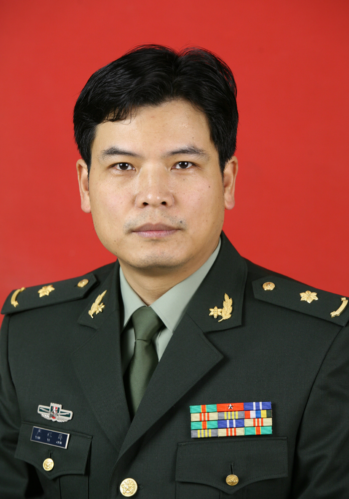 Yiqun Yan (严以群)合作论文数 Department of Hepatic Surgery I,Eastern Hepatobiliary Hospital,Second Military Medical University,Shanghai ,China50SiYuan Fu (傅思源)合作论文数49
 Zhangqing Su (苏长青)合作论文数第二军医大学东方肝胆外科医院47Lehua Shi (施乐华)合作论文数中国人民解放军第二军医大学第三附属医院肝胆外科医院综合治疗一科45Hao Xing合作论文数43
 Jie Shi合作论文数43
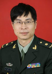 Xiangji Luo (罗祥基)合作论文数上海东方肝胆外科医院肝胆外科43Weixing Guo合作论文数Eastern Hepatobiliary Surgery Hospital, The Second Military Medical University42
 Lei Chen (陈磊)合作论文数Eastern Hepatobiliary Surgery Hospital;Fudan University Cancer Institute40Zhengfeng Yin (殷正丰)合作论文数东方肝胆外科医院分子肿瘤研究室39XiaoPing Yao (姚晓平)合作论文数38Chen Han合作论文数36Zhen-Lin Yan合作论文数36ZhenFu Cui (崔贞福)合作论文数34JianJun Yan (晏建军)合作论文数34
 Hexin Yan (鄢和新)合作论文数Shanghai Cancer Institute34MingDa Wang合作论文数34Hongyan Cheng (程红岩)合作论文数The Third Affiliated Hospital of Naval Medical University34Nan Li (李楠)合作论文数Naval Medical University33Kui Wang合作论文数33
 Yajun Guo (郭亚军)合作论文数School of Pharmaceutical Sciences and Food Engineering, Liaocheng University;School of Pharmacy, Liaocheng University32
 Chao Li合作论文数31
 Han Wu合作论文数31Junhua Lu (卢军华)合作论文数Naval Medical University31
 Han Zhang合作论文数University of Nottingham Ningbo29Hui-Fang Cao合作论文数29Shu-Hui Zhang合作论文数28Liang Huang (黄亮)合作论文数28
 Yongjie Zhang (张永杰)合作论文数中国人民解放军第二军医大学第三附属医院腹腔镜科28Wen Yang (杨文)合作论文数28
 Guangxiang Qian (钱光相)合作论文数Eastern Hepatobiliary Surgery Hospital27
 Jun Li (李俊)合作论文数Department of Hepato-Pancreato-Biliary Surgery, Tenth People's Hospital of Tongji University;School of Medicine, Tongji University27




