目的 探讨家系不宁腿综合征/Willis-Ekbom病患者NOTCH2NLC基因是否存在GGC异常重复扩增.方法 收集2016年1月至2020年10月作者医院诊治的家系不宁腿综合征/Willis-Ekbom病患者35例,对先证者提取DNA,进行重复引物聚合酶链反应PCR(RP-PCR)和富含GC的毛细管电泳的PCR(GC-PCR),以确定NOTCH2NLC基因中GGC重复序列的大小.结果 35例家系不宁腿综合征/Willis-Ekbom病患者均未检测到GGC重复序列异常扩增.结论 NOTCH2NLC基因GGC异常重复扩增可能不参与不宁腿综合征/Willis-Ekbom病的发病机制.
目的 探讨伴皮质下梗死和白质脑病的常染色体显性遗传性脑动脉病(cerebral autosomal dominant arteriop-athy with subcortical infarcts and leukoencephalopathy,CADASIL)家系内患者的临床表型差异及原因.方法 回顾性分析首都医科大学附属北京友谊医院收治的1个CADASIL家系内3例患者的临床资料,总结其临床特点、影像学变化、认知功能和神经心理改变.结果 先证者(Ⅲ2)20余岁反复头痛,31岁后多次卒中发作,伴有焦虑抑郁情绪.患者Ⅱ3(先证者母亲)于38岁意外发现脑白质病变,40岁出现情绪改变,近60岁时偏瘫后卧床,痴呆,吞咽困难,鼻饲饮食.患者Ⅱ4(先证者姨妈)偶有头晕,50岁仅感觉轻度记忆力下降.3例患者影像学表现为逐渐加重的颅内脑白质损害,颞极受累,病程中颞极信号有变化.结论 该CADASIL家系内3例患者临床表现轻重不一,疾病演变过程各有不同,影像学检查对早期诊断敏感性高,但与临床表型严重程度并不一致.
目的 总结临床表现酷似危重症性周围神经病的血管炎性周围神经病患者的临床和病理特点.方法 回顾性分析首都医科大学附属北京友谊医院收治的 1例表现为发热、多脏器功能不全和肢体无力的血管炎性周围神经病患者的临床资料.结果 患者为老年女性,重症感染、呼吸机辅助呼吸,病程中停用呼吸机意识恢复后发现四肢无自主活动,为明确诊断行腓肠神经活检.病理结果示微小血管炎性病变伴随中度活动性轴索性周围神经病理改变.结论 重症感染后发现周围神经受损的病例有可能是系统性血管炎累及周围神经的表现,疾病进程上酷似危重症性周围神经病,应该尽早行周围神经活检明确诊断.该病进展快,病死率高,早期诊断和早期治疗可改善预后.
Background: Recent studies have suggested that cerebral ischemic infarction may contribute to the development of restless legs syndrome (RLS). This study analyzed the clinical and radiological profiles of RLS with onset after acute lacunar infarction. Methods: In this retrospective study we enrolled 244 consecutive patients with acute lacunar infarction between January 2012 and June 2014. RLS was identified and evaluated based on the International RLS Rating Scale (IRLS-RS). Individual sleep quality was assessed using the Epworth Sleepiness Scale (ESS). Psychological state was also assessed using the Hamilton Depression Scale (HDS) and the Hamilton Anxiety Scale (HAS). Results: The incidence of RLS in patients with lacunar infarction was 5.33%. Our participant group consisted of nine males and four females. Three patients had symptoms in bilateral limbs, and 10 patients had symptoms only contralateral to the cerebral infarction. The infarctions were localized to the pons, centrum semiovale, thalamus, putamen, medulla, and occipital lobe. Contralateral paralysis was found in 13 patients, and contralateral sensory deficit in seven patients. The average IRLS-RS, ESS, HDS, HAS scores were 19.07 +/- 8.70, 4.69 +/- 5.82, 4.38 +/- 4.68, and 3.85 +/- 4.76, respectively. Nine patients had diabetes mellitus. After administration of dopaminergic drugs, patients' RLS significantly improved. Conclusions: The incidence of RLS after acute lacunar infarction was 5.33%. Pons, centrum semiovale, and basal ganglia were the common locations of responsible lesions. Compared to idiopathic RLS, symptoms of RLS after acute lacunar infarction appeared more unilateral and more likely involved the arm. Moreover, diabetes mellitus may be a risk factor for RLS in stroke patients. (c) 2018 Elsevier B.V. All rights reserved.
Objective To have a profound understanding of anti-N-methyl-D-aspartic receptor (anti-NMDAR) encephalitis,through the clinical analysis of 5 cases of anti-NMDAR encephalitis,and literature review.Methods This is a retrospective analysis.Five cases of anti-NMDA receptor encephalitis treated from May 2010 to June 2015,in the Department of Neurology,Beijing Friendship Hospital affiliated to Capital Medical University,were included in this study.The clinical data,including clinical manifestation,past history,radiological features,serum and cerebral spinal fluid examinations,treatment and prognosis,were analyzed.Results Among the 5 cases,3 young female and 2 middle-to old-aged male.The clinical features of the onset was mental and behavior disorder,as well as seizure and extrapyramidal features,like facial and limbic involuntary movements or tremor.Coma and hypopnea was severe in 3 young female cases,needing assistance of mechanical ventilator,while the manifestation of 2 male patients was much mild,need not assisted respiration.1 case had teratoma of ovary,1 case had Vogt-Koyanagi-Harada syndrome.The anti-NMDA receptor antibody was positive in cerebraospinal fluid of all 5 cases,but in serum of 3 cases,serum and CSF Epstein-Barr virus (EBV) IgM antibody was positive in 1 case,while herpes simplex I virus (HSV-1) IgM antibody positive in another case,and anti-myelin oligodendrocyte glycoprotein (MOG) antibody was seen in serum and CSF in 1 case.The time interval from the onset to treatment was 10-37 d (18.8 ± 9.8 d).IVIG was used in all of the 5 cases,glucocoticoid in 4 cases,and plasma exchange in 3 cases.One case with Vogt-Koyanagi-Harada syndrome,having a long time before diagnosis and treatment,died,while the other 4 cases had good prognosis,and had no relapse.Conclusions Mental and behavior disturbance is common at onset of anti-NMDAR encephalitis.The radiological and lab examination may be normal.It may be accompanied with HSV-1 or EBV infection,anti-MOG antibody may be positive in this disease.Active treatment is important.
目的 利用血管张力固定改良PE管,制作血栓栓塞大脑中动脉脑梗死(Embolic middle cerebral artery occlusion,eMCAO)大鼠模型.方法 采用清洁级Sprague-Dawley大鼠20只制作eMCAO模型,均为单人制作,利用血管张力固定改良PE管.结果 改良PE插管均成功,大鼠eMCAO造模成功率90%,制作时间每只平均30 min.结论 利用血管张力法可单人操作制作大鼠eMCAO模型,有效节省造模所需时间以及人力物力.
ObjectivePontine infarction is a common type of stroke in the cerebral deep structures, resulting from occlusion of small penetrating arteries, may manifest as hemi-paralysis, hemi-sensory deficit, ataxia, vertigo, and bulbar dysfunction, but patients presenting with restless legs syndrome (RLS) are extremely rare. Herein, we reported five cases with RLS as a major manifestation of pontine infarction.MethodsFive cases of pontine infarction related RLS were collected from July 2013 to February 2016. The diagnosis of RLS was made according to criteria established by the International RLS Study Group (IRLSSG) in 2003. Neurological functions were assessed according to the National Institutes of Health Stroke Scale (NIHSS) and modified Rankin Scale (mRS). Severity of RLS was based on the International RLS Rating Scale (IRLS-RS). Sleep quality was assessed by Epworth Rating Scale (ERS), and individual emotional and psychological states were assessed by Hamilton Depression Scale (HDS) and Hamilton Anxiety Scale (HAS).ResultsThe laboratory data at the onset including hemoglobin, serum concentration of homocysteine, blood urea nitrogen (BUN), creatinine, electrolytes, and thyroid hormones were normal. The electroencephalogram (EEG), lower-extremity somatosensory evoked potential (SEP), and nerve conduction velocity (NCV) in four limbs were normal. The average period of follow-up was 34.60 ± 12.76 months. The MRI examination showed acute or subacute pontine infarction lesions, 3 cases in the rostral inner side, 1 case in the rostral lateral and inner side, and 1 case in rostral lateral side. The neurological deficits included weakness in 4 cases, contralateral sensory deficit in 1 case, and ataxia in 2 cases. All 5 patients presented with symptom of RLS at or soon after the onset of infarction and 4 patients experienced uncomfortable sensations in the paralyzed limbs contralateral to the ischemic lesion. Their neurological deficits improved significantly 2 weeks later, but the symptoms of RLS did not resolve. Among them, 3/5 patients were treated with dopaminergic drugs. At the end of the follow-up, RLS symptom eventually resolved in 3 patients but persisted in two. The IRLS-RS, NIHSS and mRS scores were significantly lower at the onset than those at the last follow-up (P = 0.035, 0.024 and 0.049, respectively). However, there was no significant difference in the ERS, HDS and HAS scores (P = 0.477, 0.226 and 0.778, respectively).ConclusionRLS can be an onset manifestation of pontine infarction, clinicians should be aware of this potential symptom. RLS usually occurs in the paralyzed limbs contralateral to the infarction lesion. The pathogenesis still needs further investigation.
Objective To study the etiology,diagnosis and treatment of Tolosa - Hunt syndrome. Methods The data of clinical mani-festations,neuroimaging,laboratory examinations and the therapeutic effect by steroid from ten patients with Tolosa - Hunt syndrome,hospitalized from 2010 to 2015,were retrospectively analyzed. Results The ten patients of our study involve paresis of one or more of the third,fourth,fifth and sixth cranial(VI1,VI2 and VI3). One case showed complete oculomotor nerve paralysis. The ten cases of THS can be classified as benign according to neuroimaging[when no abnormal neuroimaging can be found]. Our study revealed that the rapid alleviation of pain by steroid treat-ment was a characteristic feature of THS patients. Only one patient essentially remained mild diplopia. No age and gender differences was found in response to the treatment. We noticed that the time course of symptom relief differed between pain and palsy,with pain relief occurring with a much faster time course. Conclusion THS essentially remained a diagnosis of exclusion. Neuroimaging was important in differential diagnosis. Also full blood count,ESR,C reactive protein,full biochemistry tests including thyroid and liver functional tests and Chest X ray were indicated. Brain and orbital imaging included MRI and CT scan.
Objective To investigate of clinical presentation,MR imaging and pathology features of super long intramedullary ependy-momas from the roof of fourth ventricular to the second vertebra. Methods A case of super long intramedullary ependymomas in fourth ventricular and spinal cord were reported. The clinical feature of super long intramedullary ependymomas was summarized and the related literature was re-viewed. Results MRI revealed a multinodular and multicystic lesion with enhancement in the medulla oblongala and nearly the whole spinal cord from the roof of the fourth ventricular to second vertebra. The lesion range was 33 cm × 1. 6 cm × 0. 8 cm. Intraoperative observation via cervical midline myelotomy revealed a grayish,gelatinous solid mass and part of the tumor revealed cystic formation. A pathological examination revealed the mass to be a ependymoma with mucinous papillary type( WHO gradeⅠ-Ⅱ). Conclusion This report presents the rare and the longest in-tramedullary ependymomas that has been reported in China,a review of the literature associated with super long intramedullary ependymomas,a discussion of the clinical and radiographical characteristics.
Objective To summarize the clinical, head MRI, electrophysiological and pathological characteristics of flail arm syndrome (FA) and flail leg (FL) syndrome. Methods Four patients with flail arm (leg) syndrome who visited the neurological departments of Beijing Friendship Hospital were included and their clinical manifestations, head MRI, CSF test results, the electrophysiological and the pathological changes of skeletal muscle system were analyzed. Results The age of the patients was between 44 and 60 years. The main symptoms were chronic, progressive, severe weakness and muscle atrophy of bilateral arms or legs. The head MRI suggested multiple lacunar infarctions and white matter demyelination, which were not correlated with limb weakness. The cervical (or lumber) MRI showed no remarkable symptoms of spinal cord compression. The pressure, routine, biochemical tests of the cerebral spinal fluid (CSF) were normal, no oligoclonal bands were found, the 24h IgG synthesis rate was normal and no paraneoplatic antibodies were found in the CSF. Electromyography of 4 patients showed neurogenic changes in three of the four regions of the medulla oblongata, neck, chest, and lumbosarcal region. The muscle pathology of three patients revealed neurogenic impairment. Conclusion FA (FL) is a variant subtype of ALS. It progresses slowly and presents a benign clinical course. Brain MRI and cerebrospinal fluid routine tests are generally normal and could be used to rule out other diseases. Electrophysiological and pathological changes are consistent with the characteristics of denervated skeletal muscle disorders.
目的 通过对多系统萎缩(MSA)的病例分析及文献复习加深对MSA的认识,以提高临床诊断水平.方法 对2例临床诊断为很可能的MSA的临床资料进行系统分析并复习文献.结果 例1:女性,59岁,主要以行走不稳、共济失调伴头晕为主要表现,查体发现有体位性低血压表现.头MRI表现为桥小脑脚萎缩.临床诊断为MSA-C型.例2:男性,49岁,以行走不能为主要症状.查体发现有椎体外系、锥体系、小脑受损体征,有阳痿病史.头MRI表现为桥脑小脑萎缩.临床诊断为MSA-C型.结论 MSA临床表现多样,早期容易漏诊、误诊.自主神经系统、小脑体征及帕金森综合征等多个系统的损害为该病的特点,结合头MRI检查可提高诊断率.
Objective To investigate the clinical manifestations,laboratory tests,MRI features of subacute combined degeneration of the spinal cord(SCD).Methods The clinical data of 24SCD patients were analyzed retrospectively.Results This group of patients comprised of 16males and 8females,and aged 34to 79years.Ten of them had posterior cord,lateral funiculus and peripheral nerve lesions;7of them only existed posterior cord,lateral funiculus lesions;1of them only had posterior cord lesions;3of them had lateral funiculus and peripheral nerve lesions;3of them had posterior cord and peripheral nerve lesion.Gastric disease(8/24),anemia(5/20),elevated mean corpuscular volume(MCV)(10/22),elevated mean corpuscular hemoglobin(MCH)(9/22),elevated mean corpuscular hemoglobin concentration(MCHC)(2/22),intrinsic factor antibodies positive(5/6)were found in these patients.MRI(cervical and thoracic regions were performed in 19cases,4of them have intramedullary abnormal signal,1case in thoracic cord,and 3cases in cervical cord.Abnormal signal changes were found within posterior columns and lateral columns of spinal cord.MRI of head were performed in 16cases,half of them had cerebral white matter demyelination.Conclusions Combined or individually involvement of posterior column,lateral columns of spinal cord and peripheral nerve can occur in SCD patients.Normal vitamin B12level can't exclude the diagnosis,elevated MCV may be a manifestation of vitamin B12deficiency and intrinsic factor antibody test can help confirm the etiology.Within six months of onset,spinal MRI has a high positive rate,most patients have cranial MRI white matter lesions.
目的 在临床病例分析和文献复习的基础上,加深对进行性核上性眼肌麻痹(PSP)的认识,提高诊断水平.方法 结合文献对1例PSP病例进行详细分析,总结经验教训.结果 PSP临床具有明显的异质性,有的缺乏典型临床表现,常被误诊为脑血管病、椎间盘突出等.根据头颅磁共振成像中脑萎缩呈“蜂鸟征”特征性的改变,可以做出PSP的临床诊断.结论 加深对PSP的典型及非特异性临床表现的认识,特别要重视老年人的腿脚不灵活、乏力等非特异性的临床表现,结合头磁共振成像的动态改变,可以提高PSP的临床诊断率.
Objective To improve the level of diagnosis,based on the understanding of the unusual symptoms of multiple system atrophy(MSA).Methods We analyzed the clinical characteristics of a case of MSA and reviewed the relevant literature.Results The patient was a 71 year old male,with cognitive impairment,throat singing for outstanding symptoms,rapid progress to movement disorders and vegetal nerve dysfunction.Conclusion MSA involved in multiple parts of the nervous system,The neurology physician should know the unusual symptom of throat singing.Cognitive impairment is not uncommon.Early throat singing,pseudobulbar palsy may implicate poor outcome.
Objective To summarize the clinical,pathological and genetic charactersitics of a case of riboflavin - responsive lipid myopa-thy. Methods The patient was a 33 year old man,whose initial syndrome was neck weakness,followed by the weakness of the masticatory muscle and limbs. It resembled the symptoms of polymyositis. Blood and urine organic acid analysis showed no typical changes of amino acids,organic acids and fatty acid metabolism disease. We performed nerve and muscle biopsy and ETFA,ETFB,ETFDH genetic testing. Results Muscle Bi-opsy study showed markedly increased lipid droplets in muscle fibers,accompanied by muscle fiber necrosis and regeneration. ETFA and ETFB ge-netic testing were normal,while the ETFDH gene analysis identified a c. 250G ﹥ A(Ala84Thr)heterozygous mutation. Conclusion Neck muscle weakness may be the early feature of lipid storage myopathy,which need to pay more attention. Normal urinary organic acid does not exclude the disease,further muscle biopsy and genetic testing are the gold standards for diagnosis. A heterozygous mutation for the autosomal recessive genetic disease is difficult to explain and requires further study.
<正>1病例报告患者女,75岁,因"反复手足搐搦、低血清钙30余年,右侧肢体不自主运动2周"就诊作者医院门诊。既往体健。生长发育同同龄人。否认家族遗传病史、手术及放疗史。查体:神清语利,记忆力、计算力、判断力正常,未发现脑神经异常,四肢肌张力适中,四肢肌力V级,感觉检查正常,可见头部不自主抖动及右侧肢体舞蹈样不自主运动,四肢腱反射对称存在,双侧病理征阴性。
Lewis-Sumner syndrome(LSS) is also called multifocal acquired demyelinating sensory and motor neuropathy(MADSAM),the nature of which is multiple nerve demyelination,asymmetric upper limbs distal sensorimotor involvement is the first symptom,motor nerve and sensory nerve are involved at the same time,accompanied by conduction block.In clinical practice,it is difficult to make differential diagnosis with LSS,chronic inflammatory demyelinating polyradicul-oneuropathy(CIDP) and multifocal motor neuropathy(MMN).A patient with numbness and weakness in two hands,was diagnosed with LSS in Beijing friendship hospital,2012-03-06.In order to raise the awareness of LSS,the diagnosing and treating process of the case were analyzed.
随着社会老龄化的发展,神经内科疾病患病率不断增加,需要更多的临床医生加入神经内科队伍。作为主要来源的专业型硕士研究生的培养,主要采取明确的规范化培养目标、严谨教学团队的建立、严谨教学的坚定实施等措施,从而保证培养出合格优秀的神经内科临床医生。
Objective To report the clinical manifestation and genetic characteristic of bilateral striatal necrosis(BSN) in a Chinese family.Methods A family with BSN in the remote northwest mountain area is collected and reported.Three patients of the family occurred generalized dystonia during 5-7 years old.Until about 20 years old,these patients can not walk.Head MRI showed pathological changes in bilateral putamen.The NUP62、SLC19A3、SLC25A19、PANK2 gene and mitochondrial gene were examined of all these patients and the relative.Results There is no mutation in the genes of PANK2,SLC25A19,SLC19A3,NUP62 of the proband.Forty-three single nucleotide polymorphisms(SNPs) are demonstrated in the mitochondrial gene.There is a cDNA337G-A heterozygous mutation(pG113S mutation) in the NUP62 gene of the proband.The same mutation was found in the other two patients,the normal sister,the father and the grandmother.Conclusion The pedigree of bilateral striatal necrosis may be a new type and further study need to be done.
Background Brucellosis is a multisystem disease which may present with a broad spectrum of clinical manifestations and complications. Neurobrucellosis is an uncommon complication of this infection. This article aims to present clinical manifestations and to discuss the clinical features and management of 3 neurobrucellosis cases. Methods The diagnosis, treatment, laboratory results and accessory examination findings of 3 patients with neurobrucellosis between August 2010 and March 2012 were retrospectively analyzed, and relevant literature was reviewed. Results All the 3 cases had definite history of exposure to epidemic areas or infectious diseases, and history of being infected with Brucella by drinking raw milk. During the screening because of fever for reasons unknown, they were proved to be infected with Brucella by etiological or serological tests. Initial clinical manifestations consisted of fever and headache, with meningitis symptoms and signs, spondylitis, uroschesis and constipation (which might be caused by lumbosacral nerve root lesion), or neurological manifestations in auditory nerve and abducent nerve, such as hearing loss and diplopia. All patients were treated with rifampicin, doxycycline plus trimethoprim-sulfamethoxazole or ceftriaxone. Conclusion Neurobrucellosis presents with various clinical signs and symptoms, and is often accompanied by systemic infection. Brucellosis should be kept in mind during the screening of fever for reasons unknown, and be differentiated from Mycobacterium tuberculosis infection. The combined treatment by antibotics of different pharmacological mechanisms with full dose and long range is effective, and the prognosis is favorable.
 Yun Yuan (袁云)合作论文数Peking University First Hospital21
 YaMing Zhao (赵亚明)合作论文数13
 Jimei Li (李继梅)合作论文数Beijing Friendship Hospital, Capital Medical University13
 Sheng Yao (姚生)合作论文数Department of Neurology,Navy General Hospital,Beijing ,China12
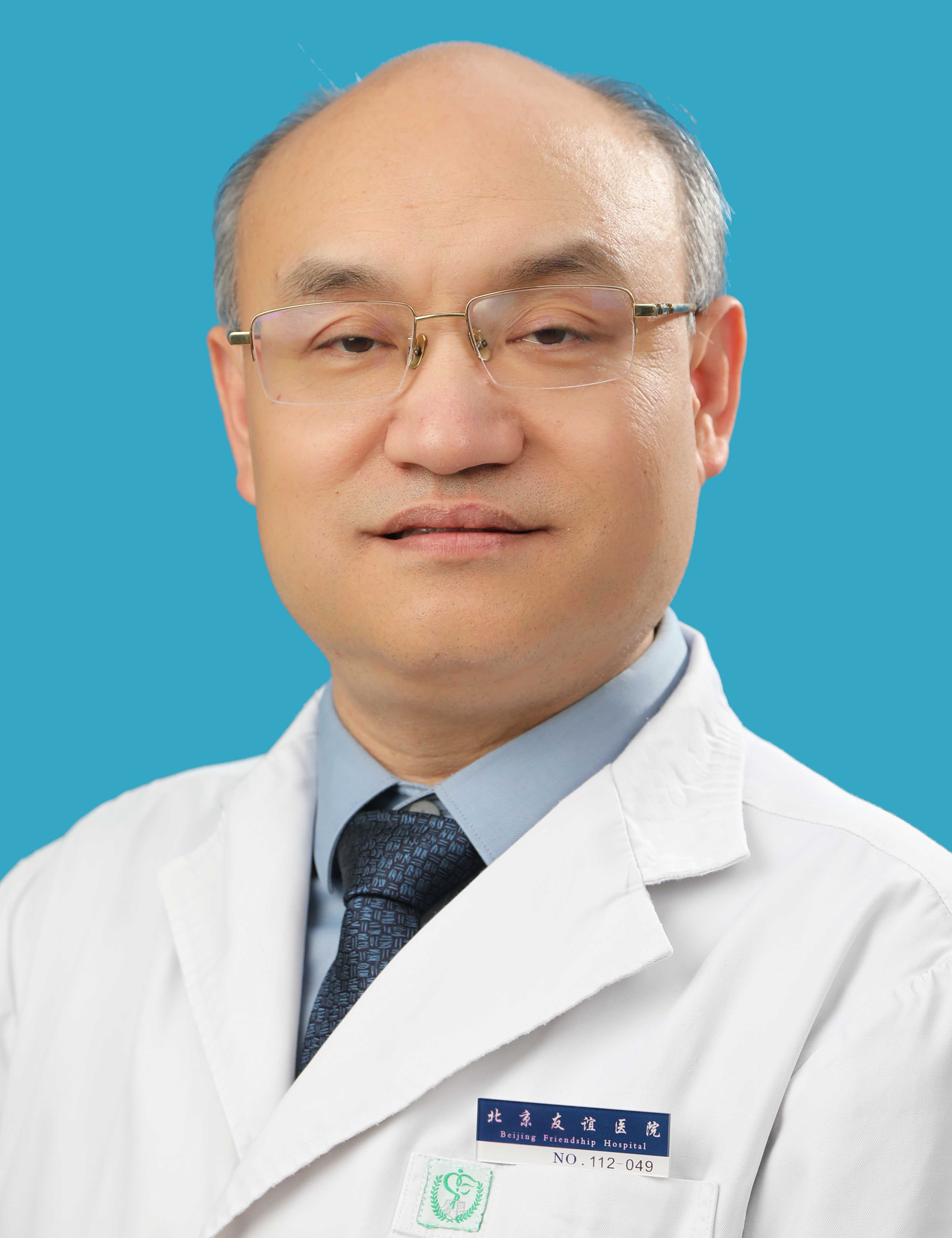 Yongbo Zhang (张拥波)合作论文数Beijing Friendship Hospital, Capital Medical University12WeiQin Zhao (赵伟秦)合作论文数11
 Houzhen Tuo (脱厚珍)合作论文数Beijing Friendship Hospital, Capital Medical University7He Lv (吕鹤)合作论文数7
 Zhaoxia Wang (王朝霞)合作论文数Peking University First Hospital;Academy for Advanced Interdisciplinary Studies, Peking University6Riliang Zheng合作论文数6
 Chunling Xu (许春伶)合作论文数Beijing Friendship Hospital, Capital Medical University5Honglian Duan合作论文数4GuangLi Shen (沈光莉)合作论文数3Yun Xue (薛云)合作论文数3XingHua Luan合作论文数3
 Yanjun Guo (郭燕军)合作论文数Beijng Friendship Hospital, Capital Medical University3Ze-Long Tian合作论文数2Yinong Cui合作论文数2Li-Yan Zhang合作论文数2WANG Yang (王洋)合作论文数2YiTong Du (杜艺彤)合作论文数2XiaoYang Ma (马小扬)合作论文数2MingRui Dong (董明睿)合作论文数2Qin Zhang (张芹)合作论文数2Lin Wang (王琳)合作论文数2XiaoJing Liu (刘晓静)合作论文数2
 Daojun Hong (洪道俊)合作论文数The First Affiliated Hospital of Nanchang University2Yan Chuanzhu合作论文数2
 Weidong Le合作论文数Sichuan Academy of Medical Sciences2
 Jiawei Wang (王佳伟)合作论文数Beijing Tongren Hospital, CMU2Yunying Gao (高云鹰)合作论文数2
 Jiong Qin (秦炯)合作论文数Peking University People's Hospital2
 Wei Zhang (张巍)合作论文数1Xiaojiao Xu合作论文数1Jingjing Che合作论文数1Kui Chen合作论文数1
 Xin Shi (史欣)合作论文数Institute of High Energy Physics, Chinese Academy of Sciences;University of Chinese Academy of Sciences1Li Yi (易立)合作论文数1William Ondo合作论文数1Ting Gao (高婷)合作论文数1Dan Gao (高丹)合作论文数1li hong (洪丽)合作论文数1yueping wang (王月平)合作论文数1
 Weiping Sun合作论文数1Guilan CAI (蔡桂兰)合作论文数1GuiMing Zang (臧贵明)合作论文数1XiangRu Sun (孙相如)合作论文数1ZhiMin Kang (康智敏)合作论文数1




