1 病例资料 65岁男性,因鞍区脊索瘤术后10年伴头痛及视物模糊1年余入院.10年前,因头痛伴左眼疼痛、视物模糊行头颅MRI示鞍区占位,行经鼻蝶入路鞍区占位切除术,术后头痛及眼部疼痛症状消失,术后病理示脊索瘤,术后行放疗(具体不详),但未定期复查头颅MRI.
1病例资料 17岁女性,因背部肿物渐进性增大半年余入院.半年前发现背部正中隆起肿物,质韧,约"花生米"大小,肿物渐进性增大.入院体格检查:背部胸6~7椎体水平可触及肿物,大小约6 cm×4 cm,质韧,边界尚清楚,与后方胸椎黏连,活动度差,触之无压痛,无皮肤红肿;四肢肌力5级,肌张力正常.
三叉-心脏反射是一种脑干反射,由Schaller等于1 999年首次报道,典型的临床表现为:三叉神经的任一感觉支受刺激,出现心率变慢,血压下降,有时伴有呼吸暂停或胃肠蠕动增强等.本文报道1例蝶骨嵴内侧型脑膜瘤术中出现三叉-心脏反射. 1病例资料 46岁女性,因头痛头晕伴恶心呕吐、视物模糊半个月入院.既往无高血压、心脏病、脑血管病等病史.入院体格检查:心律84次/min,血压125/65 mmHg,未见明确神经系统阳性体征.心电图示:窦性心律,正常心电图.头颅增强MRI示:左侧蝶骨嵴内侧型脑膜瘤,与左侧海绵窦外侧壁关系紧密.全麻下行左侧翼点入路脑膜瘤切除术,用瑞芬太尼及丙泊酚进行麻醉诱导.麻醉状态下,病人心率60次/min,有创动脉压135/80 mmHg.术中牵拉海绵窦外侧壁肿瘤时,病人心率下降至40次/min,下降比例33%;血压下降至95/55mmHg,下降比例30%.暂停手术20 s后,心率及血压恢复至正常.同样力度再次牵拉外侧壁肿瘤后,病人再次出现心率及血压急剧下降,暂停手术刺激后,心率及血压恢复正常.
目的:分析经桡动脉行冠状动脉造影或介入(PCI)术后出血性脑血管病发生的特点及诊治.方法:收集PCI术后急性出血性脑血管病4例的临床资料进行回顾性分析,并复习相关文献.结果:4例PCI术后出血性脑血管病患者,男2例,女2例;平均年龄60岁;诊断心肌梗死2例,不稳定心绞痛2例;既往服用阿司匹林鼻出血1例,PCI术后牙龈出血1例.4例既往均无糖尿病病史及长期服用抗血小板及抗凝药物者.术后使用阿司匹林100 mg、氯吡格雷75 mg 2例,使用阿司匹林100 mg、氯吡格雷75 mg及低分子肝素2例.急性出血性脑血管病发生时间为术后95 min~3 d,临床表现为头晕、头痛、烦躁、意识障碍、言语不利、肢体活动不利.脑实质出血2例,脑实质(小脑)出血后破入蛛网膜下腔1例,蛛网膜下腔出血1例.行颅内血肿钻孔引流术1例,遗留严重神经功能缺损;经内科治疗后死亡2例.结论:双联抗血小板或双联抗血小板加抗凝治疗,是PCI术后急性出血性脑血管病的最可能的因素之一.一旦术后患者出现神经系统症状,应及时完善头颅CT检查,如确诊急性出血性脑血管病,应及时给予治疗.
患者男,65岁,主因"突发左侧肢体无力2 d",于2018年2月22日收入首都医科大学附属北京友谊医院神经外科。入院体格检查:左侧肢体肌力Ⅴ - 级,肌张力偏高,神经系统其他检查未见异常。既往史:高血压病史10余年,平时血压控制在125/70 mmHg左右。既往多次发生短暂性肢体麻木。患者口服阿司匹林后有呕血症状。头部CT平扫示,右侧颞叶占位(图1),肿瘤卒中可能。头部增强MRI示,右侧颞叶占位,血肿可能,右侧基底节区近期脑梗死(图2)。
Objective To investigate the clinical effect of using anterior cervical approaches in the treatment of cervical neurogenic tumor of the peripheral nerve from different parts.Methods A retrospective review of 22 cases of cervical neurogenic tumor of peripheral nerves was performed,who were admitted to Department of Neurosurgery,Beijing Friendship Hospital,Capital Medical University from January 2009 to January 2015.All 22 patients underwent microsurgical operations with anterior approach of sternocleidomastoid muscle in 10 cases,posterior approach of sternocleidomastoid muscle in 4 cases,anterior cervical retropharyngeal approach in 3 cases,trans-oral approach in 2 cases,and supraclavicular approach in 3 cases.Results The tumors in all 22 cases were totally resected.No surgical complications such as cerebrospinal fluid leakage were observed except 2 cases which developed hoarseness postoperatively that improved that after neurotrophic treatment.All cases were followed up for 12-84 month with a mean 48.3 ± 18.7 month.Recurrence occurred in 1 case which underwent 2nd operation.Conclusion It seems safe and effective to select appropriate approaches corresponding to various tumor locations in the treatment of cervical neurogenic tumors of peripheral nerves.
目的 探讨显微手术夹闭治疗老年患者颅内动脉瘤的治疗效果.方法 回顾性分析49例60岁以上颅内动脉瘤患者的临床资料(包括破裂出血41例,未破裂出血8例).根据术前数字减影血管造影(DSA)和头颅CT血管造影(CTA)检查确认动脉瘤后,48例采用经翼点入路,1例采用经纵裂入路夹闭颅内动脉瘤,出院时根据格拉斯哥预后评分(GOS)评价预后.结果 根据GOS评分,41例破裂出血患者,Hunt-Hess分级Ⅰ~Ⅲ级28例,其中26例(93%)取得良好预后,重度残疾2例(7%);Hunt-Hess分级Ⅳ级13例,其中7例(54%)取得了良好的预后,重度残疾3例(23%),植物生存1例(8%),死亡2例(15%).未破裂动脉瘤8例,均取得良好预后(100%).结论 破裂动脉瘤的老年患者应尽可能早期手术,术前Hunt-Hess分级Ⅰ~Ⅲ级者预后良好,而Ⅳ级以上病死率和伤残率仍较高;未破裂动脉瘤老年患者应采取积极治疗.
Objective To investigate of clinical presentation,MR imaging and pathology features of super long intramedullary ependy-momas from the roof of fourth ventricular to the second vertebra. Methods A case of super long intramedullary ependymomas in fourth ventricular and spinal cord were reported. The clinical feature of super long intramedullary ependymomas was summarized and the related literature was re-viewed. Results MRI revealed a multinodular and multicystic lesion with enhancement in the medulla oblongala and nearly the whole spinal cord from the roof of the fourth ventricular to second vertebra. The lesion range was 33 cm × 1. 6 cm × 0. 8 cm. Intraoperative observation via cervical midline myelotomy revealed a grayish,gelatinous solid mass and part of the tumor revealed cystic formation. A pathological examination revealed the mass to be a ependymoma with mucinous papillary type( WHO gradeⅠ-Ⅱ). Conclusion This report presents the rare and the longest in-tramedullary ependymomas that has been reported in China,a review of the literature associated with super long intramedullary ependymomas,a discussion of the clinical and radiographical characteristics.
Objective To explore the changes in gene Sirt1 expressions in tissues of human glioma and para - glioma. Methods A 44 000 gene cDNA microarray(Affymetrix)was used to examine gene expressions in the tissues of human glioma and para - glioma. The Sirt1 genes were identified and subjected to real - time PCR analysis. Results The expression of Sirt1 in tissues of glioma was extremely lower than that in tissues of para - glioma. The results of real - time PCR were consistent with those of genechip analysis. Conclusion The expressions of Sirt1 were obviously different between tissues of glioma and para - glioma. Sirt1 may be associated with carcinogenesis of glioma.
Objective The aim of this study is to discuss the clinical characteristic and the selection of treatment of acute cerebral vein si-nus thrombosis. Methods Clinical data of 26 patients diagnosed with acute CVST were retrospectively analyzed. These patients were treated with simple anticoagulant therapy,intravenous thrombolytic therapy,and endovascular thrombolysis. Then all the patients took oral warfarin for 6~12 months. Results Among 16 patients who received systemic anticoagulant therapy,13 were cured,3 were improved. Among 6 patients who accept-ed intravenous thrombolytic therapy,4 were cured,2 were improved. Among 4 patients who accepted endovascular thrombolysis,3 were cured,1 was improved. The period of follow-up was between 6 months and 26 months( the median time was 18 months),and no recurrence happened or reported. Conclusion Personalized treatment of acute CVST should be selected in clinical practice. The influencing factors included the clinical manifestations,the degree of occlusion of cerebral vein sinus,and collateral circulation.
Objective To observe the security and effectiveness of injection methotrexate through Ommaya reservoir for the therapy of prima-ry intracranial diffuse large B - cell lymphoma. Methods A retrospective analysis was performed in 22 cases who received the injection of methotrex-ate through Ommaya reservoir to therapy primary intracranial diffuse large B - cell lymphoma. All patients were confirmed primary intracranial diffuse large B - cell lymphoma by pathologic diagnosis,whole body CT after operation or biopsy. Later,Ommaya reservoir were placed into patients′ventri-cle and injected methotrexate(MTX)12 mg + dexamethasone(DXM)5 mg through Ommaya reservoir once a week. This treatment were continued for total six weeks. Results All patients get complete response(CR)and partial response(PR). Nine(40. 9% )of twenty two patients get CR and thirteen(50. 1% )of twenty two patients get PR. Twenty one of twenty two patients accepted chemotherapy. Conclusion The injection of methotrex-ate through Ommaya reservoir during the therapy of primary intracranial diffuse large B - cell lymphoma is safe and effective.
 Cang Liu (刘藏)合作论文数Beijing Friendship Hospital, Capital Medical University8
 Xu Chen (陈旭)合作论文数Beijing Anding Hospital, Capital Medical University7
 Lei Peng (彭磊)合作论文数6Deqiu Cui (崔德秋)合作论文数4RuiGuang Hou (侯瑞光)合作论文数4GuoShun Mei (梅国顺)合作论文数2QuanShan Ai (艾泉山)合作论文数2XiangHua Zhang (张向华)合作论文数2
 Jimei Li (李继梅)合作论文数Beijing Friendship Hospital, Capital Medical University2
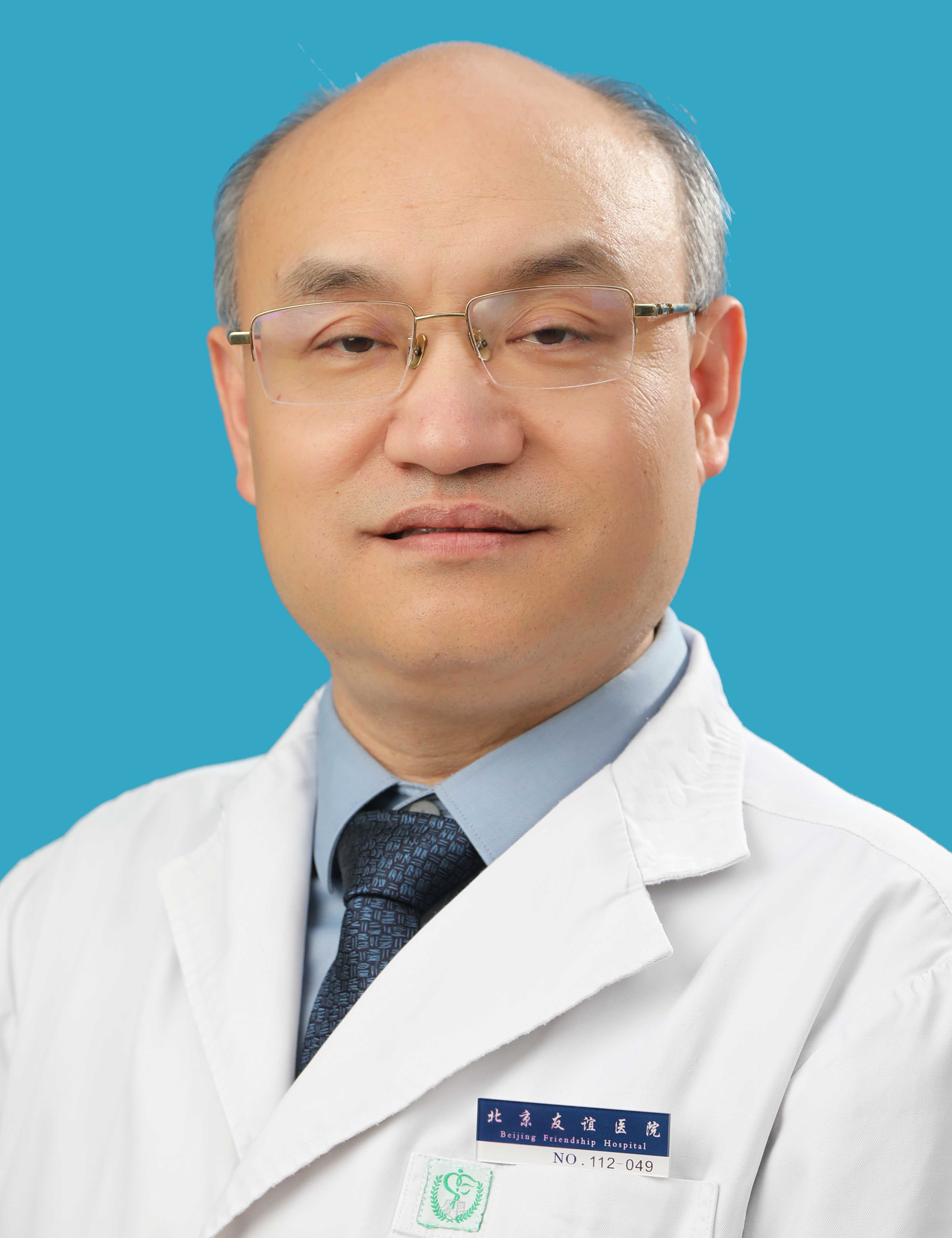 Yongbo Zhang (张拥波)合作论文数Beijing Friendship Hospital, Capital Medical University2yida lu (鲁一达)合作论文数1Yipeng Dong合作论文数1
 Qiheng He合作论文数1JiChao Ma (马骥超)合作论文数1HongYan Bi (毕鸿雁)合作论文数1WeiQin Zhao (赵伟秦)合作论文数1Yuan Zhao (赵媛)合作论文数1Bin Chen (陈彬)合作论文数1Li Zhou (周力)合作论文数1
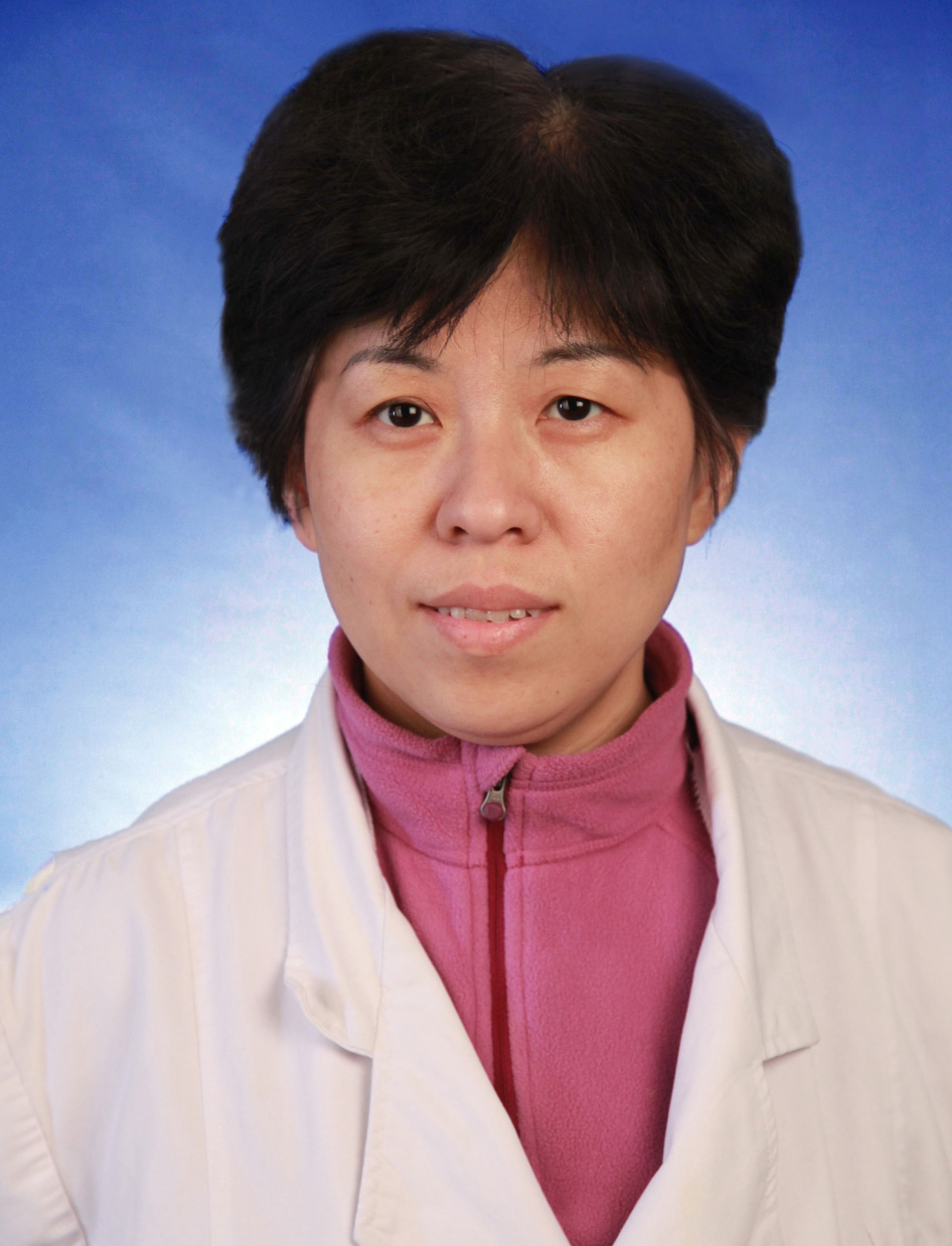 Yanjun Guo (郭燕军)合作论文数Beijng Friendship Hospital, Capital Medical University1
 Jing Zhang (张晶)合作论文数Beijng Anzhen Hospital, Capital Medical University1
 Jinqian Zhang (张锦前)合作论文数1




