目的:研究甲状腺乳头状癌(papillary thyroid carcinoma,PTC)及结节性甲状腺肿(nodular goiter,NG)患者血清中血管内皮生长因子(vascular endothelial growth factor,VEGF)的表达情况及其对PTC淋巴结转移的诊断效能.方法:收集资料完整的PTC患者69例,选择20例NG患者作为良性对照,对两组患者血清VEGF水平进行比较,分析血清VEGF对PTC淋巴结转移的诊断效能,观察血清VEGF水平与BRAFV600E基因突变在PTC中的关系,采用MACIS评分系统对PTC患者进行预后评估,并分析血清VEGF水平与预后指数的相关性.结果:VEGF在PTC患者血清中的水平明显高于NG患者,差异具有统计学意义(P<0.001);淋巴结转移患者血清VEGF水平高于无淋巴结转移患者,差异具有统计学意义(P=0.023);血清VEGF水平对于淋巴结转移的ROC曲线下面积(AUC)为0.689,灵敏度为0.698,特异性为0.750;血清VEGF水平与BRAFV600E基因突变情况无显著相关性(P=0.177);血清VEGF水平与预后指数间不具有相关关系(r=0.065,P=0.600).结论:血清VEGF能够有效区分PTC及NG,对PTC淋巴结转移具有一定的预测价值.
Objective To investigate the value of the fusion tomography(SPECT/CT)in the staging of the lesions in the pa-tients with ankylosing spondylitis.Methods The SPECT/CT data of 67 patients with ankylosing spondylitis were compared with MR images of those patients at the same period,and then,the image interpretation,staging,clinical compliance were analyzed, respectively.Results PECT/CT classification was as follows,i.e.,Grade 0 included 1 case,grade 1 included 8 cases,grade 2 included 26 cases,grade3 included 21 cases,and grade 4 included 11 cases.MR classification was as follows, i.e., Grade 0 consisted of 3 cases,grade 1 of 5 cases,grade 2 of 23 cases,grade 3 of 25 cases,and grade 4 of 11 cases.Conclusion There were no significant differences between the two different imaging methods in patients with SPECT /CT.The bone metabolism of the patients with active bone metabolism was more consistent with the clinical symptoms, which is helpful for clinical diagnosis and treatment.
Objective To analyze the relationship and the factors between the thyroid function in Graves′ disease and the cystatin C (CysC) levels and observe their correlation of different levels and treatments.Methods 185 patients with Graves hyperthyroidism who receiving treatment from January 2013 to September 2015 at Xijing Hospital Department of Nuclear Medicine and 24 healthy patients were recruited.Their serum level of thyroid hormone (FT3,FT4,TSH) and CysC were detected and compared.19 in the 185 patients′ were detected again after Iodine-131 treatment.Results Serum CysC concentration in the Graves′ Disease group was significantly higher than the normal group, and it was positively correlated with the serum concentrations of FT3 (rs=0.69, P<0.01) and FT4 (rs=0.66, P<0.05).After treatment, the CysC level decreased with thyroid hormones.The difference was statistically significant (P<0.05).Conclusion Graves′ Disease hyperthyroidism is one of the important reasons for the increasing of cystatin C.Serum CysC concentration was positively correlated with the severity of thyroid dysfunction before and after the treatments in patients with Graves′ Disease, which indicates CysC can reflect the thyroid function status when kidney disease was excluded.
目的 探讨半胱氨酸蛋白酶抑制剂C(CysC)及其相关肾小球滤过率(GFR)估计值(eGFR)计算方程在糖尿病患者肾脏功能评价中的应用价值.方法 收集110例2型糖尿病患者资料,依据mGFR将患者分为代偿组和中重度损伤组,了解不同组别患者的基本情况,分析Scr、CysC以及不同的eGFR公式与mGFR的相关性.结果 全部患者、代偿组和中重度损害组的患者在与mGFR进行相关性比较时,CysC(r全部患者=-0.661,r代偿组=-0.795,r中重度损害组=-0.420)较SCr(r全部患者=-0.593,r代偿组=-0.657,r中重度损害组=-0.380)均表现出更好的相关性,以CysC为基础的eGFR-EPICycs计算的eGFR与mGFR之间的差异不具有统计学意义(P=0.804).结论 与Scr相较,CysC在糖尿病患者肾功能评价中显示更高的应用价值,在对不同eGFR公式进行比较后本研究推荐eGFR-EPICysC对患者进行GFR的估计.
Objective:To discuss the diagnostic value of single positron emission computed tomography (SPECT/CT)of rib lesion of patients with breast cancer.Methods:We collected 21 patients with breast cancer,the whole body scan showed high uptake lesions in the ribs,all of lesions received SPECT/CT.Results:SPECT/CT results as follows:39 lesions were founded.21 lesions were founded as bone metastasis,12 lesions bone fracture,3 lesions rib benign os-teopathy,2 lesions alteration after surgery,1 lesion pleura reactivity.Conclusion:SPECT/CT can find the lesions sen-sitively,it can also identify the properties of lesions accurately.SPECT/CT can help us make therapeutic schedule,it also have high clinical value for patients with breast cancer.
目的探索血清CEA、Tg联合检测在甲状腺良、恶性肿瘤中的临床价值。方法采用化学发光法分别检测经病理证实的68例分化型甲状腺癌(DTC)术前和手术联合131I清甲治疗后、65例甲状腺腺瘤、55例正常对照的CEA、Tg水平,并将结果应用SPSS13.0软件进行统计学分析。结果术前DTC组的CEA水平明显高于腺瘤组(P<0.05)、Tg水平与腺瘤组差异无统计意义(P>0.05);DTC组患者手术联合131I清甲治疗后的Tg水平有显著性下降(P<0.05);术后I和II期DTC患者的Tg、CEA水平明显低于III和IV期的(P<0.05)。结论血清CEA、Tg联合检测对手术及131I清甲治疗后DTC患者的治疗效果及预后判断有较大价值,CEA检测对确诊中晚期的DTC患者有辅助诊断作用。
目的:探讨磁共振诊断运动员腰椎椎体缘离断症的临床意义,提高其诊断及鉴别诊断水平.方法:结合文献对15例腰椎椎体缘离断症患者的MRI表现进行回顾性分析.结果:MRI显示腰段椎体呈唇样变,相邻终板不规则,椎体前缘塌陷,Schmorl结节形成.结论:MRI能全面准确地提供运动员腰椎椎体缘离断症的早期表现,为临床早期发现和治疗提供有效的诊疗依据,是诊断及鉴别诊断的首选方法.
近年来结核病的发病率呈上升趋势,骨骼是其常累及的部位,而椎体结核在骨关节结核中居首位.本研究对51例椎体结核患者的骨显像及融合断层显像特点进行分析,现报道如下. 一、资料与方法 1.临床资料.回顾性分析本院2009年12月至2013年1月经手术及病理最终诊断为椎体结核的患者51例,其中男33例,女18例,平均年龄51.5岁.
放射免疫分析技术在操作过程中影响因素多,操作者的操作技术、测量仪器以及整个反应体系等任何步骤的不规范都会直接影响患者的检测结果,导致错误的诊断,因此找出放射免疫分析过程中引起误差的各种因素、认清误差的来源,做好误差控制方法有重要的意义。
Objective To evaluate the usefulness of electron microscopy in detection of virus in animal samples. Methods Negative staining and ultrathin sections were used for electron microscopic examination. Results Plentiful mouse poxvirus particles were observed under the electron microscope by both staining methods. Conclusion Electron microscopy is a reliable and useful technique and can be successfully used in animal virus detection.
An outbreak of poxvirus infection was occurred in twenty bought KM and BALB/c mice .The livers,spleens, kidneys, lymph nodes and cerebrum of ill mice were used to histology observation. The characteristic of pathologic change was poxvirus infection .when the tissue homogenate fluid infected passaging BHK cell, we could see apparent CPE change. A lot of poxvirus particle were found in EMP. The center of particle was nucleoid composed by DNA and protein ,The agglutination reaction was positive (HA=1:16).By ELISA kit examination the serum of ill mice expressed poxvirus antibody positive reaction, After suspension of virus infected KM and BALB/c mice .It was found that the former was easily infected, with apparent symptom .The latter had strong resistance, latent infection. The symptom always occurred slowly, but when experiment factors were exerted,the BALB/c mice quickly had symptom, and died. It proved that the pathogen was poxvirus .
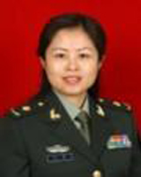 Jing Wang (汪静)合作论文数第四军医大学西京医院核医学科9
 XiQing Wang (王喜青)合作论文数5Ni Wang (王妮)合作论文数4Zhe Wang (王喆)合作论文数4Xiang ZHOU (周详)合作论文数3Guiyu Li合作论文数2Ming ZHANG (张明)合作论文数2LiuJin Li (李六金)合作论文数2XiaoYan Liang (梁晓燕)合作论文数2Hong LI (李宏)合作论文数1xiang zhou (周翔)合作论文数1JingLan Deng (邓敬兰)合作论文数1Hong Yin (印弘)合作论文数1TieQiao Chen (陈铁桥)合作论文数1XiaoWei Ma (马晓伟)合作论文数1Kang Liu (刘康)合作论文数1WeiDong Yang (杨卫东)合作论文数1Gang Chen (陈纲)合作论文数1Ping Tian (田萍)合作论文数1
 Zhanghong Shi (师长宏)合作论文数Air Force Medical University1
 Fei Kang (康飞)合作论文数Air Force Medical University1Yibin Xi合作论文数1Xiaoli An (安小利)合作论文数1Jie Tang (唐婕)合作论文数1Ming Zhang (张明)合作论文数1




