Primate-specific genes (PSGs), important contributors to the origin of adaptive evolutionary novelties, are abundantly expressed in the testis. However, the specific roles of PSGs in the male reproductive system of humans and other primates are largely unknown. Here, we employed whole-exome sequencing and identified deleterious variants of TFDP3, an X-linked PSG, in eight infertile men with oligoasthenoteratozoospermia. All of the male subjects harboring TFDP3 variants presented dramatic reductions in sperm concentration, motility, and abnormal sperm morphology. Furthermore, Tfdp3-knockdown (KD) in the testes of cynomolgus monkeys confirmed the important role of TFDP3 in normal spermatogenesis in primates. Consistently, dramatic decreases in sperm concentration, motility, and abnormal sperm morphology were also observed in Tfdp3-KD male cynomolgus monkeys. More importantly, further functional studies revealed that TFDP3 deficiency activated E2F1 induced apoptosis and thus led to decreased sperm count and motility. In addition, five of the eight couples underwent intra-cytoplasmic sperm injection treatment and achieved a successful pregnancy, indicating a potentially good outcome of assisted reproduction for those with TFDP3 deficiency-mediated oligoasthenoteratozoospermia. Collectively, our genetic analyses and experimental observations in humans and cynomolgus monkeys highlight the crucial role of TFDP3, an inhibitor of apoptosis, in normal spermatogenesis. These findings expand the spectrum of pathogenic variants for oligoasthenoteratozoospermia-associated male infertility and also reveal the special significance of primate-specific TFDP3 for the human male reproductive system, thus providing important guidance for genetic counseling and the clinical diagnosis of male infertility.
BACKGROUND:Cryptozoospermia is defined, according to WHO criteria, as the absence of spermatozoa in fresh semen preparations but the presence of rare spe matozoa after centrifugation. Identifying candidate genes and their clinical phenotypes is essential for understanding its etiology and developing targeted therapies. METHODS:Whole-exome sequencing (WES) was performed on 503 oligozoospermic and 516 azoospermic infertile patients to identify candidate variants associated with severe male infertility. Functional studies, including protein expression, subcellular localization, and interaction assays, were conducted in vitro, and assisted reproductive outcomes were analyzed. RESULTS:We identified a compound heterozygous CHTF18 variant (NM_022092: c.538G > A [p. Val180Met] and c.1434delG [p. Leu481Serfs*3]) in a cryptozoospermic patient exhibiting a high proportion of aneuploid sperm. The missense variant (c.538G > A) was rare (< 0.001% population frequency) and predicted to disrupt protein function by in silico tools. The frameshift variant (c.1434delG) was predicted to introduce premature truncation and a loss-of-function effect. In vitro studies suggested altered subcellular localization of mutant CHTF18 proteins and possible effects on interactions with known binding partners. Despite severe cryptozoospermia, the patient achieved a successful pregnancy and live birth via intracytoplasmic sperm injection (ICSI). CONCLUSIONS:These findings support CHTF18 as a candidate gene associated with cryptozoospermia and suggest that biallelic CHTF18 variants may contribute to spermatogenic failure by altering CHTF18 protein localization and interactions within the CHTF18-RLC complex. Further cases and in vivo validation are required to strengthen this gene-disease association.
The ubiquitin-proteasome system (UPS) represents an evolutionarily conserved machinery governing proteostasis through spatiotemporal regulation of protein degradation. While spermatogenesis involves multilayered regulatory mechanisms spanning translation to dynamic post-translational modifications (PTMs), the identity of UPS-associated E3 ligases orchestrating germ cell-specific protein turnover remains elusive. Here, we identify a testis-specific E3 ubiquitin ligase complex comprising elongin B/C, Cullin-2 (CUL2), RING-box protein-1 (RBX1), and SOCS box protein ASB9, designated ECSASB9. Genetic ablation of ECSASB9 in mice via ubiquitous Asb9 knockout (KO) or spermatid-specific elongin B/C conditional KO disrupts spermiogenesis and compromises fertility. Mechanistic studies reveal that ECSASB9 engages tubulin beta 4 A (TUBB4A) through substrate recognition, catalyzing K48-linked polyubiquitination at lysine 379 (K379) to promote proteasomal degradation. Notably, Tubb4aK379R knock-in (KI) mice phenocopy the spermiogenesis defects observed upon ECSASB9 deficiency. Clinically, we identify three hemizygous missense variants in X-linked ASB9 among Chinese males with idiopathic infertility. Male mice bearing orthologous ASB9 variant exhibit oligoasthenoteratozoospermia (OAT) and subfertility, mirroring human phenotypes. Taken together, our findings establish ECSASB9 as an important regulator of spermatogenic proteostasis and provide mechanistic insights into UPS-mediated tissue-specific degradation, while implicating ASB9 variants in male infertility pathogenesis.
Objective:FBXO43 is a known inhibitor of the anaphase-promoting complex/cyclosome (APC/C), a key E3 ubiquitin ligase that controls meiotic cell cycle progression. However, how different FBXO43 mutations affect APC/C inhibition and lead to divergent clinical phenotypes remains unclear. This study is aimed at clarifying how different FBXO43 mutations produce divergent clinical phenotypes in male infertility and to investigate their preliminary molecular mechanisms, thereby providing evidence-based guidance for precision-assisted reproduction in affected individuals. Methods:Two infertile patients carrying distinct compound heterozygous FBXO43 variants-missense mutations in a macrozoospermia case and truncating mutations in a nonobstructive azoospermia (NOA) case-were identified through whole-exome sequencing. Sperm morphology, chromatin status, and aneuploidy were assessed, and a testicular biopsy was performed for the NOA case. Functional consequences of each mutation were evaluated using in vitro HEK 293T cell models, including protein stability, ubiquitination, and APC/C subunit interactions. Clinical outcomes of assisted reproductive technology (ART) were also reviewed. Results:Missense variants (NM_001029860, p.Pro641Leu/p.Arg660Gln) in the macrozoospermia patient allowed completion of meiosis but led to severe sperm head enlargement, chromatin condensation defects, and markedly elevated aneuploidy, resulting in repeated ICSI failure. In contrast, the truncating variants (p.Trp532 ∗/p.Ser577Leufs ∗11) in the NOA patient abolished the C-terminal functional domain and caused meiotic arrest with complete absence of mature sperm. Mechanistically, all mutations reduced FBXO43 protein stability and disrupted its specific binding to the APC/C substrate recognition subunit APC3, whereas interactions with APC2/6/8 remained intact. Loss of APC3 binding likely impairs APC/C inhibition, disturbing meiotic chromosome segregation in a mutation-severity-dependent manner. Conclusions:Different types of FBXO43 mutations generate distinct infertility phenotypes through a dose-dependent mechanism: Truncating mutations cause profound meiotic arrest and NOA, whereas hypomorphic missense mutations permit meiotic completion but result in severe teratozoospermia with high aneuploidy. These findings link FBXO43 to a continuous phenotypic spectrum and highlight its relevance for precision reproductive counseling. Based on the identified mutation-specific risks, preimplantation genetic testing for aneuploidy (PGT-A) is recommended for affected couples to improve embryo selection and optimize ART outcomes.
STUDY QUESTION What is the role of calmodulin-regulated spectrin-associated protein 1 (CAMSAP1) in human spermatogenesis?SUMMARY ANSWER Biallelic CAMSAP1 variants cause human male infertility with multiple sperm head deformities and MMAF-like phenotypes by disrupting manchette microtubule dynamics and the microtubule-spectrin-actin scaffold essential for sperm head shaping.WHAT IS KNOWN ALREADY Sperm head shaping requires coordinated acrosome formation, chromatin condensation, and manchette-driven nuclear remodeling. Although several genes have been implicated in globozoospermia, macrozoospermia, and multiple morphological abnormalities of the sperm flagella (MMAF), the genetic causes of other forms of abnormal sperm head morphology remain largely unknown. Camsap1-deficient mice exhibit severe spermatogenic defects, including abnormal sperm head shaping and flagellar malformations resulting from disruption of the acrosome-acroplaxome-manchette complex, ultimately leading to male infertility. However, whether CAMSAP1 plays a conserved role in human sperm development has not been established.STUDY DESIGN, SIZE, DURATION The CAMSAP1 variants were identified by whole-exome sequencing in a cohort of 3757 male infertility patients, and all participants were recruited from 2015 to 2025. In vitro functional assays, immunoprecipitation-mass spectrometry, and sperm morphological analyses were performed.PARTICIPANTS/MATERIALS, SETTING, METHODS Three infertile men with severely impaired sperm morphology and motility were recruited. Bioinformatic assessment, cell-based expression assays, immunostaining, and co-immunoprecipitation (Co-IP) coupled with liquid chromatography-tandem mass spectrometry (LC-MS/MS) using human testicular extracts were used to characterize the functional impact of the variants.MAIN RESULTS AND THE ROLE OF CHANCE Homozygous CAMSAP1 variants were identified in two individuals from consanguineous families (Chinese and Pakistani), and compound heterozygous missense variants in a third individual from a non-consanguineous Chinese family through whole-exome sequencing and further confirmed by Sanger sequencing. Sperm from affected individuals displayed MMAF phenotypes and diverse head abnormalities, including pyriform, amorphous, small, and globozoospermic forms. All four CAMSAP1 variants were predicted to be damaging and exhibited markedly reduced protein stability in vitro. Proteomic analysis revealed that CAMSAP1 interacts with proteins involved in actin cytoskeleton organization, acrosomal vesicle trafficking, microtubule bundle assembly, and calmodulin regulation. Mechanistically, CAMSAP1 associates with CALM1 and SPTBN1 to couple manchette microtubules with the perinuclear spectrin-actin network, ensuring proper force transmission for nuclear shaping. Mutations disrupted these interactions, leading to defective manchette architecture, abnormal acrosome assembly, and nuclear deformation.LIMITATIONS, REASONS FOR CAUTION Although CRISPR-Cas9 knock-in cell lines carrying patient-derived CAMSAP1 variants were generated, this in vitro model could not reproduce the highly dynamic processes of manchette assembly and nuclear remodeling that occur uniquely during spermiogenesis, and therefore cannot fully capture the mechanistic sequence leading to sperm head deformation. Additionally, the number of affected individuals remains limited, and larger multi-center, multi-ethnic cohorts will be required to validate the pathogenicity of CAMSAP1 variants and further delineate the associated clinical spectrum. WIDER IMPLICATIONS OF THE FINDINGS This work identifies CAMSAP1 as a previously unrecognized cause of human asthenoteratozoospermia and expands the genetic landscape of sperm head deformities. The findings underscore the essential role of CAMSAP1 in manchette-mediated nuclear remodeling and provide new insights for genetic diagnosis, counseling, and management of male infertility.STUDY FUNDING/COMPETING INTEREST(S) This work was supported by the National Natural Science Foundation of China (grant no. 82301815), the National Key R&D Program of China (grant no. 2022YFC2702603), the Sichuan Province Science and Technology Innovation Talent Project (grant no. 2024JDRC0006), the 2024 Key Open Project of Sichuan Provincial Key Laboratory for Human Disease Gene Research (grant no. 2024kflx002), and the China Postdoctoral Science Foundation (grant nos. 2023M732468 and GZC20231835). The authors declare no competing interests.TRIAL REGISTRATION NUMBER N/A.
The precise assembly of the sperm flagellum is essential for male fertility and has long been ascribed to kinesin-2-driven intraflagellar transport (IFT) of protein cargoes. However, during late spermiogenesis, when transcription activity is largely silenced, how the spatiotemporally regulated delivery of flagellar components remains poorly understood. Here, we systematically screened kinesin genes in asthenozoospermic patients and identified two homozygous deleterious KIF6 variants in unrelated men characterized by complete sperm immotility. Mouse models carrying the corresponding mutations recapitulated the human infertility phenotypes. Multi-omics analyses revealed that most testicular mRNAs remained largely unchanged in Kif6 M1/M1 mice, whereas proteins involved in axonemal organization and energy metabolism were markedly reduced. Mechanistically, KIF6 interacts with the RNA-binding proteins (RBPs) FMRP and FXR1 to assemble mRNP transport complexes that ferry transcripts encoding flagellar structural proteins (e.g., DNALI1) and metabolic enzymes (e.g., HK1). Impaired KIF6 function compromises mRNP trafficking to the developing flagellum, reducing flagellar transcript levels and ultimately causing decreased protein abundance and defective flagellar function. Collectively, we identify KIF6 as a key regulator of mRNA transport during spermiogenesis. It interacts with RBPs through a novel IFT-like pathway to deliver mRNAs essential for flagellar biogenesis, redefining the traditional protein-centric IFT paradigm.
PURPOSE:Infertility affects about 15% of couples globally, with genetic factors contributing significantly. Advances in genomic technologies have led to the discovery of genes like MEI1, which play a crucial role in meiosis. However, the population prevalence and pathological mechanisms of human MEI1 variants remains poorly defined. METHODS:To elucidate the contribution of MEI1 to spermatogenic failure, initial screening for biallelic mutations was conducted in 626 non-obstructive azoospermia (NOA) patients, followed by targeted screening for heterozygous variants in a validation cohort comprising 1,607 well-characterized idiopathic male infertility cases (626 NOA, 799 severe oligoasthenospermia, and 182 terato/asthenozoospermia). RESULTS:Here, we identify MEI1 as a prominent gene associated with non-obstructive azoospermia (NOA), with biallelic pathogenic variants in MEI1 were identified in 12 individuals (1.9%, 12/626). Furthermore, heterozygous MEI1 mutations were identified in 28 patients (0.17%, 28/1607) from the broader infertility screening cohort implicate MEI1 in a wider phenotypic spectrum, broadening its clinical relevance. Critically, micro-TESE uniformly showed no sperm retrieval in individuals carrying biallelic MEI1 mutations, underscoring the imperative for preemptive genetic screening to avoid unnecessary surgical interventions. Mechanistically, these mutations disrupted interactions with key meiotic proteins (ANKRD31, IHO1, REC114, MEI4) in co-immunoprecipitation assays. CONCLUSION:These results not only elucidate its essential function in meiosis and DSB formation but also support its potential as a molecular marker for non-invasive diagnosis.
Oligoasthenoteratozoospermia (OAT) is a prevalent phenotype among infertile males, yet its underlying genetic etiology remains largely undefined. In this study, we found Spem2 deficiency did not affect the development of spermatogenic cells or the eventual generation of elongated spermatids within the testis, but affected the normal formation of residual bodies during the late stage of spermiation, which led to incomplete cytoplasmic removal of spermatids, abnormal sperm release and caused OAT. Affected sperm exhibited severely bent heads, cytoplasmic remnants encasing the head, and multiple sperm gathered. We further found SPEM2 may be involved in maintaining spermiation and cell polarity through interactions with cell polar molecules VANGL2, PRICKLE3, and DVL3. In vitro experiments showed that the transmembrane region of SPEM2 was its key functional domain and the binding domain for interactions between SPEM2 and VANGL2. Importantly, Spem2-deficient sperm enabled successful fertilization via intracytoplasmic sperm injection. Three novel SPEM2 heterozygous pathogenic variants were identified in four OAT patients by the whole-exome sequencing. Our findings revealed key roles of SPEM2 in spermiation, explored the potential pathogenesis of male infertility caused by SPEM2 deficiency, and provided essential information for genetic and reproductive counseling for such patients.
Non-obstructive azoospermia (NOA) and cryptozoospermia are two significant conditions contributing to male infertility. However, the underlying genetic factors in most cases remain unknown. In our study, whole exome sequencing identified novel biallelic variants in TBC1 domain family member 8 (TBC1D8) in two patients. Patient P1 with NOA harbored c.890C>T (p.A297V) and c.2461G>A (p.V821I), and patient P2 with cryptozoospermia carried c.854C>T (p.P285L) and c.1912G>A (p.D638N). Bioinformatic analyses predicted that all identified TBC1D8 variants were likely pathogenic. Compared with a normal control, patient P1 showed reduced expression of TBC1D8 in testicular tissue. Subsequently, hematoxylin-eosin staining and immunofluorescence analysis of testicular sections showed defective acrosome formation and the absence of elongated spermatids in patient P1, resulting from abnormal autophagy. Additionally, intracytoplasmic sperm injection treatment was beneficial for patient P2 with cryptozoospermia. In conclusion, our results suggest that TBC1D8 is a potentially novel candidate gene for male infertility associated with NOA or cryptozoospermia in humans.
Spermatogenesis is a highly complex cellular differentiation process. Recent advances employing knockout or knock-in mouse models have functionally characterized more than 700 genes as essential for male fertility maintenance. Paradoxically, emerging evidence reveals that a substantial proportion of the continuously expanding catalog of testis-enriched genes exhibits biological dispensability for normal sperm production. To systematically catalog non-essential testis-enriched genes in murine spermatogenesis, we performed a comprehensive PubMed literature review encompassing studies published up to 1 August 2025. Through stringent inclusion criteria, this analysis consolidates data from 83 publications that identified 261 testis-enriched genes demonstrated to be non-essential for spermatogenesis. We further categorize these genes by their familial relationships and explore potential explanations for the fertile phenotype observed in these knockout models, including genetic redundancy, compensatory mechanisms, differences in knockout strategies, and environmental influences. This review provides a valuable resource to avoid unnecessary expenditures and effort by research teams.
This study aimed to identify the genetic causes of male infertility associated with oligozoospermia/azoospermia in two unrelated Chinese families. Whole-exome sequencing (WES) and Sanger sequencing were performed on peripheral blood samples from three infertile individuals with reduced sperm counts. Semen analysis data were collected, and sperm morphology was evaluated using hematoxylin and eosin staining, along with transmission electron microscopy. Acidic aniline staining and fluorescence in situ hybridization (FISH) were employed to assess sperm nuclear maturity and chromosome aneuploidy. In vitro analyses were performed to determine the effect of the identified variants. We identified two novel homozygous variants in ZMYND15: a frameshift variant (NM_001136046.3:c.828-2_833dupAGAGAGCT) in family 1 and a missense variant (c.2051 T > A:p.Met684Lys) in family 2. Both variants were absent in public databases, and the missense variant was predicted to be deleterious. In vitro analyses confirmed that the frameshift variants likely impact protein function. Abnormal sperm head morphologies, characterized by reduced chromatin condensation and nuclear aneuploidy, were frequently observed in ZMYND15 mutant individual. Our findings reveal two novel ZMYND15 variants in three infertile patients with oligozoospermia/azoospermia, expanding the mutational spectrum of ZMYND15 and providing valuable insights for genetic counseling and the diagnosis in cases of male infertility.
Epigenetics is the link between the genome and environment, which can respond to physiological (such as age) or environmental factors (such as diet, stress, and pollution) and induce changes in epigenetic modifications (such as DNA methylation, non-coding RNA, and histone modifications). It can also serve as cellular memory transmitted from generation to generation. Sperm is highly responsive to such environmental changes and has unique epigenetic profiles. The paternal inter-/trans-generational inheritance mediated by sperm epigenetic changes is closely related to the health of offspring, which is an issue of great concern. This review has summarized the epigenetic mechanisms of paternal inter-/trans-generational inheritance and recent studies on the paternal inheritance mediated by sperm epigenetic changes in human and mice, which may facilitate understanding of the relationship between paternal epigenetic changes and the health of offspring caused by physiological or environmental changes and provide a basis for genetic counseling and clinical intervention.
Asthenoteratozoospermia is a common underlying cause of male infertility, with dynein dysfunction playing an important role in the aetiology of the condition. Dysfunction in certain dynein proteins has been implicated in asthenoteratozoospermia, while others exclusively induce asthenozoospermia in the absence of overt morphological abnormalities in the sperm. Dynein axonemal heavy chain 10 (DNAH10), an inner dynein arm heavy chain protein, was identified as being associated with asthenoteratozoospermia in our previous studies. However, the mechanism through which DNAH10 contributes to this condition remains unclear. In this study, we demonstrated that DNAH10 deficiency leads to abnormal morphology of the sperm head and flagella. Additionally, DNAH10 dysfunction leads to impaired manchette function and aberrant localisation of axonemal proteins. Mechanistically, ubiquitin carboxyl-terminal hydrolase L3 (UCHL3) binds to parkin co-regulated gene protein (PACRG) and stabilises it via deubiquitination. In this process, DNAH10 exerts a bridging effect, enhancing the interaction between the UCHL3-PACRG complex to facilitate their involvement in manchette function. Collectively, this study demonstrated the function of DNAH10 in intra-manchette transport, providing important guidance for genetic diagnosis and prognosis in patients with infertility.
PURPOSE:Cryptorchidism is one of the most prevalent male congenital abnormalities, affecting 1.6%-9% of newborn males, and it poses substantial risks to male fertility. INSL3 and its receptor RXFP2 play a major role in the first phase of the biphasic testicular descent process. The genetic etiology of cryptorchidism has long remained controversial, and the association between INSL3 gene mutations and cryptorchidism still requires robust evidence to substantiate. This study aims to clarify that the novel homozygous frameshift variants of INSL3 are the genetic cause of cryptorchidism in patients. METHODS:Whole-exome sequencing (WES) and Sanger sequencing were performed on peripheral blood samples collected from two infertile patients with cryptorchidism. The AlphaFold database and PyMOL software were used to predict the 3D structure of INSL3 protein. In vitro analyses were performed to determine the effects of the identified INSL3 variants on protein function. RESULTS:We identified two novel homozygous frameshift variants (NM_005543:c.176_182delCGACCGG:p.Ala59GlufsTer66 for F1-II-1; c.148dupC:p.Arg50ProfsTer16 for F2-II-1) in INSL3. Both variants were absent or rare from public databases. Prediction of 3D protein structure indicated that INSL3 variants caused alterations in the spatial conformation of the protein. In vitro experiments further confirmed that these variants led to the production of truncated proteins, which potentially disrupt the function of INSL3 and its interaction with RXFP2. CONCLUSION:In this study, two novel homozygous frameshift variants in INSL3 were detected in two patients with cryptorchidism. These findings strengthen the link between INSL3 mutations and male infertility caused by cryptorchidism.
BACKGROUND:Sperm head shaping, controlled by the acrosome-acroplaxome-manchette complex, represents a significant morphological change during spermiogenesis and involves numerous proteins expressed in a spatially and temporally specific manner. Defects in sperm head shaping frequently lead to teratozoospermia concomitant with oligozoospermia and asthenozoospermia, but the pathogenic mechanism underlying sperm head shaping, and its role in male infertility, remain poorly understood. OBJECTIVE AND RATIONALE:This review aims to summarize the mechanism underlying sperm head shaping, reveal the relationship between gene defects associated with sperm head shaping and male infertility in humans and mice, and explore potential clinical improvements in ICSI treatment. SEARCH METHODS:We searched the PubMed database for articles published in English using the keyword 'sperm head shaping' in combination with the following terms: 'acrosome formation', 'proacrosomal vesicles (PAVs)', 'manchette', 'perinuclear theca (PT)', 'chromatin condensation', 'linker of nucleoskeleton and cytoskeleton (LINC) complex', 'histone-to-protamine (HTP) transition', 'male infertility', 'ICSI', and 'artificial oocyte activation (AOA)'. The selected publications until 1 August 2024 were critically summarized, integrated, and thoroughly discussed, and the irrelevant literature were excluded. OUTCOMES:A total of 6823 records were retrieved. After careful screening, integrating relevant literature, and excluding articles unrelated to the topic of this review, 240 articles were ultimately included in the analysis. Firstly, we reviewed the important molecular events and structures integral to sperm head shaping, including PAV formation to fusion, acrosome attachment to the nucleus, structure and function of the manchette, PT, chromatin condensation, and HTP transition. Then, we set forth human male infertility associated with sperm head shaping and identified genes related to sperm head shaping resulting in teratozoospermia concomitant with oligozoospermia and asthenozoospermia. Finally, we summarized the outcomes of ICSI in cases of male infertility resulting from mutations in the genes associated with sperm head shaping, as well as the ICSI outcomes through AOA for infertile men with impaired sperm head. WIDER IMPLICATIONS:Understanding the molecular mechanisms of sperm head shaping and its relationship with human male infertility holds profound clinical implications, which may contribute to risk prediction, genetic diagnosis, and the potential treatment of human male infertility.
Lung adenocarcinoma (LUAD) is a leading cause of cancer mortality, with many patients facing poor prognosis, particularly those with metastatic or drug-resistant tumors. Homologous recombination genes (HRGs) are crucial in tumor progression and therapy resistance, but their clinical significance in LUAD is not well understood. In this study, we systematically characterize key HRGs in LUAD patients, identifying two distinct HR subtypes associated with different outcomes and biological functions. We establish a 5-gene scoring system (XRCC2, RAD51, BRCA1, FANCA, and CHEK1) that reliably predicts patient outcomes and immunotherapy responses in LUAD. Bioinformatics analysis and clinical validation highlight XRCC2 as a crucial biomarker in LUAD. Functional investigations through in vivo and in vitro experiments reveal the role of XRCC2 in promoting lung cancer migration and invasion. Mechanistically, XRCC2 stabilizes vimentin (VIM) protein expression through deubiquitylation. We predict c-MYC as a potential regulator of XRCC2 and demonstrate that inhibiting c-MYC with compound 10058-F4 reduces XRCC2 and VIM expression. Preclinical studies show the synergistic inhibition of metastasis in vivo when combining 10058-F4 with doxorubicin (Dox). Our findings present a potential personalized predictive tool for LUAD prognosis, identifying XRCC2 as a critical biomarker. The c-Myc-XRCC2-VIM axis emerges as a promising therapeutic target for overcoming lung metastasis. This study provides valuable insights into LUAD, proposing a prognostic tool for further clinical validation and unveiling a potential therapeutic strategy for combating lung metastasis by targeting c-Myc-XRCC2-VIM.
Lung cancer represents one of the most prevalent malignant neoplasms, commanding an alarming incidence and mortality rate globally. Non-small cell lung cancer (NSCLC), constituting approximately 80 %-90 % of all lung cancer cases, is the predominant pathological manifestation of this disease, with a disconcerting 5-year survival rate scarcely reaching 10 %. Extensive prior investigations have elucidated that the aberrant expression of X-ray repair cross-complementing gene 2 (XRCC2), a critical meiotic gene intricately involved in the DNA damage repair process, is intimately associated with tumorigenesis. Nevertheless, the precise roles and underlying mechanistic pathways of XRCC2 in NSCLC remain largely elusive. In the present study, we discerned an overexpression of XRCC2 within NSCLC patient tissues, particularly in high-grade samples, when juxtaposed with normal tissues. Targeted knockdown of XRCC2 notably impeded the proliferation of NSCLC both in vitro and in vivo. Comprehensive RNA sequencing and flow rescue assays unveiled that XRCC2 augments the proliferation of NSCLC cells through the down-regulation of FOS expression. Moreover, the c-Myc gene was definitively identified as an XRCC2 transcriptional factor by means of chromatin immunoprecipitation (ChIP) and luciferase reporter assays, whereby pharmacological attenuation of c-Myc expression, in conjunction with Doxorubicin, synergistically curtailed NSCLC cell growth both in vitro and in vivo. Collectively, our findings proffer critical insights into the novel c-Myc-XRCC2-FOS axis in promoting both proliferation and resistance to Doxorubicin in NSCLC cells, thereby extending a promising avenue for potential new diagnostic strategies and therapeutic interventions in NSCLC.
Oligoasthenoteratozoospermia (OAT) is a common type of male infertility; however, its genetic causes remain largely unknown. Some of the genetic determinants of OAT are gene defects affecting spermatogenesis. BCORL1 (BCL6 corepressor like 1) is a transcriptional corepressor that exhibits the OAT phenotype in a knockout mouse model. A hemizygous missense variant of BCORL1 (c.2615T > G:p.Val872Gly) was reported in an infertile male patient with non-obstructive azoospermia (NOA). Nevertheless, the correlation between BCORL1 variants and OAT in humans remains unknown. In this study, we used whole-exome sequencing to identify a novel hemizygous nonsense variant of BCORL1 (c.1564G > T:p.Glu522*) in a male patient with OAT from a Han Chinese family. Functional analysis showed that the variant produced a truncated protein with altered cellular localization and a dysfunctional interaction with SKP1 (S-phase kinase-associated protein 1). Further population screening identified four BCORL1 missense variants in subjects with both OAT (1 of 325, 0.31%) and NOA (4 of 355, 1.13%), but no pathogenic BCORL1 variants among 362 fertile subjects. In conclusion, our findings indicate that BCORL1 is a potential candidate gene in the pathogenesis of OAT and NOA, expanded its disease spectrum and suggested that BCORL1 may play a role in spermatogenesis by interacting with SKP1.
STUDY QUESTION Are there other pathogenic genes for asthenoteratozoospermia (AT)?SUMMARY ANSWER DNAH3 is a novel candidate gene for AT in humans and mice.WHAT IS KNOWN ALREADY AT is a major cause of male infertility. Several genes underlying AT have been reported; however, the genetic aetiology remains unknown in a majority of affected men.STUDY DESIGN, SIZE, DURATION A total of 432 patients with AT were recruited in this study. DNAH3 mutations were identified by whole-exome sequencing (WES). Dnah3 knockout mice were generated using the genome editing tool. The morphology and motility of sperm from Dnah3 knockout mice were investigated. The entire study was conducted over 3 years.PARTICIPANTS/MATERIALS, SETTING, METHODS WES was performed on 432 infertile patients with AT. In addition, two lines of Dnah3 knockout mice were generated. Haematoxylin and eosin (H&E) staining, transmission electron microscopy (TEM), immunostaining, and computer-aided sperm analysis (CASA) were performed to investigate the morphology and motility of the spermatozoa. ICSI was used to overcome the infertility of one patient and of the Dnah3 knockout mice.MAIN RESULTS AND THE ROLE OF CHANCE DNAH3 biallelic variants were identified in three patients from three unrelated families. H&E staining revealed various morphological abnormalities in the flagella of sperm from the patients, and TEM and immunostaining further showed the loss of the central pair of microtubules, a dislocated mitochondrial sheath and fibrous sheath, as well as a partial absence of the inner dynein arms. In addition, the two Dnah3 knockout mouse lines demonstrated AT. One patient and the Dnah3 knockout mice showed good treatment outcomes after ICSI.LARGE SCALE DATA N/A.LIMITATIONS, REASONS FOR CAUTION This is a preliminary report suggesting that defects in DNAH3 can lead to asthenoteratozoospermia in humans and mice. The pathogenic mechanism needs to be further examined in a future study.WIDER IMPLICATIONS OF THE FINDINGS Our findings show that DNAH3 is a novel candidate gene for AT in humans and mice and provide crucial insights into the biological underpinnings of this disorder. The findings may also be beneficial for counselling affected individuals.STUDY FUNDING/COMPETING INTEREST(S) This work was supported by grants from National Natural Science Foundation of China (82201773, 82101961, 82171608, 32322017, 82071697, and 81971447), National Key Research and Development Program of China (2022YFC2702604), Scientific Research Foundation of the Health Committee of Hunan Province (B202301039323, B202301039518), Hunan Provincial Natural Science Foundation (2023JJ30716), the Medical Innovation Project of Fujian Province (2020-CXB-051), the Science and Technology Project of Fujian Province (2023D017), China Postdoctoral Science Foundation (2022M711119), and Guilin technology project for people's benefit (20180106-4-7). The authors declare no competing interests.
 Yueqiu Tan (谭跃球)合作论文数Xiangya School of Basic Medical Sciences, Central South University71
 Lanlan Meng合作论文数53
 Ge Lin (林戈)合作论文数Institute of Reproductive and Stem Cell Engineering, Xiangya School of Basic Medicine Sciences, Central South University;Reproductive and Genetic Specialty Hospital of CITIC-Xiangya48
 Guangxiu Lu (卢光琇)合作论文数Central South University;Reproductive & Genetic hospital of CITIC Xiangya42Juan Du合作论文数37
 Huan Zhang合作论文数Department of Marine Sciences, University of Connecticut22
 Qianjun Zhang (张前军)合作论文数Xiangya School of Basic Medical Sciences, Central South University22Weili Wang合作论文数19Wen-Bin He合作论文数19Chen Tan合作论文数18Hongchuan Nie合作论文数17Shimin Yuan合作论文数17
 Liang Hu (胡亮)合作论文数11Tongyao Hu合作论文数10
 Guiyuan Li (李桂源)合作论文数Cancer Research Institute, Central South University10Dongyan Li合作论文数8
 Feng Zhang (张锋)合作论文数Institute of Metabolism & Integrative Biology, Fudan University;The Obstetrics & Gynecology Hospital of Fudan University;Institute of Medical Genetics and Genomics, Fudan University;Soong Ching Ling Institute of Maternity and Child Health8
 Chunyu Liu (刘春宇)合作论文数Department of Psychiatry and Behavioral Sciences, SUNY Upstate Medical University;Department of Neuroscience and Physiology, SUNY Upstate Medical University8
 Wen Li (李汶)合作论文数Reproductive & Genetic Hospital of Citic-Xiangya7
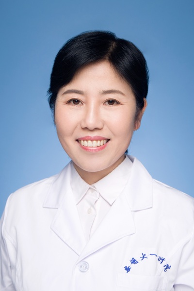 Yunxia Cao (曹云霞)合作论文数The First Affiliated Hospital of Anhui Medical University7Yong Li合作论文数6Xiaojin He合作论文数6Lilan Su合作论文数6Fei Gong合作论文数6Aixiang Luo合作论文数6Haiyu Li合作论文数6Huan Wu (吴欢)合作论文数6
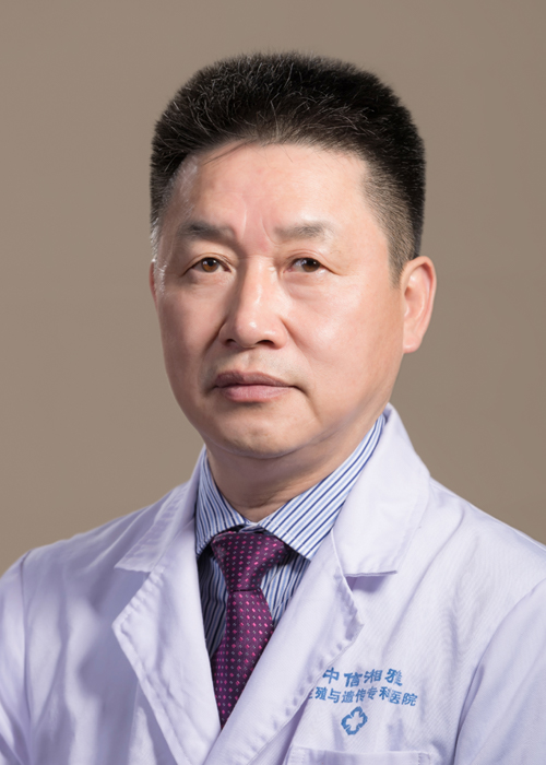 Liqing Fan (范立青)合作论文数School of Basic Medical Sciences, Central South University6
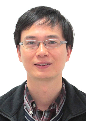 Wenming Xu (许文明)合作论文数West China Second University Hospital, Sichuan University6C Luo合作论文数5
 Shixiong Tian合作论文数Shanghai Key Laboratory of Female Reproductive Endocrine Related Diseases, Shanghai, China.5
 Mingrong Lv合作论文数5Peng Qi合作论文数5Jiaxiong Wang合作论文数5
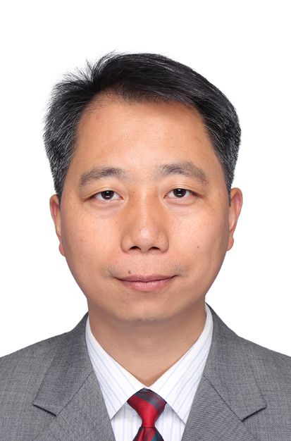 Wei Xiong (熊炜)合作论文数Cancer Research Institute, School of Basic Medical Sciences, Central South University5Lixia Yu (余丽霞)合作论文数5
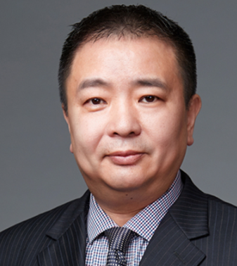 Li Jin (金力)合作论文数Department of Anthropology and Human Genetics, School of Life Sciences, Fudan University;Shanghai College of Medicine, Fudan University5
 Zhaoyang Zeng (曾朝阳)合作论文数School of Basic Medical Sciences, Central South University5
 Xiaoling Li (李小玲)合作论文数Key Laboratory of Cancer Carcinogenesis and Invasion of Chinese Ministry of Education, Cancer Research Institute, Central South University5Chunbo Xie合作论文数4Jiaxin He合作论文数4Gao, Yang合作论文数4Shuyan Tang合作论文数Human Phenome Institute, Fudan University4Rui Zheng合作论文数4Xiaoxuan Yang合作论文数4DeHua Cheng (程德华)合作论文数4
 Minghua Wu (武明花)合作论文数Xiangya School of Medicine, Central South University4Pierre F. Ray合作论文数3




