非瓣膜性心房颤动(房颤)所致的脑栓塞是缺血性卒中最为常见的类型之一,且短期内栓塞复发风险较高,病情容易进展,尤其是抗凝药物应用不当时。复发性栓塞多以脑栓塞为主,肠系膜上动脉栓塞(superior mesenteric artery embolism, SMAE)少见。SMAE是严重的急腹症,病情凶险,当其进展为严重的缺血坏死性肠病并发罕见的肝门静脉积气(hepatic portal vein gas, HPVG)时往往预后极差,病死率很高。以往SMAE的影像诊断主要依靠腹部增强CT显示肠系膜上动脉充盈缺损来诊断,但对于碘对比剂禁忌者,非对比增强CT上出现的肠系膜上动脉内的“动脉致密征”也是一个有重要价值的诊断征象,以往罕见报道。现本文结合文献,报道1例经非对比剂增强腹部CT确诊的非瓣膜性房颤性卒中患者短期内并发急性SMAE并继发HPVG的罕见致死性案例。.
患者男性,15岁,2019年4月劳累后出现腰痛,活动后加重,休息可缓解,行走、起床困难,无昼夜规律,2020年4月上述症状加重,查血常规:白细胞、血小板正常,HGB 119g/L;CRP 7.17mg/dl(0~0.8),ESR 54mm/h (0~15).随后四肢及头部出现散在鲜红色皮疹.2020年7月无明显诱因出现右侧胸痛及背痛,伴间断发热,最高体温38.6℃.2020年10月因肺部感染就诊于中日友好医院呼吸与危重症医学科.入院后查外周血,结核感染的T淋巴细胞斑点试验T-SPOT(-),抗核抗体谱+类风湿关节炎抗体谱+血管炎抗体谱均为(-),HLA-B27(-).
目的 探讨基底平行解剖扫描磁共振成像(BPAS-MRI)在颅内椎-基底动脉病变诊断中的应用价值.方法 回顾性连续纳入2017年8月至2019年3月于中日友好医院神经科就诊主诉为"头晕或眩晕"并常规行头部三维时间飞跃法MR血管成像(3D TOF-MRA)及BPAS-MRI检查的80例患者,对双侧颅内段椎动脉、基底动脉在3D-TOF-MRA的基础上进行BPAS-MRI检查.对3D TOF-MRA血管显影正常的双侧椎动脉及基底动脉管径进行3D TOF-MRA与BPAS-MRI检查结果的比较,以观察BPAS-MRI检查对正常管径轮廓结构的显示情况.对3D TOF-MRA血管显影不正常的双侧椎动脉及基底动脉进行BPAS-MRI与3D TOF-MRA显影结果的对比分析,以探讨BPAS-MRI在颅内后循环血管病变中的诊断及鉴别作用.结果 (1)80例患者共240支椎-基底动脉中,123支(51.2%)3D TOF-MRA显影正常,其左、右侧椎动脉及基底动脉的管径平均值3D TOF-MRA与BPAS-MRI比较,差异均无统计学意义(均P>0.05).(2)117支(48.8%)血管在3D TOF-MRA上显影异常,其中39支(16.2%)血管仅通过MRA易漏诊或误诊,包括椎动脉发育不全15支(6.2%),椎动脉闭塞9支(3.8%),椎-基底动脉重度狭窄7支(2.9%),椎动脉发育不良5支(2.1%),椎-基底动脉扩张延长2支(0.8%)及椎动脉夹层1支(0.4%).结论 BPAS-MRI能够清晰显示颅内后循环系统血管外部轮廓结构,联合3D TOF-MRA提高了颅内椎-基底动脉病变诊断的准确性.
Objective To analyze the CT features of cavitation between primary lung adenocarcinoma and squamous cell cancer.Methods The CT findings of cavity of primary lung adenocarcinoma and squamous cell carcinoma were evaluated in 57 patients,including 33 of squamous cell carcinoma and 24 of adenocarcinoma.The clinical data and CT features were analyzed retrospectively using the independent samples t-test,Pearson Chi-square test or Fisher's exact test.Results The mean age of ptients with squamous cell carcinoma was higher than that of patients with adenocarcinoma (65.57-4-9.26 vs 58.75 ± 11.12,P =0.015).Statistical differences were found in distribution of gender and smoking habit between the two kinds of carcinomas (P =0.014 and P =0.029).The T stages were also different between the two carcinomas (P=0.003).In addition,the maximum diameter of tumor (P =0.003),the maximum diameter of cavity (P =0.029) and the maximum thickness of the cavity wall (P=0.001) of squamous cell carcinoma were higher than those of adenocarcinoma.Moreover,the presence of ground-glass opacity (P =0.010),vessel passing through the cavity (P =0.001),septum inside the cavity (P<0.001) and tumoral bronchogram (P =0.027) in adenocarcinoma were higher than those in squamous cell carcinoma.Conclusion There are significant differences between adenocarcinoma and squamous cell carcinoma in the population distribution and image features,comprehensive analysis helps the differential diagnosis.
Objective:To evaluate multi-slice CT(MSCT)features of peripheral lung adenocarcinomas with cavities.Methods:Twenty four cases of pathologically confirmed lung adenocarcinomas containing cavities were retrospectively analyzed with regard to clinical data and MSCT features between Jan.2012 and April 2016.According to the thickness of the cavity walls (<4mm or ≥4mm),the lesions were divided into thin-walled or thick-walled cavity groups.Fisher exact test and ANOVA test were used for the statistical analysis.Results:There were 24 cases (10 with thin walled cavities,14 with thick-walled cavities).These features accounted for more than 50% of all MSCT signs in 24 cases,including irregular shapes in 22 cases (91.6%),lobulations in 18 cases(75%),irregular inner walls in 13 cases(54.2%).The frequency of septum in airspace,the blood vessel passing through the airspace and ground-glass opacity around the cavity in thin-walled cavities were higher than thick-walled cavities (P<0.05).The frequency of lobulations and pleura1 in lesions with thick-walled cavities were higher than those in thin-walled cavity adenocarcinomas (P<0.05).Conclusion:Radiological characteristics of the thin-walled cavities were different from the thick-walled cavity adenocarcinomas.
Objective:To compare MR imaging with ultrasonography performed at the same day during different phases of menstrual cycle in the evaluation of dimensions of ovaries and number of follicles,and thus to investigate morphological changes of the normal ovaries and follicles in healthy child-bearing age women during the menstrual cycle.Methods:MR examinations of the pelvis were performed in 26 healthy women between 20 to 40 years old (mean,29 yrs).Each volunteer underwent totally four MR and ultrasound scans in menstrual phase (MP),follicular phase (FP),ovulatory phase (OP) and luteal phase (LP).Axial,coronal and sagittal T2 weighted images and axial T1 weighted images were obtained on a 3.0T MR scanner.Ovarian and follicular measurements were acquired on T2 WI.T1WI were used for diagnosis of ovarian anomaly.One-way analysis of variance,paired student t test and liner mixed models were preformed using SPSS statistics 19.0.Results:26 volunteers were included in our study (mean age:29.1 years,average BMI:20.3kg/m2 and average height:1.61m).Mean value of ovary dimensions,number of follicles and dimension of the biggest follicles were compared between MR imaging and ultrasonography.Both left and right ovarian volume and number of follicles acquired on MR were larger than that on ultrasound (P =0.000 ~ 0.003).Cyclic changes were found on left,right and overall maximum follicle diameter with the biggest diameter in LP.The maximum follicle diameter acquired by MR during menstrual cycle was as follows:MP (0.90 ± 0.28)cm,FP (1.24 ± 0.31)cm,OP (1.71 ± 0.54)cm,and LP (1.20 ± 0.66)cm;however,the maximum follicle diameter obtained by ultrasound was MP (1.01 ± 0.40)cm,FP (1.26 ± 0.46)cm,OP (1.47 ± 0.47)cm,and LP (0.89 ± 0.37)cm.The ovarian volume change was not obvious.Conclusion:There was statistical significant difference in the measurement of ovary and follicular structure between using MR and ultrasound,with bigger values obtained by MR.The changes of maximum follicle diameter during menstrual cycle were observed on both MR and ultrasound.
 Jianghui Duan合作论文数2HaiXu Zhu (朱海旭)合作论文数2
 Hongliang Sun (孙宏亮)合作论文数China-Japan Friendship Hospital2
 Sheng Xie (谢晟)合作论文数China-Japan Friendship Hospital2Yanyan Xu (徐妍妍)合作论文数China-Japan Friendship Hospital2Li-fang HAO (郝丽芳)合作论文数1ZHANG Zhiyong (张志勇)合作论文数1Yi-ru NIU (牛一茹)合作论文数1tianzhi yang (杨天智)合作论文数1YuRun Yang (杨雨润)合作论文数1YaNan Qiao (乔亚男)合作论文数1He Liu (刘赫)合作论文数1ZhiYong Zhang (张志勇)合作论文数1Ning Ding (丁宁)合作论文数1
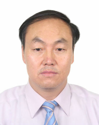 Guolin Ma (马国林)合作论文数China-Japan Friendship Hospital;Peking University Health Science Center;Peking Union Medical College;Capital Medical University1
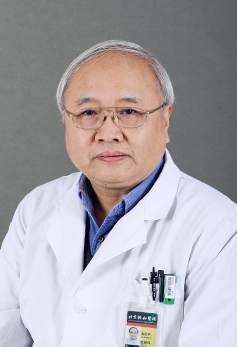 Zhengyu Jin (金征宇)合作论文数Peking Union Medical College;Peking Union Medical College Hospital1
 Huadan Xue (薛华丹)合作论文数Peking Union Medical College Hospital1
 Wu Wang (王武)合作论文数Shanghai Sixth People's Hospital Affiliated to Shanghai Jiao Tong University School of Medicine1
 Dantao Peng (彭丹涛)合作论文数China-Japan Friendship Hospital;Graduate School, Capital Medical University1
 Jinsong Jiao (焦劲松)合作论文数China-Japan Friendship Hospital1
 Yonglan He (何泳蓝)合作论文数Chinese Academy of Medical Sciences Peking Union Medical College1




