Background Zinc finger and bric-a-brac/tramtrack/broad (ZBTB) domain-containing proteins have been reported to be associated with many tumors’ development. However, in tumor initiation and progression, the role of ZBTB9, one of the protein family, and its prognostic value were yet to be elucidated in Liver Hepatocellular Carcinoma (LIHC). Methods We used R software and online bioinformatics analysis tools such as GEPIA2, cBioPortal, TIMER2, Metascape, UALCAN, STRING, TISIDB, and COSMIC to investigate ZBTB9’s characteristics and function in LIHC, including abnormal expression, carcinogenic role, related signaling pathways and prognostic value. Furthermore, cell experiments (such as formation, wound healing, and transwell assays) and analyses based on clinical samples (such as immunohistochemistry (IHC) and promoter methylation analysis) were conducted to verify pivotal conclusions. Results ZBTB9 was overexpressed in LIHC samples compared to adjacent normal tissues. Through the analysis of genomic alteration and promoter hypomethylation, the clinical value and etiology of abnormal expression of ZBTB9 were preliminarily exlpored. Subsequent evidence showed that it could result in tumor progression and poor prognosis via activating cell cycle, DNA repair, MYC, and KRAS-associated signaling pathways as well as rendering immune dysregulation. After the knockdown of ZBTB9, evidently inhibited capacities of tumor cells proliferation and migration were observed. These results together indicated that ZBTB9 could be a promising prognostic biomarker and had the potential value to offer novel therapeutic targets for LIHC treatment. Conclusions ZBTB9 was identified as a novel biomarker to predict the prognosis and tumor progression in LIHC, and a promising therapeutic target to invert tumor development.
近年来,随着放射治疗技术的发展,手术联合放射治疗病理性瘢痕取得了显著疗效.本文从放射治疗在病理性瘢痕应用原理、常用放射源的比较和放疗方案的设计,以及放射治疗新技术应用及瘢痕放射治疗的不良反应防范措施等方面,对病理性瘢痕的放射治疗技术和临床应用进展进行综述.
放射治疗可有效利用放射线对瘢痕局部进行照射,是治疗瘢痕的方法之一.近年来,应用手术加放射治疗瘢痕的综合疗法取得了一定的突破和进展.本文从病理性瘢痕的放射治疗原理,常用放射源和方法的选择,电子线及X射线放射治疗技术的应用,以及瘢痕放射治疗的不良反应及防范措施等方面,对病理性瘢痕的放射治疗技术和临床研究进展进行综述.
AIM To assess the value of combined acoustic radiation force impulse (ARFI) imaging, serological indexes and contrast-enhanced ultrasound (CEUS) in distinguishing between benign and malignant liver lesions. METHODS Patients with liver lesions treated at our hospital were included in this study. The lesions were divided into either a malignant tumor group or a benign tumor group according to pathological or radiological findings. ARFI quantitative detection, serological testing and CEUS quantitative detection were performed and compared. A comparative analysis of the measured indexes was performed between these groups. Receiver operating characteristic (ROC) curves were constructed to compare the diagnostic accuracy of ARFI imaging, serological indexes and CEUS, alone or in different combinations, in identifying benign and malignant liver lesions. RESULTS A total of 112 liver lesions in 43 patients were included, of which 78 were malignant and 34 were benign. Shear wave velocity (SWV) value, serum alpha-fetoprotein (AFP) content and enhancement rate were significantly higher in the malignant tumor group than in the benign tumor group (2.39 ± 1.20 m/s vs 1.50 ± 0.49 m/s, 18.02 ± 5.01 ng/mL vs 15.96 ± 4.33 ng/mL, 2.14 ± 0.21 dB/s vs 2.01 ± 0.31 dB/s; P < 0.05). The ROC curve analysis revealed that the areas under the curves (AUCs) of SWV value alone, AFP content alone, enhancement rate alone, SWV value + AFP content, SWV value + enhancement rate, AFP content + enhancement rate and SWV value + AFP content + enhancement rate were 85.1%, 72.1%, 74.5%, 88.3%, 90.4%, 82.0% and 92.3%, respectively. The AUC of SWV value + AFP content + enhancement rate was higher than those of SWV value + AFP content and SWV value + enhancement rate, and significantly higher than those of any single parameter or the combination of any two of parameters. CONCLUSION The combination of SWV, AFP and enhancement rate had better diagnostic performance in distinguishing between benign and malignant liver lesions than the use of any single parameter or the combination of any two of parameters. It is expected that this would provide a tool for the differential diagnosis of benign and malignant liver lesions.
早期诊断是降低肝癌患者病死率的重要方法[1].目前临床上针对肝肿瘤常用的早期检测方法有常规超声、声学造影(CEUS)、血清学检测等.血清学检测是肝脏恶性肿瘤的重要参考指标[2].超声技术简便、无创,CEUS能动态观察肝脏的血流动力学情况,这2种技术应用比较广泛但主要靠检查者肉眼判断,定性诊断肝脏有无占位性病变,受仪器设备和检查者的主观因素影响较大[3].
Objective To establish a novel Monte Carlo simulation method for reconstruction of medical accelerator model and X?ray energy spectrum based on IAEA Varian 6 MV X?ray phase?space file and photon energy spectrum of the target accelerator. Methods The verified 6 MV X?ray phase?space files were preprocessed to elevate the energy of each particle. Particles were saved in different Phase?Space?Let ( PSL) files according to their position and energy, yielding an initial photon energy spectrum for Monte Carlo simulation of accelerator under an initial target energy. The initial photon energy spectrum was fit to a photon energy spectrum of an accelerator ( Elekta Precise 10 MV X?ray accelerator) under an unknown target energy to yield a fitting coefficient, which was the weight of each PSL. Finally, an accelerator model under an unknown target energy was reconstructed using the initial PSL files and the weight information. The percentage depth dose ( PDD) distribution was calculated in different square open fields. The effectiveness of this method was verified using one dimensional gamma passing rate. Results The peak position and overall distribution of the reconstructed 10 MV photon energy spectrum were in accordance with those of the verified 10 MV photon energy spectrum. The PDD calculated from the reconstructed 10 MV accelerator model agreed well with the measured PDD. The one?dimensional gamma passing rate was above 96%( 1%/1 mm, threshold=0%) . Conclusion The Monte Carlo reconstruction method proposed in this study is reliable, accurate, and effective.
 Jiehua WANG (汪杰华)合作论文数2Peihua Gu (顾培华)合作论文数2Xiaolan SUN (孙晓岚)合作论文数2LiWei Liang (梁立维)合作论文数2KeZhu Hou (侯克柱)合作论文数2Yun Wang (王芸)合作论文数2Qiong Men (门琼)合作论文数2Zhenshan Zhang合作论文数1Jiayan Chen合作论文数1Yonggang OU (区永刚)合作论文数1Juan Li合作论文数1Zhen CHEN (陈真)合作论文数1Zhen Chen合作论文数1Chao YIN (尹超)合作论文数1Lei-Lei Wu合作论文数1
 Xiaoli Wu (武晓莉)合作论文数Shanghai Ninth People’s Hospital, Shanghai Jiao Tong University School of Medicine1
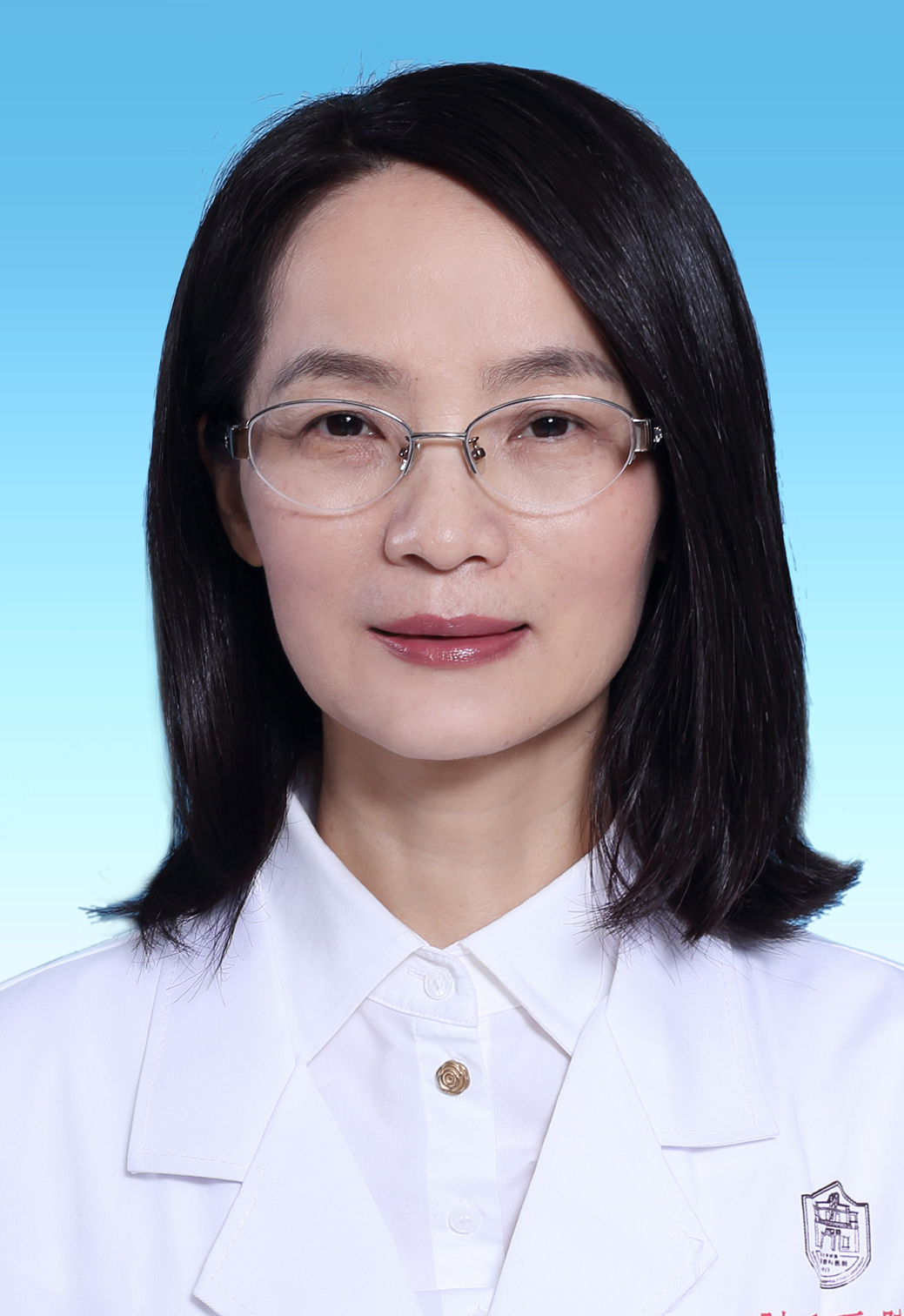 Yaping Xu (许亚萍)合作论文数School of Medicine, Tongji University1
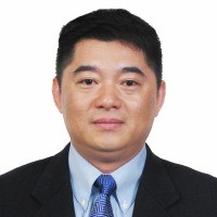 Yixin Zhang (章一新)合作论文数Department of Plastic and Reconstructive Surgery, Shanghai Ninth People's Hospital, School of Medicine, Shanghai Jiao Tong University1
 Yu Qi (齐宇)合作论文数1Liang Liu合作论文数1




