To investigate the characteristics of pharyngeal and laryngeal structure in patients with acquired brain injury (ABI), who were long time wearing nasogastric tube (NGT) with or without tracheostomy. 103 ABI patients with NGT indwelled for more than 1 month were retrospectively studied and divided into two groups by whether or not undergoing tracheostomy. Age, gender, types of brain injury, course of the disease, disorders of consciousness, activities of daily living (ADL) and fiberoptic endoscopic examination of swallowing (FEES) were evaluated. The structure and function of pharyngeal and laryngeal were assessed by FEES, focusing on the morphology of the arytenoid cartilage, epiglottis, vocal folds, tongue base, and pharyngeal cavity. Prolonged indwelling nasogastric tubes and tracheostomy tubes might lead to abnormal alterations of the structure and function in the arytenoid cartilage, epiglottis, tongue base, and pharyngeal cavity. Epiglottis shape abnormality, glossoptosis and pharyngeal stenosis were present in a larger proportion of the NGT–TRACH (nasogastric tube with tracheostomy) group than the NGT group (p < 0.05). This study highlights potential physiological changes associated with prolonged placement of nasogastric tubes and tracheostomy tubes, which could impede the recovery of swallowing function and decannulation. We hope to provide valuable evidence to develop effective management strategies for ABI patients with NGT or tracheostomy.
BACKGROUND:The Theta-burst Transcranial Magnetic Stimulation (TBS) is an emerging modality of Repetitive Transcranial Magnetic Stimulation (rTMS). However, the efficacy of TBS on post-stroke recovery-stage patients with dysphagia remains unclear.AIM:To investigate the effect of intermittent theta burst stimulation (iTBS) and continuous theta burst stimulation (cTBS) in post-stroke dysphagia patients within the recovery stage.DESIGN:Randomized controlled double blinded trial.SETTING:Inpatient.POPULATION:Ninety patients with dysphagia after stroke within 1 to 6 months.METHODS:Patients were divided into the supratentorial group and the brainstem group, and both of groups were further divided into three subgroups, including the sham subgroups, the iTBS subgroups, and the cTBS subgroups. Each of subgroups received 30 min of traditional swallowing rehabilitation treatment every day for 4 weeks. In addition, the iTBS subgroups received iTBS over the cortex of the suprahyoid muscles on the affected hemisphere, the cTBS subgroups received cTBS on the unaffected hemisphere, and the sham subgroups received sham stimulation on unilateral hemisphere. Standardized swallowing assessment (SSA), Oral Motor Function Scale (OMFS) and fiberoptic endoscopic examination of swallowing (FEES) were assessed before and after treatments.RESULTS:In the supratentorial group, compared with the sham and cTBS subgroups, the iTBS subgroups showed significant improvement in SSA, OMFS, vocal folds movement, laryngeal sensation, and Rosenbek Penetration-Aspiration Scale (PAS) (P<0.05). In the brainstem group, compared with the sham subgroup, the iTBS subgroup significantly improved SSA, OMFS, Yale Pharyngeal Residue Severity Rating Scale (YPR-SRS) and PAS (P<0.05), and the cTBS subgroup significantly improved SSA, YPR-SRS and PAS (P<0.05).CONCLUSIONS:This study demonstrated that iTBS might be an effective stimulation pattern to improve the overall swallowing function whether in supratentorial stroke patients or brainstem stroke patients.CLINICAL REHABILITATION IMPACT:iTBS seems to be a promising approach for rehabilitation of overall swallowing function in post-stroke patients.
目的:采用功能性近红外光谱技术(fNIRS)研究吞咽相关任务时大脑皮质激活及偏侧化情况.方法:纳入22名健康成年被试,在fNIRS采集过程中执行咀嚼及舌尖滑动任务,对任务激活脑区进行分析,并计算不同脑区的偏侧化指数.结果:在执行咀嚼任务时,左侧初级运动皮层(primary motor cortex,PMC)、左侧初级躯体感觉皮层(primary so-matosensory cortex,PSC)、左侧运动前区/运动辅助区(pre-motor and supplementary motor cortex,pSMC)、左侧缘上回(supramarginal gyrus,SMG),右侧背外侧前额叶(dorsolateral prefrontal cortex,DLPFC)、右侧额叶眼动区(frontal eye fields,FEF)、右侧颞上回(superior temporal gyrus,STG)及颞中回(middle temporal gyrus,MTG)、右侧额极区(frontopolar area,FPA)及右侧额下回三角区(pars triangularis,PTG)脑区明显激活.舌尖滑动任务则表现为双侧PMC、双侧PSC、双侧pSMC及左侧SMG脑区明显激活.同时在全脑及PMC、pSMC、PSC等脑区的激活呈现偏侧化模式.咀嚼任务主要表现为局部脑区的左侧偏侧化激活,舌尖滑动任务表现为局部脑区的右侧偏侧化激活.全脑左右半球间的偏侧化变异性较大,且与利手无关.结论:fNIRS可应用于吞咽相关的脑功能研究.PMC、pSMC、PSC、SMG、STG、MTG、PTG等脑区与吞咽特定过程的神经调控有关,并表现为偏侧化激活模式,并且随着吞咽活动的进行,相关脑区的激活可能存在从左侧逐渐向右侧偏侧化的转变.
目的:基于软管喉镜吞咽功能评估(flexible endoscopic evaluation of swallowing,FEES),比较经鼻留置胃管管饲(nasal indwelling gastric tube feeding,NGT)与间歇性经口至食管管饲法(Intermittent oro-esophageal tube feed-ing,IOE)在减少鼻胃管综合征发生率、改善咽期吞咽功能和气道保护功能方面是否存在疗效差异.方法:选取脑卒中后吞咽障碍患者54例,根据营养摄入方式分为NGT组28例,IOE组26例.其中NGT组中有14例患者由NGT转变为IOE,将这些患者归为NGT-IOE亚组,另外14例继续沿用NGT,为NGT-NGT亚组.采用FEES评定各组患者的咽喉部生理结构情况,包括会厌形状、杓状软骨黏膜水肿程度、声带运动能力;咽喉部分泌物情况,包括分泌物严重程度分级(Murry secretion severity scale,MSS);咽期吞咽功能-喉部感觉功能、吞咽反射能力、食物残留等级(Yale pharyngeal residue severity rating scale,YPR-SRS)、Rosenbek渗漏/误吸等级(penetration-aspi-ration scale,PAS),并使用吞咽功能性交流测试评分(functional communication measure swallowing,FCM)评定患者整体的吞咽能力.结果:NGT组与IOE组患者的各项评分比较,NGT组患者的MSS、PAS、FCM评分显著低于IOE组(P<0.05),且会厌形状异常和杓状软骨黏膜水肿程度显著高于IOE组(P<0.05).NGT-IOE亚组患者拔管后MSS、YPR-SRS(会厌谷)、YPR-SRS(梨状窝)、PAS、FCM评分较拔管前显著提高(P<0.05),提示存在鼻胃管综合征的会厌形状异常情况、杓状软骨黏膜水肿和声带外展受限均较拔管前显著降低(P<0.05),另外,喉部感觉功能也较拔管前显著改善(P<0.05).NGT-NGT亚组两周前、后的各项评分比较均无显著性意义(P>0.05).结论:与NGT相比,IOE可以减少脑卒中后吞咽障碍患者鼻胃管综合征的发生率,并改善咽期吞咽和气道保护功能,故对于长期留置鼻饲胃管的患者,有必要尽早考虑转变为IOE的可能性.
Objectives The aims of the study were to investigate the effect of tongue-pressure resistance training in poststroke dysphagia patients with oral motor dysfunction and to examine the therapeutic value of tongue-pressure resistance training in the oral and pharyngeal phases. Design Patients were divided into an experimental and a control group. Both groups received 30 mins of traditional swallowing rehabilitation treatment every day for 4 wks. In addition, the experimental group received tongue-pressure resistance training for an extra 20 mins/d. Maximum tongue pressure and fiberoptic endoscopic examination of swallowing were assessed before and after treatments. Results Compared with the control group, the experimental group showed significant improvement in Functional Communication Measure for swallowing, Oral Motor Function Scale, maximum tongue pressure, Murray Secretion Scale, Rosenbek Penetration–Aspiration Scale, and food residue in pyriform sinuses ( P < 0.05). There was no significant difference in food residue in epiglottic vallecula between both groups ( P > 0.05). Conclusions This study demonstrated that tongue-pressure resistance training is an effective approach to improve the overall swallowing function in patients with oral motor dysfunction. The improvement of oral motor function could facilitate the recovery of pharyngeal motor function. Tongue-pressure resistance training seems to have more clearance of residue in piriform sinus than epiglottic vallecula.
Dysphagia is one of the most common functional disorders after stroke, with an incidence of up to 30%-50%, resulting in malnutrition and aspiration pneumonia, which could be life-threatening. The pathogenic mechanism of dysphagia after stroke involves multiple brain functional areas and neural networks. It is generally believed that dysphagia after stroke is related to the lesions of cerebral cortex, subcortical structures and brainstem swallowing center. There are significant differences in the mechanism and clinical manifestations of dysphagia caused by different lesion sites. Understanding the mechanism is conducive to the development of more targeted and individualized rehabilitation therapy in clinical practice, which has an important impact on the prognosis of patients. Except for the traditional swallowing treatment programs commonly used, such as swallowing motion-sensory training, peripheral neuromuscular electrical stimulation, acupuncture, postural training, and dietary adjustment, people have become more and more interested in central nervous regulation in the treatment of swallowing dysfunction. Transcranial Magnetic Stimulation(TMS), as a non-invasive neuroregulation technique, achieves the purpose of neuroregulation by regulating the excitability of the cerebral cortex. With the development of research, different pulse stimulation patterns have been developed and experimentally applied in the treatment of post-stroke dysphagia. Repetitive transcranial magnetic stimulation(rTMS)is a repeated, continuous and constant intensity stimulation of selected brain regions. Theta burst stimulation(TBS)is based on rTMS with the addition of cluster rhythmic stimulation, which has lower stimulus intensity and shorter stimulus duration. Paired associative stimulation(PAS)combines central TMS with peripheral electrical stimulation at certain intervals to activate brain networks in pairs. Each mode of treatment has its own characteristics. This article reviews the application of different stimulation modes in healthy people and patients with dysphagia after stroke, analyzes the current research progress and limitations in this field, and provides a new idea for the clinical research and treatment of deglutition disorder after stroke from three points.
认知障碍是指学习、记忆、推理和判断等大脑高级功能出现异常,进而引起一系列症状,影响日常和社会生活能力。功能性近红外光谱技术(fNIRS)作为一种新兴的脑功能检测技术,因其价格低廉、便携性好,且具有实时连续动态捕捉的功能,在认知障碍的研究领域内展现出巨大的潜力。
Silver nanoparticle (nAg), which is one of the most common manufactured nanomaterials, has a wide range of biomedical applications. The human oncogenic γ-herpesviruses, Kaposi’s sarcoma-associated herpesvirus (KSHV) and Epstein–Barr Virus (EBV), are etiologically linked to many malignancies. Currently, there are no efficient or specific treatments for these types of tumors, and most patients die because of resistance to conventional cytotoxic chemotherapy. Despite nAg having antitumor and antiviral activities, its effects on oncogenic herpesvirus-related cancer cells remain largely unknown. Here, we reveal that nAg presents higher cytotoxicity against KSHV- or EBV-latently infected cells via reactivating viral lytic replication, which relies on the induction of reactive oxygen species (ROS) generation and autophagy. Moreover, nAg blocks KSHV primary infection by directly destroying virion particles, as well as effectively inhibits colony formation and moderately represses the growth of KSHV-associated tumors in xenograft mouse model. Taken together, these results demonstrate the therapeutic potential of nAg for use in the antiviral infection and treatment of oncogenic herpesvirus-related cancers.
 tingwei wang (王婷玮)合作论文数6JunFa Wu (吴军发)合作论文数6
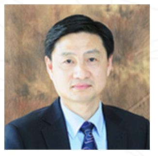 Yi Wu (吴毅)合作论文数Department of Rehabilitation Medicine, Huashan Hospital, Fudan University, Shanghai, China5RuiPing Hu (胡瑞萍)合作论文数4Shunjuan Fan合作论文数3yulian zhu (朱玉莲)合作论文数3Haozheng LI (李浩正)合作论文数2
 Caixia Zhu (朱彩霞)合作论文数School of Basic Medical Sciences, Fudan University1Yi Guo合作论文数1Feng Gu合作论文数1Shan Tian (田闪)合作论文数1
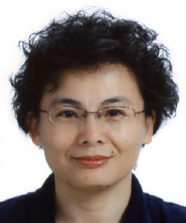 Yiwen Shen (沈忆文)合作论文数School of Forensic Medicine and Science, Fudan University;Department of Forensic Medicine, School of Basic Medical Sciences, Fudan University1Li Shen (沈莉)合作论文数1
 Qiliang Cai (蔡启良)合作论文数School of Basic Medical Sciences, Fudan University;Shanghai Institute of Infectious Disease and Biosecurity, Fudan University1Ding Ling合作论文数MOE&Shanghai Medical College, Fudan University;Shanghai Medical College, Fudan University1Zhu Qing合作论文数MOE&Shanghai Medical College, Fudan University1
 Jin Gan合作论文数MOE&Shanghai Medical College, Fudan University;Shanghai Medical College, Fudan University1
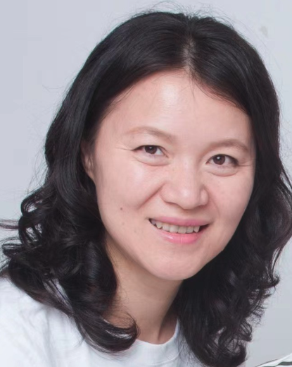 Fang Wei (魏芳)合作论文数School of Life Science and Technology, Shanghai Jiaotong University1Jiayi Liu (刘佳怡)合作论文数1
 Yuyan Wang (王玉艳)合作论文数Beijing Cancer Hospital1
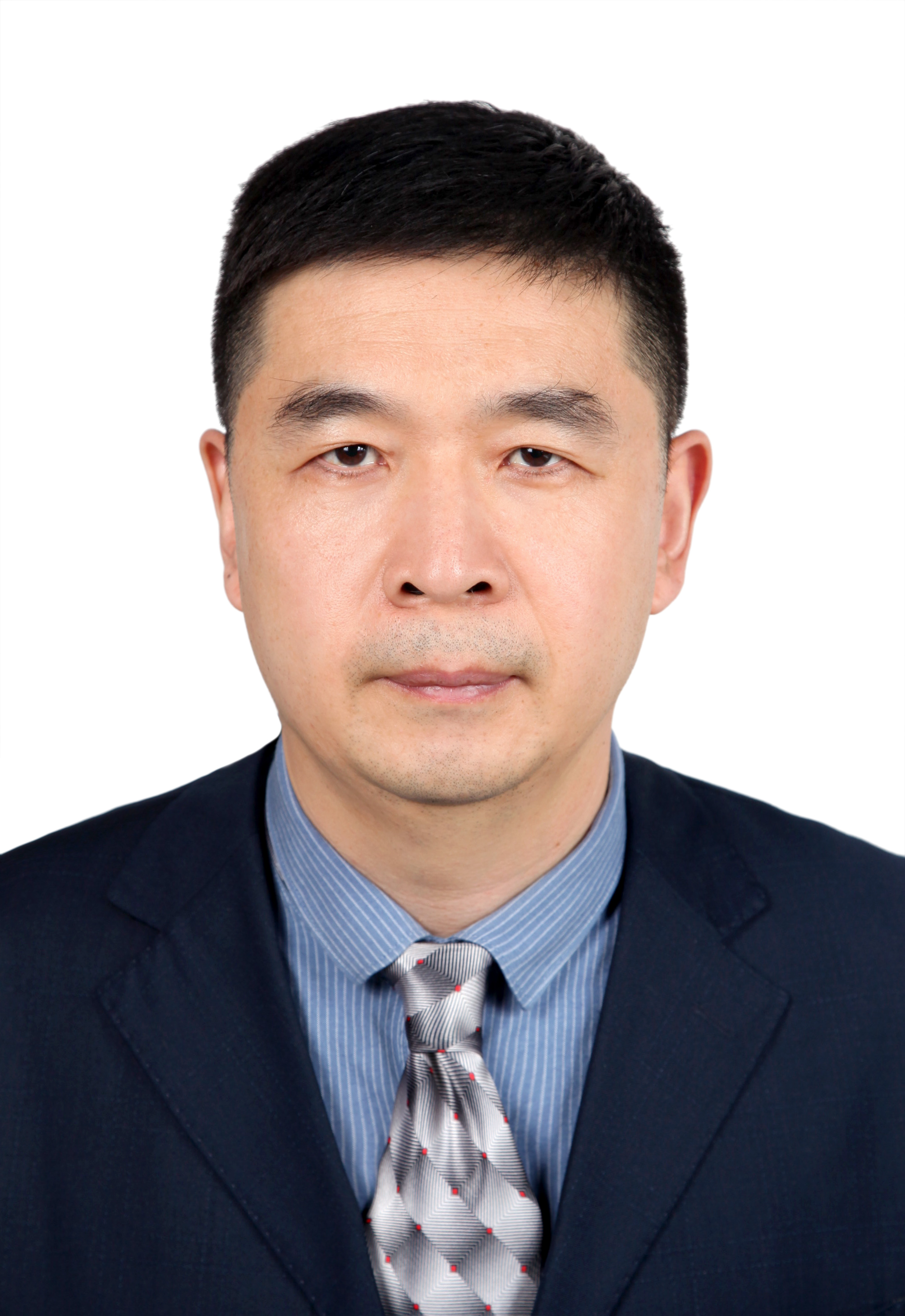 Sijin Liu (刘思金)合作论文数Research Center for Eco-Environmental Sciences, Chinese Academy of Sciences;University of Chinese Academy of Sciences1
 Jie Zhang合作论文数Department of Cell and Molecular Pharmacology and Experimental Therapeutics, College of Medicine, Medical University of South Carolina1




