目的:探讨经额底纵裂入路切除经鼻蝶术后复发或残留颅咽管瘤的技术要点并分析治疗效果。方法:回顾性分析2015年4月至2020年6月首都医科大学三博脑科医院诊治的10例首次经鼻蝶手术后复发或残留的颅咽管瘤患者再次手术治疗后的临床资料。男6例,女4例,年龄13 ~ 44岁。其中5例患者首次手术采用经鼻显微镜手术,5例患者采用经鼻内镜手术。10例患者再次手术中全部采用经额底纵裂入路切除肿瘤,其中6例患者在手术中用磨钻磨除鞍结节或蝶骨平台骨质。结果:肿瘤全切除者6例,近全切除3例,大部分切除者1例。术后视力改善者3例,无显著变化者6例,视力下降者1例。围手术期无死亡病例。术后脑脊液鼻漏2例,分别行经鼻内镜手术修补脑脊液鼻漏,得以治愈。颅内感染者1例,该例患者并发脑积水,在颅内感染治愈后行脑室-腹腔分流术。随访时间22 ~ 83个月,中位随访时间49个月。随访期内无死亡病例,2例患者复发。9例患者有垂体前叶功能低下,7例有尿崩症。按Karnofsky生活质量量表评分,90分7例,80分3例。结论:经额底纵裂入路手术切除经鼻蝶手术后复发或残留的颅咽管瘤可以取得良好效果。
Objective:To evaluate the clinical effect of anterior clinoid process grinding in the treatment of ophthalmic / superior clinoid process aneurysms and sellar tumors.Methods:The clinical data of 16 patients who underwent anterior clinoid process grinding in Sanbo Brain Hospital, Capital Medical University from January 2015 to July 2021 were analyzed retrospectively. There were 1 patient with recurrent craniopharyngioma, 1 patient with recurrent pituitary adenoma, 13 patients with aneurysms, and 1 patient with suprasellar granulosa cell tumor combined with ophthalmic aneurysm of right internal carotid artery. The Modified Rankin Scale (mRS) score was used to evaluate the situation at discharge and in the medium-and-long term.Results:Sixteen patients underwent anterior clinoidprocess grinding. At discharge and the latest follow-up, the mRS scores of the patients were 0-2. A total of 15 aneurysms were treated, and there were no symptoms of visual loss or visual field defect after operation. No cerebrospinal fluid leakage occurred in all patients.Conclusions:The grinding of anterior clinoid process can effectively and fully stretch the optic nerve and internal carotid artery, and can observe the tumor neck at the lower end of pituitary stalk and the ocular segment/superior clinoid process of internal carotid artery under direct vision. It is one of the important auxiliary methods for the treatment of sellar lesions.
目的 评价颌内动脉搭桥治疗颅内复杂动脉瘤的临床中远期治疗效果.方法 回顾性分析91例颅内复杂动脉瘤行颌内动脉搭桥病人的临床资料,使用mRS评分评价病人中远期预后.结果 出院时mRS评分0~2分67例(73.6%),3~5分23例(25.3%),死亡1例.83例随访31~136个月,平均(82.0±25.0)个月,mRS评分0~2分67例(80.7%),3~5分6例(7.2%);死亡10例(12.0%),其中死于肺部感染5例,大面积脑梗死2例,颅内出血1例,胰腺癌1例,对侧动脉瘤破裂1例.结论 颌内动脉搭桥是治疗颅内复杂动脉瘤的有效方式之一,其术式选择可忽略动脉瘤的个体化差异.
目的 评估症状性颈内动脉或大脑中动脉慢性闭塞病人经颌内动脉搭桥治疗后的血流动力学改变和长期临床效果.方法 回顾性研究25例症状性颈内动脉或大脑中动脉慢性闭塞病人的临床资料,均行颌内动脉-桡动脉-大脑中动脉搭桥治疗.采用超声测量术中血流量,MRI灌注(perfusion weighted imaging,PWI)或CT灌注(CT perfusion,CTP)评估局部脑血流量(regional cerebral blood flow,rCBF)、局部脑血容量(regional cerebral blood volume,rCBV)、平均通过时间(mean transit time,MTT)和达峰时间(time to peak,TTP).结果 术中搭桥血管血流量为(81.36±30.41)ml/min.术后3 d CTA证实搭桥血管通畅率100%.术后血流动力学指标rCBF改善13例,rCBV改善7例,MTT改善18例,TTP改善22例.术后病人症状改善16例,无明显变化9例.随访3个月搭桥血管通畅率96%,6个月时通畅率84%.术后2年死亡2例.20例随访满5年,均无新发神经功能缺失.结论 对症状性颈内动脉或大脑中动脉慢性闭塞病人,颌内动脉搭桥可改善其血流动力学,是治疗上的一种有效选择.
The use of the internal maxillary artery (IMA) in intracranial artery bypass or subcranial-intracranial (SC-IC) bypass has recently been described as an alternative to traditional bypass. This study explores cerebral glucose metabolism characteristics of SC-IC bypass. Ten crescendo transient ischemic attack (TIA) patients with chronic occlusion of the middle cerebral artery (MCA) received bypass surgery of IMA with the radial artery graft (RAG) to the branch of MCA. The graft’s flow volume (FV) was measured by operative intraoperative duplex ultrasonography. Positron emission tomography (PET)/computed tomography (CT) was used to calculate the preoperational and postoperational average of the standard uptake value (SUVavg) of the 18-fluoro-2-deoxy-d-glucose (18F-FDG) in the region of interest (ROI). The asymmetric index (AI) is recommended to reflect the SUVavg changes, and subsequently, cerebral glucose metabolism changes are supposedly clarified. Patent IMA-RAG-MCA bypass in ten chronic ischemia patients was confirmed by angiography after surgery. The intraoperative FV measurement value was 65.64 ± 10.52 (58.11–73.17) ml/min. Before the operation, the SUVavg of the ROI in the ischemic hemisphere (4.76 ± 2.35 (3.08–6.04)) clearly decreased compared to the one (5.99 ± 2.63 (4.11–7.87)) in the contralateral mirror region (P = 0.003). The result of AI of preoperation minus AI of postoperation was more than 10% (P = 0.031), which indicated suspicious significant changes in cerebral metabolism. All symptoms of study patients having crescendo ischemia were resolved in 1 month after the operation. In the cerebral hypoperfusion territory, uptake of 18F-FDG deceased. Improving the flow volume via SC-IC bypass makes available an elevated uptake of 18F-FDG.
Objective To investigate the factors affecting long-term quality of life in patients with craniopharyngiomas following surgical resection.Methods A retrospective analysis was conducted on the clinical data of 18 patients with primary craniopharyngiomas who underwent surgical resection at Department of Neurosurgery,Fuxing Hospital,Capital Medical University and 61 cases undergoing operations at Department of Neurosurgery,Sanbo Brain Hospital,Capital Medical University.Quality of life was assessed based on a short-form health survey (SF-36 version 2) for all patients.The mean value of physical component summary (PCS) and mental component summary.(MCS) were calculated according to the results of SF-36 scores accomplished by all 79 patients.The patients whose PCS and MCS were both over 50 were categorized into good group (60 cases);those with either or both scores less than 50 were regarded as fair life quality (19 cases).T test and chi-square test were utilized to identify the factors influencing the quality of life in patients before and after surgery.Results In all the 79 cases,gross total resection was achieved in 74 patients,partial resection in 5 patients.The average follow-up time was 6.1 years (range:4-9 years).Cortisol hormone deficits (P =0.004),thyroid hormone deficits (P =0.010) and obesity (P =0.023) during follow-up significantly affected the outcomes.Conclusions The long-term quality of life of patients with craniopharyngiomas following surgical resection seems to be affected by the cortisol hormone deficits,thyroid hormone deficits and obesity.Attention should thus be paid to the protection of hypothalamus structure during surgery.
Objective To explore the risk factors for formation of deep vein thrombosis (DVT)in lower extremity (LE)in order to avoid the occurrence of this complication. Methods A total of 42 patients with LE DVT in this hospital during March 2013 to May,2014 were entered in this study. Their lower limbs were preoperatively examined by color Doppler ultrasonography (CDU)on the 3rd,7th and 14th postoperative days,and every 7 days thereafter. If clinical signs and symptoms of LE DVT appeared,CDU will be performed immediately. The gender,age,di-agnosis,past medical history,alcohol drinking,intraoperative blood loss,pathological examination,postoperative consciousness,application of dehydrating agent,and occurrent time of LE DVTwere all recorded. Results Among 42 patients,there were 18 males (42. 9%)and 24 females (57. 1%),and the age of patients ranged from 15 to 80 years old (with mean of 58. 54 ± 13. 55 years),3 (7. 1%)patients with diabetes melli-tus and 9 (21. 4%)patients with alcohol drinking. The average amount of intraoperative blood loss was 232. 8 ml (5 ~ 1200 ml). There were 12 cases (28. 6%)with vascular diseases,21 (50. 0%)with tumors,and 9 (21. 4%)with other neurosurgical diseases. The levels of conscious-ness in postoperative patients ranged from clearness to coma [clearness in 29 cases (69%),sleepiness in 11 (26. 2%)and coma in 2 (4. 8%)] respectively. Mannitol had been administrated in 7 patients with an average dosage of 457. 1 g (200 ~ 800 g)and an average period of 5. 6 days (4 ~ 8 days)per person,glycerol fructose had been administrated in 9 patients with an average dosage of 161. 1 g (50 ~ 300 g)and an average period of 2. 9 days (1 ~ 7 days)per person. The DVT in LE was found between 1st to 16th postoperative days (with average of 6. 9 days). Con-clusion Diabetes mellitus,alcohol drinking,more intraoperative blood loss,tumor entity,consciousness disturbance and administration of de-hydrating agent can promote the occurrence of post operative LE DVT in neurosurgical patients. The DVT of LE occurred mostly in the 4th to 7th postoperative days in neurosurgical patients.
OBJECTIVE:A retrospective review of the surgical outcome for patients with craniopharyngioma (CP) treated in a single neurosurgical center with surgical resection using visualization to ensure hypothalamic preservation.METHODS:The study included 1054 patients. Before 2003, a pterional cranial approach was preferred for 78% of patients; after 2004, the unifrontal basal interhemispheric approach was performed in 79.1% of patients.RESULTS:Complete tumor resection was achieved in 89.6% of patients; vision improved in 47.1% of patients who had preoperative vision impairment. However, diabetes insipidus worsened in 70.4% of patients and new-onset diabetes insipidus occurred in 29.7% of the remaining patients. Pituitary stalk preservation occurred in 48.9% of cases. There were 89.6% of patients with total tumor removal; 13.3% of patients showed tumor recurrence within an average of 2.8 years. Of 69 follow-up patients with a subtotal or partial resection, 94.2% showed tumor recurrence within an average of 4.3 months. Of the total patients, 82.3% fully recovered.CONCLUSIONS:This study has shown that radical surgical resection of CP using microsurgical excision can be effective with a good patient outcome without more limitations on each individual tumor of distinct features despite the impact of recent endoscopic techniques on CP surgery. The surgical approach depends on a direct and wider visualization of CP located in the midline with preserving hypothalamic structures by identifying some hypothalamic landmark structures. After surgery, most patients can resume their normal activities even after aggressive tumor removal, although patients require postoperative hormonal replacement.
Background: Ischemic cerebrovascular diseases are traditionally treated using an extracranial-intracranial (EC-IC) bypass. The use of the internal maxillary artery (IMA) in the subcranial-intracranial (SC-IC) bypass was recently described as an alternative treatment. However, the haemodynamics of this new approach have not been defined. Methods: The haemodynamic parameters (flow volume [FV], internal diameter [ID], time-averaged mean velocity [TAM], pulsatility index [PI] and resistance index [RI]) of the IMA-radial artery graft (RAG)-middle cerebral artery (MCA) (n = 12) bypass and superficial temporal artery (STA)-MCA bypass (n = 18) were measured using intraoperative duplex ultrasonography and compared. Results: The FV was 81.36 +/- 30.41 (62.05-100.70) ml/min for the IMA-RAG-MCA bypass. This was significantly higher than that of the STA-MCA bypass (27.25 +/- 9.32 (22.62-31.88) ml/min; P<.01). The ID and TAM in the IMA-RAG-MCA bypass were higher than in the STA-MCA bypass (P<.01; P<.01). No significant differences were observed in PI (P approximate to .21) and RI (P approximate to .08). The early patency rate (one month after the operation) was 100% for the IMA-RAG-MCA bypass and 94% for the STA-MCA bypass. Conclusions: The IMA-RAG-MCA bypass provides moderate to high blood flow to the revascularized territory and blood flow was higher by this method than the STA-MCA bypass.
Objective: To evaluate the effectiveness of internal maxillary artery (IMA) - radial artery graft (RAG) - middle cerebral artery (MCA) bypass surgery for chronic arterial-sclerotic severe stenosis or occlusion of the internal carotid artery (ICA) or MCA.Methods: A retrospective study was conducted in 31 patients with ischemic cerebrovascular disease who underwent bypass surgery of the IMA with a RAG of the MCA. Twenty-seven patients had complete occlusion of the ICA or MCA, and four patients had severe stenosis of the M1 segment of the MCA.Results: Patent IMA-RAG-MCA in 30 (96.8%) patients was confirmed by angiography after surgery. One case developed a new motor aphasia due to unsuccessful bypass. Eleven transient ischemic attacks (TIA) and five ischemic strokes resolved following surgery. The other 14 cases showed some improvement without ischemic events at one month following surgery. Prior to surgery, mean +/- SD National Institute of Health Stroke Scale (NIHSS) score was 5.4 +/- 1.1 in the ischemic stroke group. In the first month post-procedure, the NIHSS score decreased significantly to 3.8 +/- 1.2, (p < 0.01). Perfusion weighted imaging (PWI) or computed tomography perfusion (CTP) indicated improved hemodynamics in 30 patients. In addition, seven patients demonstrated improved glucose metabolism on 18-fluoro-2-deoxy-D-glucose (F-18-FDG) positron emission tomography (PET) postoperatively. No new neurological deficit occurred in the 30 patients during a 2.19 +/- 1.59 years of follow-up.Conclusions: By supplying an adequate flow to a larger flow territory of chronically stenotic/occlusive major cerebral arteries, IMA bypass surgery is efficient for restoring hemodynamics in selected patients and improving their neurological deficits.
This study explored the hemodynamic characteristics of a subcranial–intracranial bypass from the internal maxillary artery by measuring blood flow on intraoperative duplex sonography. The hemodynamic parameters of the internal maxillary artery (n = 20), radial artery (n = 20), internal maxillary artery–middle cerebral artery bypass (n = 42), and internal maxillary artery–posterior cerebral artery bypass (n = 9) were measured by intraoperative duplex sonography. There was no significant difference in the internal diameters of the internal maxillary and radial arteries (mean ± SD, 2.51 ± 0.34 versus 2.56 ± 0.22 mm; P = .648). The mean radial artery graft length for subcranial–intracranial bypasses was 88.5 ± 12.78 mm (95% confidence interval [CI], 80.8–90.2 mm). Internal maxillary artery–middle cerebral artery bypasses required a shorter radial artery graft than internal maxillary artery–posterior cerebral artery bypasses (77.8 ± 2.47 versus 104.8 ± 4.77 mm; P = .001). The mean flow volumes were 85.3 ± 18.5 mL/min (95% CI, 76.6–93.9 mL/min) for the internal maxillary artery, 72.6 ± 26.4 mL/min (95% CI, 64.3–80.9 mL/min) for internal maxillary artery–middle cerebral artery bypasses, and 45.4 ± 6.7 mL/min (95% CI, 40.7–50.0 mL/min) for internal maxillary artery–posterior cerebral artery bypasses. All grafts were opened after the success of the salvage procedures had been established, and the early patency rates (1 month after the operation) were 95% for internal maxillary artery–middle cerebral artery bypasses and 100% the internal maxillary artery–posterior cerebral artery bypasses. Measurement of blood flow by intraoperative sonography can be helpful in decision making and predicting graft patency and success after neurosurgical bypass procedures.
Objective To report results of the internal maxillary artery bypass (MA)to the intracranial arteries with radial artery (RA) graft for complex aneurysms.Methods 22 patients with complex aneurysms underwent MA bypass with radial artery graft.Aneurysms were located in the cavernous segment of the internal carotid artery (C3) in 8 cases,middle cerebral artery (MCA) in 6 cases,ophthalmic segment in 4 cases,supraclinoid segment (C4) in 1 case,vertebrobasilar junction in 2 cases,and low basilar trunk in 1 case.Vital branches or perforating arteries arose at the origin of aneurysm bodies in 9 cases.4 patients had recurrent filling aneurysms or migration after coiling treatment.A 7 cm RA harvest was gathered from the forearm.An end-to-end anastomosis was first performed between the proximal radial artery graft and the proximal MA.In internal carotid aneurysms,the distal end of the RA graft was then anastomosed to the side of the temporal-occipital branch of the MCA,while the distal end of the RA graft was anastomosed to the side of the P2 segment of the PCA for vertebrobasilar aneurysms.After the completion of all anastomosis,the aneurysms were trapped in 16 cases,and proximal artery patent arteries was occluded only for creating reversal flow bypasses to aneurysms related to the small perforating arteries in 6 cases.Results 20 patients obtained excellent recovery; furthermore,patency of the bypass graft with elimination of aneurysms was confirmed in 20 patients.Patency of the bypass was unidentifiable in the remaining 2 patients,however,both patients did not have any symptoms without patent bypass in spite of the occlusion.22(100%)patients were followed-up between 3 months and 3 years after surgery.21 patients recovered their normal lives,while 1 patient still needed assistance for daily life for a disease unrelated to the surgery.Conclusion The selection of maxillary artery bypass to the intracranial artery distal to the aneurysm with subsequent reversal flow to the aneurysm body or trapping of the aneurysm should be an effective management in patients with unclipped giant aneurysm.
 Xiangen Shi (石祥恩)合作论文数首都医科大学三博脑科医院12
 Zhongqing Zhou (周忠清)合作论文数Sanbo Brain Hospital, Capital Medical University8
 Yuming Sun (孙玉明)合作论文数首医大三博脑科医院(北京)有限公司6
 Fangjun Liu (刘方军)合作论文数Sanbo Brain Hospital, Capital Medical University4
 Hai Qian合作论文数Google Inc.4
 Jie Zhang (张杰)合作论文数Beijing Tiantan Hospital, Capital Medical University3
 Yang Yang合作论文数3Shuaibin Lu合作论文数2Pengbo Li (李鹏波)合作论文数2
 Yongli Zhang (张永力)合作论文数Sanbo Brain Hospital,Capital Medical University2Yipeng Dong合作论文数1QuanShan Ai (艾泉山)合作论文数1RuiGuang Hou (侯瑞光)合作论文数1WeiMin Ni (倪伟民)合作论文数1ZhiWei Tang (汤志伟)合作论文数1Lei Peng (彭磊)合作论文数1XiangHua Zhang (张向华)合作论文数1
 Shams Raza Brohi合作论文数1
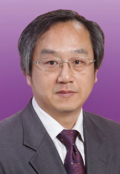 Yuqi Zhang (张玉琪)合作论文数Yuquan Hospital, Tsinghua University1
 Bin Wu (吴斌)合作论文数Sanbo Brain Hospital Capital Medical University1
 Cang Liu (刘藏)合作论文数Beijing Friendship Hospital, Capital Medical University1




