Mechanical coupling and chemical communication between cardiomyocytes are facilitated through a specialized adhesive structure called the intercalated disc (ICD). The ICD is essential for heart organization and contraction. Yet, the network of adhesion, adaptor and signaling proteins that form the ICD remains poorly defined. Here, we combined proximity labeling and quantitative mass spectrometry to identify proteins associated with the desmosomal cadherin desmoglein 2 (DSG2), in cultured neonatal cardiomyocytes. We identified over 300 proteins in the DSG2 interactome, half of which are shared with the N-cadherin (CDH2) interactome in cardiomyocytes. Proteins unique to DSG2 include connexin 43 and the plakin family of cytolinker proteins. Comparison of the cardiomyocyte DSG2 interactome with the interactomes of desmosomal proteins from epithelia revealed few shared proteins. In cardiomyocytes, plakoglobin and plakophilin 2 (PKP2) were the most abundant shared proteins between the DSG2 and CDH2 interactomes. We show that PKP2 is a dynamic protein whose membrane recruitment in cardiomyocytes is dependent on tension. Our analysis of the DSG2 interactome provides a crucial new dimension to the proteomic atlas of the essential molecular complexes required for cardiomyocyte adhesion.
Mechanical strain substantially influences tissue shape and function in various contexts from embryonic development to disease progression. Disruptions in these processes can result in congenital abnormalities and short-circuit mechanotransduction pathways. Manipulating strain in live tissues is crucial for understanding its impact on cellular and subcellular activities, unraveling the interplay between mechanics and cells. Existing tools, such as optogenetic modulation of strain, are limited to small strains over limited distances and durations. Here, a high-strain stretcher system, the TissueTractor, is introduced to enable simultaneous high-resolution spatiotemporal imaging of live cells and tissues under strain applications varying from 0% to over 100%. We use the system with organotypic explants from Xenopus laevis embryos, where applied tension reveals cellular strain heterogeneity and remodeling of intracellular keratin filaments. To highlight the device's adaptability, the TissueTractor is also used to study two other mechanically sensitive cell types with distinct physiological roles: human umbilical vein endothelial cells and mouse neonatal cardiomyocytes, revealing cell morphological changes under significant strain. The results underscore the potential of the TissueTractor for investigating mechanical cues that regulate tissue dynamics and morphogenesis.
In the heart, cardiomyocyte cell-matrix and cell-cell adhesions reorganize in response to increased cardiac demand and growth. Vinculin (VCL), a mechanosensitive adaptor protein, links filamentous actin to cell-matrix and cell-cell adhesions. Yet how VCL regulates remodeling of the two adhesion systems is poorly understood. Here, we investigate the role of phosphorylation at VCL tyrosine residue 822 (pY822) in cardiomyocyte adhesion and heart function. VCL Y822 phosphorylation levels peaked during adhesion remodeling in the developing heart and were reduced as adhesions matured postnatally. VCL pY822 levels also increased in the adult heart following injury. We mutated Vcl Y822 to phenylalanine (Y822F) in the mouse to determine the in vivo function of pY822. Homozygous mutant Vcl Y822F mice were viable but exhibited cardiac dysfunction at 28 weeks. We found that VCL pY822 regulated cardiomyocyte cell-matrix and cell-cell adhesions during postnatal heart development. Defects in cell-cell adhesion organization were also observed in cultured Vcl Y822F cardiomyocytes. Our results demonstrate that VCL Y822 phosphorylation regulates adhesion organization in cardiomyocytes, highlighting the importance of post-translational modification in modulating VCL function in the heart.
Desmosomes are essential cell-cell adhesion organelles that enable tension-prone tissues, like the skin and heart, to withstand mechanical stress. Desmosomal anomalies are associated with numerous epidermal disorders, cardiomyopathies, and cancer. Despite their critical importance, how desmosomes sense and respond to mechanical stimuli is not understood. Here, we combine super-resolution imaging in epithelial cells and primary cardiomyocytes, FRET-based tension sensors, atomistic computer simulations, and biochemical assays to demonstrate that actomyosin forces induce a conformational change in desmoplakin, a key cytoplasmic desmosomal protein. We show that in human breast cancer MCF7 cells, keratin-19 couples F-actin filaments to desmosomes and regulates the level of actomyosin forces integrated into the desmosomal complex. We demonstrate that actomyosin contractility reorients keratin intermediate filaments and directs force to desmoplakin along the keratin network, plausibly converting the N-terminal plakin domain from a folded to an extended conformation. We also show that desmoplakin undergoes a similar actomyosin force-dependent conformational change in primary cardiomyocytes, with the extent of the change affected by myofibril orientation. Our findings establish that desmoplakin is mechanosensitive and its structural states reflect the level of forces transmitted through the actin network across cell types.
In the heart, cell-matrix and cell-cell adhesions reorganize in response to increased cardiac demand and growth to promote cardiomyocyte maturation. Vinculin, a mechanosensitive adaptor protein, links filamentous actin to cell-matrix and cell-cell adhesions and is thus positioned to regulate adhesion reorganization. However, how the two adhesion systems are coordinated in the heart, and the role of vinculin in this process is poorly understood. Here, we define the role of vinculin phosphorylation at tyrosine residue 822 (pY822) in cardiomyocyte adhesion and heart function. We found that pY822 correlated with dynamic junction remodeling in the developing heart but was lost as junctions matured postnatally. We then mutated Y822 to phenylalanine (Y822F) in the mouse to determine pY822 function in vivo. Homozygous mutant Vcl Y822F mice were viable and exhibited normal cardiac function at ten weeks of age; however, cardiac dysfunction was observed at 28 weeks. Vinculin and adherens junction proteins were reduced at cardiomyocyte junctions in Y822F hearts. In contrast, α5/β1 integrin and fibronectin increased along the lateral border of Y822F cardiomyocytes. Our results demonstrate that vinculin Y822 phosphorylation regulates the balance between cadherin and integrin adhesion organization, highlighting the importance of post-translational modification in modulating vinculin function in heart physiology.
Spatial and temporal tracking of fluorescent proteins (FPs) in live cells permits visualization of proteome remodeling in response to extracellular cues. Historically, protein dynamics during trafficking have been visualized using constitutively active FPs fused to proteins of interest. While powerful, such FPs label all cellular pools of a protein, potentially masking the dynamics of select subpopulations. To help study protein subpopulations, bioconjugate tags, including the fluorogen activation proteins (FAPs), were developed. FAPs are comprised of two components: a single-chain antibody (SCA) fused to the protein of interest and a malachite-green (MG) derivative, which fluoresces only when bound to the SCA. Importantly, the MG derivatives can be either cell-permeant or-impermeant, thus permitting isolated detection of SCA-tagged proteins at the cell surface and facilitating quantitative endocytic measures. To expand FAP use in yeast, we optimized the SCA for yeast expression, created FAP-tagging plasmids, and generated FAP-tagged organelle markers. To demonstrate FAP efficacy, we coupled the SCA to the yeast G-protein coupled receptor Ste3. We measured Ste3 endocytic dynamics in response to pheromone and characterized cis- and trans-acting regulators of Ste3. Our work significantly expands FAP technology for varied applications in S. cerevisiae .
Recent advantages of genetically encoded fluorescent probes have led to the development of fluorogen-activating proteins (FAPs). This technology has two components: a non-fluorescent single chain antibody (SCA) that can be fused to a protein of interest and fluorogens, which in our case are derivatives of malachite green that are non-fluorescent when free in solution. When the SCA and fluorogen bind, there is a 20,000-fold fluorescent increase relative to unbound dye. The FAP-technology has two major advantages to standard fluorescent probes: i) the fluorogen exists as either a membrane-permeant or impermeant form and this allows for selective labeling of cell surface proteins and (2) since SCA does not fluoresce when it is not bound by dye and so there is no background fluorescence during the imaging of other markers until after the fluorogen is added. Although developed in yeast, this technology surprisingly has not been applied in this model system until our recent studies. In order to make this technology more available to the yeast community, we have optimized the SCA sequence for yeast expression, created a series for FAP-tagged plasmid constructs, and generated a suite of intracellular localization markers to aid in cell biology studies in yeast. We further employ the FAP technology to study the trafficking of the G-protein coupled receptor Ste3 and its regulation by the alpha-arrestins, an important class of trafficking adaptor.
α-Catenin binds directly to β-catenin and connects the cadherin-catenin complex to the actin cytoskeleton. Tension regulates α-catenin conformation. Actomyosin-generated force stretches the middle (M)-region to relieve autoinhibition and reveal a binding site for the actin-binding protein vinculin. It is not known whether the intramolecular interactions that regulate epithelial (αE)-catenin binding are conserved across the α-catenin family. Here, we describe the biochemical properties of testes (αT)-catenin, an α-catenin isoform critical for cardiac function and how intramolecular interactions regulate vinculin-binding autoinhibition. Isothermal titration calorimetry showed that αT-catenin binds the β-catenin-N-cadherin complex with a similar low nanomolar affinity to that of αE-catenin. Limited proteolysis revealed that the αT-catenin M-region adopts a more open conformation than αE-catenin. The αT-catenin M-region binds the vinculin N-terminus with low nanomolar affinity, indicating that the isolated αT-catenin M-region is not autoinhibited and thereby distinct from αE-catenin. However, the αT-catenin head (N- and M-regions) binds vinculin 1000-fold more weakly (low micromolar affinity), indicating that the N-terminus regulates the M-region binding to vinculin. In cells, αT-catenin recruitment of vinculin to cell-cell contacts requires the actin-binding domain and actomyosin-generated tension, indicating that force regulates vinculin binding. Together, our results show that the αT-catenin N-terminus is required to maintain M-region autoinhibition and modulate vinculin binding. We postulate that the unique molecular properties of αT-catenin allow it to function as a scaffold for building specific adhesion complexes.
ABSTRACT α-catenin binds directly to β-catenin and connects the cadherin-catenin complex to the actin cytoskeleton. Tension regulates α-catenin conformation: actomyosin-generated force stretches the middle(M)-region to relieve autoinhibition and reveal a binding site for the actin-binding protein vinculin. Here we describe the biochemical properties of αT(testes)-catenin, an α-catenin isoform critical for cardiac function, and how intramolecular interactions regulate vinculin binding autoinhibition. Isothermal titration calorimetry (ITC) showed that αT-catenin binds the β-catenin/N-cadherin complex with a similar low nanomolar affinity to that of αE-catenin. Limited proteolysis revealed that the αT-catenin M-region adopts a more open conformation than αE-catenin. The αT-catenin M-region binds the vinculin N-terminus with low nanomolar affinity, indicating that the isolated αT-catenin M-region is not autoinhibited and thereby distinct from αE-catenin. However, the αT-catenin head (N- and M-regions) binds vinculin 1000-fold more weakly (low micromolar affinity), indicating that the N-terminus regulates M-region binding to vinculin. In cells, αT-catenin recruitment of vinculin to cell-cell contacts requires the actin-binding domain and actomyosin-generated tension, indicating that force regulates vinculin binding. Together, our results indicate that the αT-catenin N-terminus is required to maintain M-region autoinhibition and modulate vinculin binding. We postulate that the unique molecular properties of αT-catenin allow it to function as a scaffold for building specific adhesion complexes.
The adherens junction (AJ) couples the actin cytoskeletons of neighboring cells to allow mechanical integration and tissue organization. The physiological demands of intercellular adhesion require that the AJ be responsive to dynamic changes in force while maintaining mechanical load. These demands are tested in the heart, where cardiomyocyte AJs must withstand repeated cycles of actomyosin-mediated contractile force. Here we show that force-responsive cardiomyocyte AJs recruit actin-binding ligands to selectively couple actin networks and promote contact maturation. We employed a panel of N-cadherin-αE-catenin fusion proteins to rebuild AJs with specific actin linkages in N-cadherin-null cardiomyocytes. In this system, vinculin recruitment was required to rescue myofibril integration and desmosome assembly at nascent contacts. In contrast, loss of vinculin disrupted junction morphology and blocked myofibril integration. Our results identify vinculin as a critical link to contractile actomyosin and offer insight to how actin integration at the AJ is regulated to provide mechanical stability and cellular organization.
αT (Testes)-catenin, a critical factor regulating cell–cell adhesion in the heart, directly couples the cadherin-catenin complex to the actin cytoskeleton at the intercalated disk (ICD), a unique cell–cell junction that couples cardiomyocytes. Loss of αT-catenin in mice reduces plakophilin2 and connexin 43 recruitment to the ICD. Since αT-catenin is subjected to mechanical stretch during actomyosin contraction in cardiomyocytes, its activity could be regulated by mechanical force. To provide insight in how force regulates αT-catenin function, we investigated the mechanical stability of the putative, force-sensing middle (M) domain of αT-catenin and determined how force impacts vinculin binding to αT-catenin. We show that 1) physiological levels of force, <15 pN, are sufficient to unfold the three M domains; 2) the M1 domain that harbors the vinculin-binding site is unfolded at ∼6 pN; and 3) unfolding of the M1 domain is necessary for high-affinity vinculin binding. In addition, we quantified the binding kinetics and affinity of vinculin to the mechanically exposed binding site in M1 and observed that αT-catenin binds vinculin with low nanomolar affinity. These results provide important new insights into the mechanosensing properties of αT-catenin and how αT-catenin regulates cell–cell adhesion at the cardiomyocyte ICD.
The adherens junction couples the actin cytoskeletons of neighboring cells to provide the foundation for multicellular organization. The core of the adherens junction is the cadherin-catenin complex that arose early in the evolution of multicellularity to link actin to intercellular adhesions. Over time, evolutionary pressures have shaped the signaling and mechanical functions of the adherens junction to meet specific developmental and physiological demands. Evolutionary rate covariation (ERC) identifies proteins with correlated fluctuations in evolutionary rate that can reflect shared selective pressures and functions. Here we use ERC to identify proteins with evolutionary histories similar to the Drosophila E-cadherin (DE-cad) ortholog. Core adherens junction components α-catenin and p120-catenin displayed positive ERC correlations with DE-cad, indicating that they evolved under similar selective pressures during evolution between Drosophila species. Further analysis of the DE-cad ERC profile revealed a collection of proteins not previously associated with DE-cad function or cadherin-mediated adhesion. We then analyzed the function of a subset of ERC-identified candidates by RNAi during border cell (BC) migration and identified novel genes that function to regulate DE-cad. Among these, we found that the gene CG42684, which encodes a putative GTPase activating protein (GAP), regulates BC migration and adhesion. We named CG42684 raskol (“to split” in Russian) and show that it regulates DE-cad levels and actin protrusions in BCs. We propose that Raskol functions with DE-cad to restrict Ras/Rho signaling and help guide BC migration. Our results demonstrate that a coordinated selective pressure has shaped the adherens junction and this can be leveraged to identify novel components of the complexes and signaling pathways that regulate cadherin-mediated adhesion.
The junctional complexes that couple cardiomyocytes must transmit the mechanical forces of contraction while maintaining adhesive homeostasis. The adherens junction (AJ) connects the actomyosin networks of neighboring cardiomyocytes and is required for proper heart function. Yet little is known about the molecular composition of the cardiomyocyte AJ or how it is organized to function under mechanical load. Here we define the architecture, dynamics and proteome of the cardiomyocyte AJ. Mouse neonatal cardiomyocytes assemble stable AJs along intercellular contacts with organizational and structural hallmarks similar to mature contacts. We combine quantitative mass spectrometry with proximity labeling to identify the N-cadherin (CDH2) interactome. We define over 350 proteins in this interactome, nearly 200 of which are unique to CDH2 and not part of the E-cadherin (CDH1) interactome. CDH2-specific interactors are comprised primarily of adaptor and adhesion proteins that promote junction specialization. Finally, we find evidence of dynamic interplay between AJ and Z-disc proteins. Together, our results provide novel insight into the cardiomyocyte AJ and provide a proteomic atlas for defining the molecular complexes that regulate cardiomyocyte intercellular adhesion. Summary Statement Proximity proteomics reveals a specific and specialized N-cadherin (CDH2) interactome along the cell-cell contacts of primary cardiomyocytes.
Protein composition at the plasma membrane is tightly regulated, with rapid protein internalization and selective targeting to the cell surface occurring in response to environmental changes. For example, ion channels are dynamically relocalized to or from the plasma membrane in response to physiological alterations, allowing cells and organisms to maintain osmotic and salt homeostasis. To identify additional factors that regulate the selective trafficking of a specific ion channel, we used a yeast model for a mammalian potassium channel, the K+ inward rectifying channel Kir2.1. Kir2.1 maintains potassium homeostasis in heart muscle cells, and Kir2.1 defects lead to human disease. By examining the ability of Kir2.1 to rescue the growth of yeast cells lacking endogenous potassium channels, we discovered that specific -arrestins regulate Kir2.1 localization. Specifically, we found that the Ldb19/Art1, Aly1/Art6, and Aly2/Art3 -arrestin adaptor proteins promote Kir2.1 trafficking to the cell surface, increase Kir2.1 activity at the plasma membrane, and raise intracellular potassium levels. To better quantify the intracellular and cell-surface populations of Kir2.1, we created fluorogen-activating protein fusions and for the first time used this technique to measure the cell-surface residency of a plasma membrane protein in yeast. Our experiments revealed that two -arrestin effectors also control Kir2.1 localization. In particular, both the Rsp5 ubiquitin ligase and the protein phosphatase calcineurin facilitated the -arrestin-mediated trafficking of Kir2.1. Together, our findings implicate -arrestins in regulating an additional class of plasma membrane proteins and establish a new tool for dissecting the trafficking itinerary of any membrane protein in yeast.
 Frank B. Gertler合作论文数Department of Biology, MIT19
 Chelsea D. Merkel合作论文数10
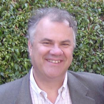 William Nelson合作论文数Departments of Biology, School of Humanities and Sciences, Stanford University;School of Medicine, Stanford University9
 William Weis合作论文数School of Medicine, Stanford University9Sabine Pokutta合作论文数8Douglas A. Rubinson合作论文数7Jonathon A Heier合作论文数5Y. Li合作论文数5J. A. Heier合作论文数4JJ Loureiro合作论文数4Xuemei Zeng合作论文数4
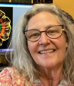 Donna Beer Stolz合作论文数McGowan Institute for Regenerative Medicine, University of Pittsburgh;Center for Biologic Imaging, School of Medicine, University of Pittsburgh;Departments of Cell Biology and Pathology, University of Pittsburgh4
 Leslie Mebane合作论文数3Je Bear合作论文数3Ian W Dale合作论文数3Ga Baltus合作论文数3
 Natalie A Hager合作论文数3
 Allyson F O'Donnell合作论文数Department of Biology, Stanford University3
 Jeffrey L. Brodsky合作论文数Center for Protein Conformational Diseases, Department of Biological Sciences, Kenneth P. Dietrich School of Arts and Sciences, University of Pittsburgh3
 Yang Li合作论文数3
 James Bear合作论文数Department of Cell Biology and Physiology, School of Medicine, The University of North Carolina at Chapel Hil3
 Simon C. Watkins合作论文数Department of Cell Biology, School of Medicine, University of Pittsburgh;Pittsburgh Center for Interdisciplinary, University of Pittsburgh;Department of Immunology, McGowan Institute for Regenerative Medicine, University of Pittsburgh3
 Erik W. Dent合作论文数Department of Anatomy, University of Wisconsin-Madison3
 Marcel Bruchez合作论文数The Center for Nucleic Acids Science and Technology, Carnegie Mellon University;Lane Center for Computational Biology, Carnegie Mellon University;Molecular Biosensor and Imaging Center, Carnegie Mellon University;Sharp Edge Labs, Inc.3
 James Shapleigh合作论文数Department of Microbiology, College of Agriculture and Life Sciences, Cornell University3
 Ed Van Veen合作论文数3Craig Furman合作论文数2
 Qanber Raza合作论文数2Jacqueline M. Benjamin合作论文数2L Van Parijs合作论文数2Mai Sun合作论文数2
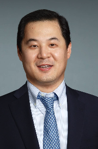 Jiangyang Zhang合作论文数Department of Radiology, Grossman School of Medicine, New York University2
 Hee Jung Choi合作论文数2Ceara K McAtee合作论文数2Sun Kyung Kim合作论文数2Christopher P Dillon合作论文数2
 Lili Yang合作论文数Yang Engineering Immunity Lab, Department of Microbiology, Immunology & Molecular Genetics, Uiversity of California, Los Angeles;Eli & Edythe Broad Center of Regenerative Medicine and Stem Cell Research, Uiversity of California, Los Angeles2Radice Glenn L合作论文数Ctr Translat Med, Thomas Jefferson Univ2
 Spiliotis Elias T合作论文数Dept Biol, Drexel Univ2Hardin Jeff合作论文数2
 Michael T. McManus合作论文数Diabetes Center, University of California, San Francisco;Department of Microbiology and Immunology, University of California, San Francisco2Susumu Mori合作论文数2Joel Vandekerckhove合作论文数2Christopher Szent-Gyorgyi合作论文数2Gretchen A. Baltus合作论文数2Gianluca Cestra合作论文数2
 Andrew P. Hinck合作论文数Molecular Structural;Biology Core;National Institute of Dental Research;National Institutes of Health;National Institute of Dental Research, National Institutes of Health2
 Nathan Yates合作论文数2




