BACKGROUND AND AIMS:The risk of atrial fibrillation (AF) is higher in endurance athletes. Pulmonary vein isolation (PVI) is effective in this group, implicating pulmonary vein (PV) remodelling, but underlying mechanisms are unclear. This study investigated if endurance training remodels PV sleeves and the PV-left atrial (LA) junction to promote PV triggers and a permissive peri-antral substrate for AF. METHODS:In canine and murine endurance-running models, in vivo PV-LA mapping, ex vivo PV electrophysiology, intracellular action potential (AP) profiling with machine learning classification, histology, bulk RNA-seq, and subcellular-resolution spatial transcriptomics of PV-LA tissue were performed. These findings were incorporated into biophysically detailed computer models of human PV cardiomyocytes and a 3D human LA. RESULTS:Training produced an athlete's heart phenotype and increased AF inducibility. In vivo, trained animals showed PV-LA conduction slowing and increased rotational activity. Ex vivo, trained PVs showed enhanced β-adrenergically evoked firing, prolonged burst activity, and a higher proportion of pacemaker-like APs. Spatial transcriptomics revealed discrete PV myocyte subpopulations with training upregulated Hcn4, Cacna1d, and Cacna1g (enhancing automaticity), downregulated Scn5a and Gja1 (slowing conduction), and enriched profibrotic/inflammatory signalling (Tnfα, Il6) alongside fibroblast expansion and extracellular matrix deposition. In silico, these changes reproduced faster spontaneous PV firing and sustained re-entry. CONCLUSIONS:Endurance training drives coupled electrical, structural, and inflammatory PV-LA remodelling that provides both trigger and substrate for AF. These data support why PV-targeted strategies can be effective in athletic AF and nominate modifiable pathways including HCN4-linked automaticity and TNFα-associated signalling.
Chronic heart failure constitutes a clinical syndrome characterized by substantial attenuation of repolarization reserve resulting from electrical remodeling. The small-conductance Ca2+-activated K+ channel (SK) has been reported to undergo upregulation in animal heart failure models and in human preparations; however, its exact function is not fully understood. This study aims to elucidate the functional role of SK channels in end-stage human heart failure. SK-protein expression of undiseased and failed human ventricular tissue was investigated by Western blot technique. Action potentials were measured by the standard microelectrode technique from right ventricular papillary muscles of undiseased hearts and from right and left papillary muscles and from left midmyocardial tissue slices of failing hearts. Ionic currents were recorded by the whole cell configuration of the patch-clamp technique on isolated cells obtained from left ventricles of failing hearts. Failing hearts exerted consistent action potential lengthening and lacked spike-and-dome compared with undiseased hearts. Western blot revealed identical SK expression between undiseased and failing hearts. Apamin (100 nM), a commonly used selective SK channel inhibitor, failed to alter action potential duration values of the failing hearts in left and right endocardial preparations and in left midmyocardium. Furthermore, no apamin-sensitive current was identified in isolated cells. Week coupling between SK2 channels and L-type Ca2+ channels was found. These results do not confirm the results of previous studies claiming an important role of SK channels in the repolarization of the human failing heart. NEW & NOTEWORTHY This study re-evaluates the role of small-conductance Ca2+-activated K+ (SK) channels in ventricular repolarization in terminal human heart failure. Although SK protein is present and pharmacological activation causes only minor AP shortening, apamin does not affect action potentials or membrane currents, even with enhanced intracellular Ca2+. Partial I-Kr block alone induces repolarization failure, indicating severely reduced repolarization reserve and negligible compensatory SK current, questioning the therapeutic relevance of SK channels in end-stage heart failure.
The ultrarapid delayed rectifier potassium current (IKur) has long been considered an atrial-specific current with no functional role in the ventricles, despite evidence of its expression in ventricular myocytes. In the present study we challenged this prevailing concept and investigated the potential role of IKur in ventricular repolarization. Kv1.5 protein expression was determined by immunocytochemistry in canine and undiseased human cardiac tissue samples. Action potentials (APs) were recorded using conventional microelectrode techniques in canine and human cardiac preparations, and ion currents were measured using the whole-cell patch-clamp technique in isolated cardiomyocytes. In silico simulations were performed to investigate the role of the IKur current using a human ventricular model (T-WorldIKur). Kv1.5 protein expression was detected in canine atrial, ventricular and Purkinje fibre cells, as well as in undiseased human left ventricular myocardium. In canine atrial preparations IKur inhibition shifted the plateau phase of the AP into the positive voltage direction and consequently shortened AP duration. Application of 100 µM 4-aminopyridine (4-AP) revealed ionic currents of similar magnitude in ventricular myocytes and Purkinje fibres, which were attributed to IKur. Inhibition of IKur caused prolongation of the APs recorded from endocardial and midmyocardial preparations, as well as from Purkinje fibres. Under conditions of attenuated repolarization reserve IKur inhibition induced augmented repolarization lengthening and frequent early afterdepolarizations (EADs). In silico modelling data regarding the IKur inhibition-evoked EAD formations are in good agreement with the experimental findings. IKur is present and functionally active in ventricular myocardium and may represent a significant contributor to ventricular repolarization reserve. KEY POINTS: The ultrarapid delayed rectifier potassium current (IKur) has long been considered an atrial-specific current with no functional role in the ventricles. In the present study, we challenged this prevailing concept and investigated the potential role of IKur in ventricular repolarization. Cellular electrophysiology and molecular cardiological experiments were performed on cardiac preparations obtained from dog and undiseased human hearts. We present clear evidence that IKur current is present in the ventricular myocardium at both molecular and functional levels. We also confirm that IKur current plays a significant role in ventricular repolarization reserve. We concluded that IKur is present and functionally active in ventricular myocardium and may represent a significant contributor to ventricular repolarization reserve.
Computer models of the human ventricular cardiomyocyte action potential (AP) have reached a level of detail and maturity that has led to an increasing number of applications in safety pharmacology, as exemplified by the Comprehensive in Vitro Proarrhythmia Assay (CiPA) initiative. However, interfacing the models with experimental data can become a substantial computational burden when considering that each simulation includes hundreds up to thousands of beats to reach the limit cycle. To address this issue, an emulator based on a neural network (NN emulator) was developed that rapidly predicts the limit cycle AP for given maximum conductances of crucial ion channels, pumps, and exchangers (publicly available at https://github.com/thomgrand/cardiomyocyte_emulator). The training was performed on simulation data produced by the ToR-ORd-dynCl model (publicly available at https://zenodo.org/records/10640339) and the evaluation was done for forward (find drugged AP for given pharmacological scaling factors of maximum conductances) and inverse problems (find pharmacological scaling factors for given APs before and after drug administration) of quantitative systems pharmacology studies on synthetic and experimental data. First and foremost, it has been shown that the NN emulator potentially enables massive speed-ups compared to regular simulations (e.g. 171× when using Myokit for the simulation of 100 beats on a CPU). Furthermore, the forward problem on synthetic data could be solved with average root-mean-square errors (RMSE) of 0.47 mV in normal APs and of 14.5 mV in abnormal APs exhibiting early afterdepolarisations while, in the latter case, 72.5 % of the predicted APs were aligning with the abnormality and most of the remaining APs demonstrated pronounced proximity. The inverse problem on synthetic data could be solved with a maximum RMSE of 0.22 in the inferred pharmacological scaling factors. However, notable mismatches were observed between pharmacological scaling factors inferred from experimental APs, and corresponding data obtained from the CiPA initiative. This reveals larger inaccuracies, which can be attributed particularly to the fact that recordings were performed in small tissue preparations while the emulator was trained on single cardiomyocyte data. Overall, our study highlights the potential of NN emulators as tool for an increased efficiency in quantitative systems pharmacology studies.
BACKGROUND: Atrial fibrillation (AF) susceptibility is heightened in endurance athletes but the underlying mechanisms are incompletely understood. Because pulmonary vein (PV) myocyte triggers are critical determinants of AF, we investigated PV electrophysiological remodelling in animal models of the athlete's heart. METHODS: The following experiments were performed in canines and mice after 16 or 6 weeks, respectively, of daily exercise training (ExT), and compared to sedentary (Sed) controls: ECG recording, echocardiography, pharmacological autonomic block, extrastimulus pacing, multielectrode array mapping, monophasic and intracellular action potential (AP) recording with custom-designed pattern recognition analysis, histology, RNAseq and spatial in situ transcriptomics. RESULTS: AF propensity was significantly increased in ExT animals. Mapping studies identified heightened rotational activity in the PV-left atrial (LA) junction of ExT vs. Sed canines in vivo, and enhanced automaticity, triggered activity and AP duration variability ex vivo in ExT canines and mice. Intracellular recordings in mouse PV cardiomyocytes determined at least six AP subtypes with increased frequency of pacemaker-like APs in ExT PV, concomitant with increased expression of pacemaking HCN4, Cav1.3 and Cav3.1 channels. PV spontaneous excitability was also significantly enhanced. Subcellular resolution spatial transcriptomics in mouse PV-LA identified diffuse ion channel remodelling and activation of established AF-promoting pro-inflammatory and pro-fibrotic cytokines and chemokines in ExT PV cardiomyocytes. Conduction slowing in the ExT PV-LA junction was attributable to: gap junction remodelling, reduced Na+ channel expression and increased extracellular matrix deposition with enhanced myofibroblast number and proximity to PV cardiomyocytes. CONCLUSIONS: Endurance exercise elicits proarrhythmic electro-anatomical remodelling of the PV-LA junction with enhanced pacemaking ion channel expression and immune-inflammatory pathway activation in PV myocytes as prominent contributors. ### Competing Interest Statement The authors have declared no competing interest.
Computer models of the human ventricular cardiomyocyte action potential (AP) have reached a level of detail and maturity that has led to an increasing number of applications in the pharmaceutical sector. However, interfacing the models with experimental data can become a significant computational burden. To mitigate the computational burden, the present study introduces a neural network (NN) that emulates the AP for given maximum conductances of selected ion channels, pumps, and exchangers. Its applicability in pharmacological studies was tested on synthetic and experimental data. The NN emulator potentially enables massive speed-ups compared to regular simulations and the forward problem (find drugged AP for pharmacological parameters defined as scaling factors of control maximum conductances) on synthetic data could be solved with average root-mean-square errors (RMSE) of 0.47 mV in normal APs and of 14.5 mV in abnormal APs exhibiting early afterdepolarizations (72.5% of the emulated APs were alining with the abnormality, and the substantial majority of the remaining APs demonstrated pronounced proximity). This demonstrates not only very fast and mostly very accurate AP emulations but also the capability of accounting for discontinuities, a major advantage over existing emulation strategies. Furthermore, the inverse problem (find pharmacological parameters for control and drugged APs through optimization) on synthetic data could be solved with high accuracy shown by a maximum RMSE of 0.22 in the estimated pharmacological parameters. However, notable mismatches were observed between pharmacological parameters estimated from experimental data and distributions obtained from the Comprehensive in vitro Proarrhythmia Assay initiative. This reveals larger inaccuracies which can be attributed particularly to the fact that small tissue preparations were studied while the emulator was trained on single cardiomyocyte data. Overall, our study highlights the potential of NN emulators as powerful tool for an increased efficiency in future quantitative systems pharmacology studies.
Recent experimental data shows that hesperetin, a citrus flavonoid, affects potassium channels and can prolong the QTc interval in humans. Therefore, in the present study we investigated the effects of hesperetin on various transmembrane ionic currents and on ventricular action potentials. Transmembrane current measurements and action potential recordings were performed by patch-clamp and the conventional microelectrode techniques in dog and rabbit ventricular preparations. At 10 µM concentration hesperetin did not, however, at 30 µM significantly decreased the amplitude of the IK1, Ito, IKr potassium currents. Hesperetin at 3–30 µM significantly and in a concentration-dependent manner reduced the amplitude of the IKs current. The drug significantly decreased the amplitudes of the INaL and ICaL currents at 30 µM. Hesperetin (10 and 30 µM) did not change the action potential duration in normal preparations, however, in preparations where the repolarization reserve had been previously attenuated by 100 nM dofetilide and 1 µg/ml veratrine, caused a moderate but significant prolongation of repolarization. These results suggest that hesperetin at close to relevant concentrations inhibits the IKs outward potassium current and thereby reduces repolarization reserve. This effect in certain specific situations may prolong the QT interval and consequently may enhance proarrhythmic risk.
To understand the large inter-species variations in drug effects on repolarization, the properties of the rapid (IKr) and the slow (IKs) components of the delayed rectifier potassium currents were compared in myocytes isolated from undiseased human donor (HM), dog (DM), rabbit (RM) and guinea pig (GM) ventricles by applying the patch clamp and conventional microelectrode techniques at 37 °C. The amplitude of the E-4031-sensitive IKr tail current measured at −40 mV after a 1 s long test pulse of 20 mV, which was very similar in HM and DM but significant larger in RM and GM. The L-735,821-sensitive IKs tail current was considerably larger in GM than in RM. In HM, the IKs tail was even smaller than in DM. At 30 mV, the IKr component was activated extremely rapidly and monoexponentially in each studied species. The deactivation of the IKr component in HM, DM, and RM measured at −40 mV. After a 30 mV pulse, it was slow and biexponential, while in GM, the IKr tail current was best fitted triexponentially. At 30 mV, the IKs component activated slowly and had an apparent monoxponential time course in HM, DM, and RM. In contrast, in GM, the activation was clearly biexponential. In HM, DM, and RM, IKs component deactivation measured at −40 mV was fast and monoexponential, while in GM, in addition to the fast component, another slower component was also revealed. These results suggest that the IK in HM resembles that measured in DM and RM and considerably differs from that observed in GM. These findings suggest that the dog and rabbit are more appropriate species than the guinea pig for preclinical evaluation of new potential drugs expected to affect cardiac repolarization.
AbstractThe antiarrhythmic and cardiac electrophysiological effects of SZV-2649 that contains a 2,6-diiodophenoxy moiety but lacks the benzofuran ring system present in amiodarone, were studied in mammalian cell line, rat and dog cardiac preparations. SZV-2649 exerted antiarrhythmic effects against coronary artery occlusion/reperfusion induced ventricular arrhythmias in rats and in acetylcholine- and burst stimulation induced atrial fibrillation in dogs. SZV-2649 inhibited hERG and GIRK currents in HEK cells (IC50: 342 and 529 nM, respectively). In canine ventricular myocytes, SZV-2649 (10 µM) decreased the densities of IKr, and Ito outward and INaL and ICaL inward currents. The compound (2.5–10 µM) elicited Class IB type Vmax reducing and Class III type action potential duration prolonging effects in dog right ventricular muscle preparations. In canine atrial muscle, SZV-2629 (2.5–10 µM) moderately prolonged action potential duration and this effect was greatly augmented in preparations pretreated with 1 µM carbachol. In conclusion, SZV-2649, has antiarrhythmic effects based on its multiple ion channel blocking properties. Since its chemical structure substantially differs from that of amiodarone, it is expected that SZV-2649 would exhibit fewer adverse effects than the currently used most effective multichannel inhibitor drug amiodarone and may be a promising molecule for further development.
Abstract Funding Acknowledgements Type of funding sources: None. Background The influence of testosterone on ventricular ion channels is widely investigated. Despite the legally stipulated availability of different androgen anabolic steroids (AAS), these agents are very popular among young adults, however, the direct effects of supra-physiological testosterone level are still unclear. Supposedly, the chronic use of AASs may result in structural and functional remodeling of the heart. Purpose The aim of our study was to investigate the potential electrophysiological effects of chronic administration of testosterone-undecanoate in a canine model in in vivo and in vitro studies. Methods Eight male beagle dogs were randomized into control (’Cont’) and treated (’Tr’) groups (n = 4; n = 4). The latter group received 15 mg/kg of long-lasting testosterone-undecanoate intramuscular injections weekly for 3 months. Blood samples were taken for monitoring testosterone levels. To investigate the altered repolarization in conscious dogs electrocardiography studies were performed. Ventricular myocytes were enzymatically dissociated via retrograde perfusion. The transmembrane ionic currents were recorded using the whole-cell configuration of the patch-clamp technique and the action potential duration (APD) was measured by the perforated patch-clamp technique. Results Testosterone level was significantly higher in the ‘Tr’ group compared to the ‘Cont’ group (47.02 nm/L vs. 15.23 nm/L; p=0.0002). The chronic treatment did not affect the heart rate between the examined groups (93.7±23.5 vs.108.8±21.4 beats/min), however, it resulted in significantly shortened QT (226±49 vs. 244.9±26.6 ms; p<0.05) and QTc (26.06±2.5 vs. 29.6±3.01 ms, p<0.05) intervals. ECG recordings also presented prolonged PQ (112.1±15.5 vs. 61.65±12.7ms; p<0.05), QRS (72.37±15.4 vs. 61.65±12.7 ms; p<0.05), and Tp-Te (32.95±7.45 vs. 53.46±16.6 ms; p<0.05) intervals in the ‘Tr’ group. Additionally, the APD of isolated left ventricular myocytes significantly shortened in the ‘Tr’ group compared to the ‘Con’ group (235.2±26.7 vs. 283.6±28.5 ms; p<0.05). Patch-clamp experiments revealed increased magnitude of transient outward potassium current (Ito), the inward rectifier potassium current (Ik1), and the slowed delayed rectifier potassium current (Iks) in the ‘Tr’ group. Conclusion The repolarization of the canine ventricular myocardium was significantly modified by constantly high level of testosterone. Many beneficial effects are attributed to physiological testosterone levels; however, the supra-physiological testosterone level may lead to potentially harmful alternations in the cardiac repolarization that may promote arrhythmogenesis, especially in the presence of various heart diseases. Presumably, the constantly high testosterone level induced electrophysiological changes may contribute to the development of life-threatening arrhythmias under certain circumstances.
A közelmúltban több tanulmány is rámutatott arra, hogy az intenzív sporttevékenység szívritmuszavarok kialakulására hajlamosíthat és akár hirtelen szívhalálhoz is vezethet, azonban a háttérben húzódó elektrofiziológiai mechanizmusok kevéssé ismertek. Munkánk során a tartós állóképességi edzés indukálta szívizom-átépülést (ún. „remodelling”) és aritmiaérzékenységet vizsgáltuk kutya sportszív-modellben. Beagle-kutyákat véletlenszerűen „kontroll” és „edzett” csoportokba soroltunk (n=12/csoport), ez utóbbi egy 4 hónapos intenzív edzésprogramban vett részt. A szívizom-remodellációt és a ritmuszavar-érzékenységet számos in vivo és in vitro technikával vizsgáltuk (elektrokardiográfia, echokardiográfia, közvetlen kamrai „burst” ingerlés, patch-clamp, immunocitokémia, szövettani vizsgálatok). Az edzésprogram hatására nőtt a szeptális falvastagság (8,1±0,2 mm vs. 7,4±0,2 mm; p<0,05), a bal kamrai végdisztolés átmérő (32,0±0,7 mm vs. 30,4±0,7 mm; p<0,05) és a bal kamrai tömegindex (125,8±4,3 g/m2 vs. 97,7±6,4 g/m2 ; p<0,05). Megnyúlt kamrai repolarizációt figyeltünk meg in vivo és in vitro körülmények között (QTc: 237,1±3,4 ms vs. 213,6±2,8 ms; APD90: 472,8±29,6 ms vs. 370,1±32,7 ms; p<0,05), amelyek a repolarizáció emelkedett rövid távú variabilitásával társultak. Az edzett állatok szívéből izolált bal kamrai szívizomsejtek tranziens kifelé irányuló K+-áramának amplitúdója csökkent (6,4±0,5 pA/pF vs. 8,8±0,9 pA/ pF, 50 mV; p<0,05), mindemellett a bal kamrai fibrózis mértéke, illetve a HCN4-fehérje expressziója is emelkedett. Fokozott ektópiás aktivitást és aritmiaérzékenységet figyeltünk meg az edzett állatokban. Modellünkben egyértelmű strukturális és elektrofiziológiai szívizom-átalakulás jelentkezett. Szívmorfológiai megfigyeléseink összhangban állnak a humán állóképességi élsportolókban megfigyeltekkel. Tanulmányunkban a repolarizáció diszperziójának fokozódása, a fibrózis emelkedett mértéke és a HCN4-túlexpresszió a modell aritmiák iránti fokozott érzékenységét jelzik.
The health benefits of regular physical exercise are well known. Even so, there is increasing evidence that the exercise regimes of elite athletes can evoke cardiac arrhythmias including ventricular fibrillation and even sudden cardiac death (SCD). The mechanism of exercise-induced arrhythmia and SCD is poorly understood. Here, we show that chronic training in a canine model (12 sedentary and 12 trained dogs) that mimics the regime of elite athletes induces electrophysiological remodeling (measured by ECG, patch-clamp, and immunocytochemical techniques) resulting in increases of both the trigger and the substrate for ventricular arrhythmias. Thus, 4 months sustained training lengthened ventricular repolarization (QTc: 237.1±3.4 ms vs. 213.6±2.8 ms, n=12; APD90: 472.8±29.6 ms vs. 370.1±32.7 ms, n=29 vs. 25), decreased transient outward potassium current (6.4±0.5 pA/pF vs. 8.8±0.9 pA/pF at 50 mV, n=54 vs. 42), and increased the short-term variability of repolarization (29.5±3.8 ms vs. 17.5±4.0 ms, n=27 vs. 18). Left ventricular fibrosis and HCN4 protein expression were also enhanced. These changes were associated with enhanced ectopic activity (number of escape beats from 0/hr to 29.7±20.3/hr) in vivo and arrhythmia susceptibility (elicited ventricular fibrillation: 3 of 10 sedentary dogs vs. 6 of 10 trained dogs). Our findings provide in vivo, cellular electrophysiological and molecular biological evidence for the enhanced susceptibility to ventricular arrhythmia in an experimental large animal model of endurance training.
The present study was designed to test the hypothesis that the selectivity of blocking the late Na+ current (INaL) over the peak Na+ current (INaP) is related to the fast offset kinetics of the Na+ channel inhibitor. Therefore, the effects of 1 µM GS967 (INaL inhibitor), 20 µM mexiletine (I/B antiarrhythmic) and 10 µM quinidine (I/A antiarrhythmic) on INaL and INaP were compared in canine ventricular myocardium. INaP was estimated as the maximum velocity of action potential upstroke (V+max). Equal amounts of INaL were dissected by the applied drug concentrations under APVC conditions. The inhibition of INaL by mexiletine and quinidine was comparable under a conventional voltage clamp, while both were smaller than the inhibitory effect of GS967. Under steady-state conditions, the V+max block at the physiological cycle length of 700 ms was 2.3% for GS967, 11.4% for mexiletine and 26.2% for quinidine. The respective offset time constants were 110 ± 6 ms, 456 ± 284 ms and 7.2 ± 0.9 s. These results reveal an inverse relationship between the offset time constant and the selectivity of INaL over INaP inhibition without any influence of the onset rate constant. It is concluded that the selective inhibition of INaL over INaP is related to the fast offset kinetics of the Na+ channel inhibitor.
Late sodium current (INa,late) is an important inward current contributing to the plateau phase of the action potential (AP) in the mammalian heart. Although INa,late is considered as a possible target for antiarrhythmic agents, several aspects of this current remained hidden. In this work, the profile of INa,late, together with the respective conductance changes (GNa,late), were studied and compared in rabbit, canine, and guinea pig ventricular myocytes using the action potential voltage clamp (APVC) technique. In canine and rabbit myocytes, the density of INa,late was relatively stable during the plateau and decreased only along terminal repolarization of the AP, while GNa,late decreased monotonically. In contrast, INa,late increased monotonically, while GNa,late remained largely unchanged during the AP in guinea pig. The estimated slow inactivation of Na+ channels was much slower in guinea pig than in canine or rabbit myocytes. The characteristics of canine INa,late and GNa,late were not altered by using command APs recorded from rabbit or guinea pig myocytes, indicating that the different shapes of the current profiles are related to genuine interspecies differences in the gating of INa,late. Both INa,late and GNa,late decreased in canine myocytes when the intracellular Ca2+ concentration was reduced either by the extracellular application of 1 µM nisoldipine or by the intracellular application of BAPTA. Finally, a comparison of the INa,late and GNa,late profiles induced by the toxin of Anemonia sulcata (ATX-II) in canine and guinea pig myocytes revealed profound differences between the two species: in dog, the ATX-II induced INa,late and GNa,late showed kinetics similar to those observed with the native current, while in guinea pig, the ATX-II induced GNa,late increased during the AP. Our results show that there are notable interspecies differences in the gating kinetics of INa,late that cannot be explained by differences in AP morphology. These differences must be considered when interpreting the INa,late results obtained in guinea pig.
Even though rodents are accessible model animals, their electrophysiological properties are deeply different from those of humans, making the translation of rat studies to humans rather difficult. We compared the mechanisms of ventricular repolarization in various animal models to those of humans by measuring cardiac ventricular action potentials from ventricular papillary muscle preparations using conventional microelectrodes and applying selective inhibitors of various potassium transmembrane ion currents. Inhibition of the IK1 current (10 µmol/L barium chloride) significantly prolonged rat ventricular repolarization, but only slightly prolonged it in dogs, and did not affect it in humans. On the contrary, IKr inhibition (50 nmol/L dofetilide) significantly prolonged repolarization in humans, rabbits, and dogs, but not in rats. Inhibition of the IKur current (1 µmol/L XEN-D0101) only prolonged rat ventricular repolarization and had no effect in humans or dogs. Inhibition of the IKs (500 nmol/L HMR-1556) and Ito currents (100 µmol/L chromanol-293B) elicited similar effects in all investigated species. We conclude that dog ventricular preparations have the strongest translational value and rat ventricular preparations have the weakest translational value in cardiac electrophysiological experiments.
Aim Long QT syndrome (LQTS) is a cardiac channelopathy predisposing to ventricular arrhythmias and sudden cardiac death. Since current therapies often fail to prevent arrhythmic events in certain LQTS subtypes, new therapeutic strategies are needed. Docosahexaenoic acid (DHA) is a polyunsaturated fatty acid, which enhances the repolarizing I-Ks current. Methods and results We investigated the effects of DHA in wild type (WT) and transgenic long QT Type 1 (LQT1; loss of I-Ks), LQT2 (loss of I-Kr), LQT5 (reduction of I-Ks), and LQT2-5 (loss of I-Kr and reduction of I-Ks) rabbits. In vivo ECGs were recorded at baseline and after 10 mu M/kg DHA to assess changes in heart-rate corrected QT (QTc) and short-term variability of QT (STVQT). Ex vivo monophasic action potentials were recorded in Langendorff-perfused rabbit hearts, and action potential duration (APD(75)) and triangulation were assessed. Docosahexaenoic acid significantly shortened QTc in vivo only in WT and LQT2 rabbits, in which both alpha- and beta-subunits of I-K(s)-conducting channels are functionally intact. In LQT2, this led to a normalization of QTc and of its short-term variability. Docosahexaenoic acid had no effect on QTc in LQT1, LQT5, and LQT2-5. Similarly, ex vivo, DHA shortened APD(75) in WT and normalized it in LQT2, and additionally decreased AP triangulation in LQT2. Conclusions Docosahexaenoic acid exerts a genotype-specific beneficial shortening/normalizing effect on QTc and APD(75) and reduces pro-arrhythmia markers STVQT and AP triangulation through activation of I-Ks in LQT2 rabbits but has no effects if either alpha- or beta-subunits to I-Ks are functionally impaired. Docosahexaenoic acid could represent a new genotype-specific therapy in LQT2.
Abstract Funding Acknowledgements Type of funding sources: Public Institution(s). Main funding source(s): Economic Development and Innovation Operative Programme GINOP-2.3.2-15-2016-00012, the National Research Development and Innovation Office (NKFIH K 135464 and NKFIH K 128851), the Ministry of Human Capacities Hungary (20391-3/2018/FEKUSTRAT and EFOP-3.6.2-16-2017-00006), and from the Eötvös Loránd Research Network Background Cannabidiol (CBD), a major active phytogenic cannabinoid, is one of the main constituents of cannabis. Cannabis has been widely used as recreational drug over the decades and its use is constantly increasing as hallucinogenic and/or medicinal agent. However, significant cardiovascular side effects can accompany its use ranging from arrhythmia to sudden cardiac death. Purpose The aim of the present work was to investigate the possible cardiac adverse electrophysiological effects of cannabidiol (CBD) on action potentials and various transmembrane potassium currents, such as the rapid (IKr) and slow (IKs) delayed rectifier, the transient outward (Ito) and inward rectifier (IK1) potassium currents in rabbit and dog cardiac preparations to assess the cardiac safety profile and proarrhythmic risk. Methods In the current study, conventional microelectrode and voltage clamp techniques were used to record the action potential and transmembrane ionic currents in dog and rabbit ventricular tissue preparations and enzymatically isolated myocytes, respectively. Results The results show that CBD lengthens APD90 significantly at the concentration of 5 µM both in dog and rabbit ventricular tissues without changing other action potential parameters significantly. To further investigate the APD90 lengthening effect of CBD, transmembrane potassium currents (IKr, IKs, Ito and IK1) were investigated in dog and/or rabbit ventricular myocytes using voltage clamp technique. CBD significantly inhibited IKr and IKs currents in rabbit ventricular myocytes with an estimated EC50 values of 4.9 and 3.1 µM, respectively. The effect of CBD on rabbit’s Ito current was not significant while it was significant on dog’s Ito current with an estimated EC50 value of 5 µM. IK1 was not responsive to CBD even at high concentration. Conclusion In conclusion, looking at the inhibitory effects of CBD on repolarizing potassium currents, despite of the fact that these calculated EC50 values are higher than pharmacokinetics based Cmax values of CBD recorded after smoking and oral intake, it can be speculated that in the presence of certain cardio active drugs or co-morbidity where CBD metabolism or cardiac repolarization reserve is impaired CBD can have an additive and proarrhythmic effect.
 Laszlo Virag合作论文数Orvosi Vegytani Intezet, DE Altalanos Orvostudomanyi Kar;Faculty of Medicine, University of Debrecen150
 Julius Gyula Papp合作论文数Division of Cardiovascular Pharmacology, Hungarian Academy of Sciences;Department of Pharmacology & Pharmacotherapy, Albert Szent-Györgyi Medical Center, University of Szeged122
 Graham Dockray合作论文数Institute of Systems, Molecular & Integrative Biology, University of Liverpool;Faculty of Health and Life Sciences, University of Liverpool109
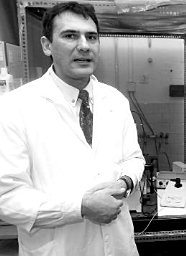 Norbert Jost合作论文数Division of Cardiovascular Pharmacology, Hungarian Academy of Sciences108
 Istvan Baczko合作论文数106
 Norbert Nagy合作论文数53
 Juulius Gyula Papp合作论文数52
 R Dimaline合作论文数48
 David A Lathrop合作论文数44
 Peter Pal Nanasi合作论文数Department of Physiology, University of Debrecen;Division of Physiology and Pharmacology, Department of Basic Medical Sciences, Faculty of Dentistry, University of Debrecen43
 András Farkas合作论文数Faculty of Economics;Budapest Tech;Budapest Tech, Faculty of Economics41Julius Gy Papp合作论文数38
 Tamas Banyasz合作论文数Department of Physiology, University of Debrecen36Kohajda Zsófia合作论文数34
 János Magyar合作论文数Department of Physiology, University Medical School of Debrecen34
 Janos Prorok合作论文数Department of Pharmacology and Pharmacotherapy, University of Szeged33P P Nánási合作论文数32
 Andras J. Toth合作论文数Department of Manufacturing Engineering, Budapest University of Technology and Economics31
 Norbert Szentandrássy合作论文数Department of Physiology, University of Debrecen;Institute for General Medical Knowledge, Faculty of Dentistry, University of Debrecen;Department of Basic Medical Sciences, Faculty of Dentistry, University of Debrecen30Szuts Viktoria合作论文数29Károly Acsai合作论文数29
 Attila Farkas合作论文数28
 Stanley Nattel合作论文数Département de Médecine, Faculté de Médecine, Université de Montréal28Norbert Iost合作论文数27
 David Mark Pritchard合作论文数School of Clinical Sciences, University of Liverpool27
 Tamás Forster合作论文数Medical Faculty, University of Szeged27
 Balazs Ordog合作论文数26Péter Biliczki合作论文数25Janos Pataricza合作论文数24Zoltan Husti合作论文数23
 Timothy Cragin Wang合作论文数Herbert Irving Comprehensive Cancer Center, Columbia University;Division of Digestive and Liver Diseases, Columbia University23
 Miklos Bitay合作论文数22C. Lengyel合作论文数21
 Balazs Horvath合作论文数21Csaba Lengyel合作论文数University of Szeged21
 Erich Wettwer合作论文数21
 Leila Topal合作论文数19Alexandra Polyák合作论文数Dept Internal Med, Univ Szeged19
 Istvan Koncz合作论文数Dept Pharmacol & Pharmacotherapy, Univ Szeged19Janos Lonovics合作论文数19Gyorgy Rabloczky合作论文数19
 Attila Tóth合作论文数Centre for Neuroscience, University of Pécs19K Acsai合作论文数18O Hala合作论文数18
 Tamas Takacs合作论文数18
 Piero Pollesello合作论文数Cardiovascular Research, Orion Pharma18István Leprán合作论文数18
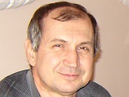 Gyorgy Seprenyi合作论文数Dept Anat Histol & Embryol, Univ Szeged17
 Rakonczay Zoltán合作论文数Dept Med 1, Univ Szeged17




