Change in peripheral blood cytokines. Panel A shows the median change from baseline in peripheral blood cytokine concentration over time by dose of mRNA-2752 in Arm A and Arm B, and panel B shows cytokine fold change relative to C1D1 over time by dose of mRNA-2752 in Arm A and Arm B. Data are shown from samples from 125 patients. C1D1, cycle 1 day 1; IFN-γ, interferon gamma; IL, interleukin; IP-10, IFN-γ–induced protein 10; MCP-1, monocyte chemoattractant protein 1.
TPS9613 Background: The high response rate of neoadjuvant anti-PD-1 has changed the management of locally advanced cutaneous squamous cell carcinoma. However, little is known about the benefit of neoadjuvant immunotherapy for basal cell carcinoma (BCC). Anti-PD-1 is approved second line for advanced BCC where it has modest response rates compared to the first line setting. High LAG-3 expression within the BCC tumor microenvironment may function as a subdominant checkpoint to synergize with anti-PD-1. The current randomized prospective multicenter phase 2 trial evaluates the safety and efficacy of neoadjuvant nivolumab (anti-PD-1) + relatlimab (anti-LAG-3) versus nivolumab alone in resectable high-risk BCC. Methods: This multicenter study conducted across the University of California Melanoma and Skin Cancer Consortium will enroll up to 30 patients with resectable high-risk BCC. High-risk BCC is defined by size 2.0 cm or greater in the head and neck region or 4.0 cm or greater for the trunk and extremities. All patients must have surgically resectable disease that is at increased risk for cosmetic disfigurement, functional defects, poor oncologic control, or anticipated to require skin grafting or free flap reconstruction. Patients must also be checkpoint and hedgehog inhibitor naïve with ECOG performance status of <2. Key exclusion criteria include recent radiation therapy and any prior history of myocarditis. Patients will be randomized in 2:1 fashion to receive nivolumab 480 mg + relatlimab 160 mg or nivolumab 480 mg IV every 4 weeks for up to 4 doses. Patients may forgo surgery and continue beyond 4 doses for up to 1 year in cases of ongoing clinical benefit. Tumor tissue and peripheral blood will be interrogated for immune cell subsets and checkpoint expression before and after neoadjuvant treatment. The trial’s primary endpoint is the combined rate of pathological complete response, major pathologic response, and clinical response following neoadjuvant therapy with secondary endpoints including surgical de-escalation, safety, duration of response, and recurrence-free survival. This trial uses a Simon’s minimax two-stage design for the nivolumab + relatlimab cohort. Eleven patients will be enrolled in the first stage. If ≥2 patients respond to nivolumab + relatlimab, the study will continue enrollment for a total of 20 patients. This design yields a type I error rate of 0.1 and power of 0.8. This study was approved by UC San Diego’s Institutional Review Board; approval number is 810617. Trial Registration NCT06624475. Clinical trial information: NCT06624475 .
Mucosal and acral melanomas are rare subtypes of melanoma with unique clinical and biological features. A dearth of well-established and characterized melanoma cell lines that reflect rare subtypes impedes research on these lethal diseases. We present a protocol developed by exploring various combinations of established techniques that we used to generate mucosal and acral cell lines from patient tissues and patient derived xenografts.
Change in proinflammatory cytokine gene expression over time by dose of mRNA-2752 in Arm A and Arm B. Data are shown from 22 patients, and are log2 fold change in gene expression relative to baseline. C1D2, cycle 1 day 2; C1D15, cycle 1 day 15; C2D1, cycle 2 day 1; IL, interleukin; Scr, screening.
PURPOSE:Messenger RNA (mRNA)-2752, a lipid nanoparticle‒encapsulated, mRNA-based therapeutic encoding OX40L, interleukin 36γ (IL-36γ), and IL-23, has demonstrated modulation of the tumor microenvironment (TME) and antitumor efficacy in combination with immune checkpoint inhibitors (CPI) in CPI-resistant models. PATIENTS AND METHODS:In this phase I study (NCT03739931) of intratumoral mRNA-2752 monotherapy (arm A) or mRNA-2752 plus durvalumab (arm B) in advanced solid tumors, the primary objectives were safety, tolerability, determination of maximum tolerated dose (MTD), and objective response rate (ORR) as per Response Evaluation Criteria in Solid Tumors version 1.1 in CPI-resistant melanoma (arm B only). RESULTS:Among 134 patients (arm A, n = 19 and arm B, n = 115), the MTD was not reached; a recommended dose for expansion of ≤8 mg was selected. Dose-limiting toxicities included two grade II cytokine-release syndrome events in arm B. Treatment-related adverse events (AE) were mostly grade I/II; grade III mRNA-2752-related AEs occurred in 1 patient (5.3%) in arm A and 29 patients (25.2%) in arm B. In the CPI-resistant melanoma cohort (n = 28), across doses, the confirmed ORR was 17.9% (95% CI, 6.1%‒36.9%), and the disease control rate was 42.9% (95% CI, 24.5%‒62.8%). Increased peripheral cytokine levels and sustained inflammatory responses in the TME were observed, particularly in patients with an objective response. CONCLUSIONS:mRNA-2752 monotherapy or in combination with durvalumab demonstrated antitumor activity with manageable safety in patients with heavily pretreated, relapsed, or resistant solid tumors, particularly in patients with CPI-resistant melanoma. In addition, biomarker analyses demonstrated a sustained inflammatory response within the TME. Together, these findings support the investigation of mRNA-based therapeutics for patients with advanced cancer.
Change in plasma IFN-γ and IL-23 cytokine and IL-23 mRNA concentration over time in (A) Arm A and (B) Arm B, and IL-23 and IFN-γ AUC at cycle 1 and cycle 2 in (C) Arm A and (D) Arm B, by dose of mRNA-2752. Panels A and C show the change in cytokine (IFN-γ and IL-23) and mRNA (IL-23) concentration (pg/mL) for each participant. Panels B and D show the cycle 1 and cycle 2 AUC of IFN-γ and IL-23 cytokines, where each dot is representative of an individual patient. For panels A and B, the IL-23 analyses dataset included 109 patients, and the IFN-γ analyses included 108 patients. AUC, area under the curve; IFN-γ, interferon gamma; IL, interleukin; mRNA, messenger RNA.
Pharmacokinetic Exposures of mRNA-2752 components (IL-23, IL-36γ, and OX40L) by Dose of mRNA-2752.
TPS2675 Background: Interleukin 2 (IL-2) is a cytokine involved in lymphocyte expansion and differentiation during the anticancer immune response. Aldesleukin, an approved recombinant high-dose IL-2 therapy, has shown complete and durable responses in some cases; however, high-dose IL-2 is associated with severe toxicity, including vascular leak syndrome and pulmonary edema. Despite clinical advances with checkpoint inhibitor therapies, many advanced solid tumors continue to respond poorly to these treatments. REGN10597 is an antibody–cytokine fusion protein comprising a human anti–programmed cell death-1 antibody fused with a receptor-masked cytokine, IL2Rα-IL2. REGN10597 has demonstrated tumor inhibition, enhanced specificity, and reduced toxicity versus recombinant high-dose IL-2 in preclinical mouse models (Wu et al. Cell Rep Med 2024;5:101747). Here we describe BrILliance (NCT06413680), a study evaluating the safety, tolerability, pharmacokinetics, and preliminary antitumor activity of REGN10597 in patients with advanced solid tumors. Methods: This is an open-label, Phase 1/2, dose escalation/expansion, first-in-human, multicenter study evaluating REGN10597 in advanced or metastatic solid tumors. Patients must be aged ≥18 years with histologically or cytologically confirmed locally advanced or metastatic tumors and confirmed disease progression on standard-of-care therapy. During the dose escalation phase, patients will be enrolled to receive REGN10597 by intravenous infusion at the assigned dose level and schedule (additional dose schedules are available for further exploration). When the recommended Phase 2 dose level and schedule are determined, additional patients will be enrolled across two dose expansion cohorts: patients with locally advanced or metastatic melanoma (Cohort 1) and those with advanced or metastatic clear cell renal cell carcinoma (Cohort 2). Dose escalation primary endpoints include incidence of dose-limiting toxicities, incidence of treatment-emergent adverse events (including those leading to treatment discontinuation or death), incidence of serious adverse events, and the number of patients with Grade ≥3 laboratory abnormalities. The dose expansion primary endpoint is objective response rate per Response Evaluation Criteria in Solid Tumors version 1.1 (RECIST 1.1) by investigator assessment. Secondary endpoints for both phases include additional efficacy measures (best overall response, duration of response, disease control rate, time to response, and progression-free survival, all per RECIST 1.1), and pharmacokinetics and immunogenicity of REGN10597. Trial enrollment for the dose escalation phase began on October 1, 2024; as of January 27, 2026, 22 patients have been enrolled. Clinical trial information: NCT06413680 .
Liver metastases (LM) in melanoma are linked to poor prognosis, reduced immunotherapy efficacy, and survival, likely due to a suppressive liver immune microenvironment. We hypothesized that LM induces distinct, detectable immune changes in blood and tumor biopsies, supported by our prior preclinical findings. We set to immunophenotype tissues from stage IV melanoma patients to identify systemic and local immune changes, uncover immune tolerance mechanisms, and define biomarkers distinguishing LM-associated suppression. These insights aim to inform strategies to overcome liver-specific immune tolerance, enhance immunotherapy predictability and efficacy, and improve clinical outcomes for LM+ melanoma patients. Peripheral blood and tumor samples were longitudinally collected from advanced melanoma patients pre, during, and post treatment clinic visits and/or surgeries. Clinical data, including imaging, treatment, and bloodwork, were obtained. Stage IV patients were stratified into two cohorts: LM+ and LM-. Fresh tumor and blood tissues were immunophenotyped by 12-24 color flow cytometry. In tumor specimens, ICOS expression on CD8+ subsets were significantly reduced in LM+ patients. Three distinct CD8+ T cell subsets were identified correlating with LM status. In peripheral blood, LM+ patients exhibited significantly lower frequencies of HLA-DR+CD8+ and HLA-DR+CD11c+CD11b-CD8+ subsets compared to LM- patients, while the CD11c+CD11b-CD8+ subset was significantly higher. Similarly, in tumor tissue, LM+ patients showed an increased frequency of CD11c+CD11b-CD8+ subset and a decreased frequency of HLA-DR+CD8+ subset. These observations underscore potential functional differences in immunity between LM+ and LM- patients. These subsets warrant further studies on the immune dynamics of melanoma LM. Our parallel immunophenotyping of melanoma clinical samples is a foundational step towards developing predictive/prognostic biomarkers to distinguish between LM+ and LM- patients and their outcomes. We identified distinct T-cell subsets with differential representation between cohorts, with reduced expression of ICOS in CD8+ subsets and enrichment of CD11c+CD11b-CD8+ subset in LM+ patients. The functional roles of these subsets require further investigation. Given the immune-regulatory nature of the liver tumor microenvironment and clinical manifestations in LM+ patients, we hypothesize that the CD11c+CD11b-CD8+ subset exhibits a regulatory phenotype, contributing to immune suppression and tumor progression. We aim to characterize this subset at phenotypic, transcriptional, and functional levels, elucidating its role in LM-mediated immune modulation. These findings will inform the development of novel biomarkers and therapeutic strategies for melanoma patients with metastatic disease. Shao Tao, Zang Han, Asha Bunyan, Adil Daud, Ajay Maker, James Lee. Distinct CD8+T cell subsets in advanced melanoma patients with liver metastasis [abstract]. In: Proceedings of the American Association for Cancer Research Annual Meeting 2025; Part 1 (Regular Abstracts); 2025 Apr 25-30; Chicago, IL. Philadelphia (PA): AACR; Cancer Res 2025;85(8_Suppl_1):Abstract nr 7125.
BACKGROUND:There is an unmet need for novel immunotherapies to overcome immune evasion in patients with advanced skin cancers resistant to programmed death (PD)-1 / PD-ligand 1 (PD-L1) blockade. Cavrotolimod is a novel spherical nucleic acid configuration of a toll-like receptor 9 agonist oligonucleotide, designed to trigger innate and adaptive immune responses to tumors. PATIENTS AND METHODS:The safety, pharmacokinetics, pharmacodynamics and preliminary efficacy of intratumoral cavrotolimod, first dosed alone and then in combination with anti-PD-1 antibodies (pembrolizumab or cemiplimab), were assessed in a combined Phase 1b/2 dose escalation/dose expansion study in patients with advanced skin cancers, including melanoma, Merkel cell carcinoma and cutaneous squamous cell carcinoma (www. CLINICALTRIALS:gov; NCT03684785). RESULTS:A total of 58 patients (20 in dose-escalation and 38 in expansion cohorts) were enrolled. 55 (95%) of the 58 patients experienced progressive disease on prior anti-PD-(L)1 therapy. Cavrotolimod, in combination with anti-PD-1 therapy, produced objective responses in 6 (12%) and stable disease (SD) in 8 (16%) of 51 evaluable patients on this study, leading to a disease control rate of 27% (14/51). 5 of 6 (83%) patients with an objective response and 13 of 14 (93%) patients with disease control had progressed on prior anti-PD-(L)1 therapy. Disease control was durable, with median duration of 54 (range 24-88+) weeks for responses and 24 (range 11-35+) weeks for SD. Regression of both injected and non-injected tumors was observed. Cavrotolimod, alone and in combination with anti-PD-1 therapy, had a manageable safety profile with mostly transient adverse events (AEs). The most frequent Grade 3/4 cavrotolimod-related AEs were fatigue and injection site reactions. Cavrotolimod dosing was associated with robust chemokine/cytokine induction and lymphocyte activation in peripheral blood. Serial tumor biopsies of injected tumors suggested upregulation of genes associated with the interferon pathway, antiviral proteins, immune checkpoints, chemokines, granzymes and costimulatory proteins, along with increases in certain immune cell populations. CONCLUSIONS:Cavrotolimod had a manageable safety profile and showed clinical activity in anti-PD-(L)1 refractory cutaneous malignancies, suggesting potential for further development as an antitumor immunotherapy in combination with other agents. TRIAL REGISTRATION NUMBER:NCT03684785.
TPS9596 Background: Prior studies demonstrated use of neoadjuvant plus adjuvant immune checkpoint inhibitors (ICIs) improves event-free survival (EFS) compared with upfront surgery and adjuvant ICI therapy alone, supporting that peri-operative (neoadjuvant plus adjuvant) ICI therapy improves survival outcomes in patients (pts) with clinical stage III and resectable IV melanoma (Mel). Initial efforts in Mel trials to explore combined blockade by anti-programmed cell death-1 (anti–PD-1) and anti-lymphocyte activation gene 3 (anti–LAG-3) antibodies produced incrementally better efficacy than blockade of the PD-1 pathway alone. However, these efforts may not have provided optimal blockade of the two pathways. We have utilized VelocImmune technology to create potentially best-in-class, high-affinity, fully human immunoglobulin G4-blocking antibodies, fianlimab (FIAN; anti–LAG-3) and cemiplimab (CEMI; anti–PD-1). In a multicohort study (NCT03005782), FIAN + CEMI demonstrated reproducibly high clinical activity (objective response rate [ORR]: 57%; median progression-free survival: 24 months; N=98) in three independent cohorts of pts who were naïve to anti–PD-1 treatment in the advanced Mel setting, with an acceptable safety profile. Thus, the combination of FIAN + CEMI warrants an investigation as a peri-operative regimen in resectable, clinically detectable, high-risk, stage III and IV cutaneous Mel. Methods: This is a randomized Phase 2 peri-operative study (NCT06190951) in pts with clinical stage III/IV Mel with resectable disease. Pts will receive 3 cycles of neoadjuvant therapy followed by complete surgical resection, and continue with an optional 15 cycles of adjuvant therapy, based on pathological response. The primary objective is to compare the effect of FIAN + CEMI versus CEMI alone as measured by the pathological complete response (pCR) rate. Approximately 150 pts will be randomized 1:1:1 to three arms (intravenously once every 3 weeks): Arm A, CEMI 350 mg + placebo; Arm B, High Dose FIAN + CEMI 350 mg; Arm C, Low Dose FIAN + CEMI 350 mg. Pts will be stratified based on tumor, node, metastasis (TNM) stage and geographical region. Key inclusion criteria: age ≥18 years; resectable clinical stage III/IV histologically confirmed Mel; pts with stage III Mel must have clinically detectable disease; Eastern Cooperative Oncology Group performance status 0 or 1; adequate bone marrow, hepatic, and kidney function. The primary endpoint is pCR rate by blinded independent pathological review performed centrally. The secondary endpoints are pCR rate (by local assessment), major pathological response (by local and central review), EFS, overall survival, distant metastasis-free survival, relapse-free survival, ORR, safety, pharmacokinetics, immunogenicity, and patient-reported outcomes. Clinical trial information: NCT06190951 .
BACKGROUND:New treatment options are needed for patients with metastatic anti-programmed cell death 1 (PD-1)-resistant melanoma. The final analysis of a phase 1b study evaluating the Toll-like receptor 9 agonist vidutolimod is reported here. METHODS:This two-part, open-label, multicenter, phase 1b study in adults with metastatic/unresectable anti-PD-1-resistant melanoma evaluated the safety and clinical activity of intratumoral vidutolimod plus systemic pembrolizumab (part 1) or vidutolimod alone (part 2). Two vidutolimod formulations were evaluated with different concentrations of polysorbate (PS20-A, 0.005%-0.01% polysorbate 20; PS20-B, 0.00167% polysorbate 20). Key end points were safety and investigator-assessed objective response rate (ORR; Response Evaluation Criteria in Solid Tumors, version 1.1). RESULTS:A total of 159 patients were treated in part 1 (PS20-A, n = 98; PS20-B, n = 61), and 40 patients were treated in part 2. Any-grade treatment-emergent adverse events (TEAEs) occurred in 100.0% of patients. Grade ≥3 TEAEs occurred in 55.3% (part 1) and 37.5% (part 2) of patients. No treatment-related deaths occurred. Best ORR was 23.5% (95% CI, 15.5%-33.1%; complete response [CR], 7.1%) for vidutolimod PS20-A plus pembrolizumab, 11.5% (95% CI, 4.7%-22.2%; CR, 1.6%) for vidutolimod PS20-B plus pembrolizumab, and 20.0% (95% CI, 9.1%-35.6%) for vidutolimod monotherapy. Median duration of response was 25.2 months with vidutolimod PS20-A plus pembrolizumab, 11.4 months with vidutolimod PS20-B plus pembrolizumab, and 5.6 months with vidutolimod monotherapy. CONCLUSIONS:Vidutolimod PS20-A alone or in combination with pembrolizumab had an acceptable safety profile and promising clinical activity in patients with PD-1 blockade-resistant melanoma.
Finding effective therapeutic targets to treat NRAS-mutated melanoma remains a challenge. Long non-coding RNAs (lncRNAs) recently emerged as essential regulators of tumorigenesis. Using a discovery approach combining experimental models and unbiased computational analysis complemented by validation in patient biospecimens, we identified a nuclear-enriched lncRNA (AC004540.4) that is upregulated in NRAS/MAPK-dependent melanoma, and that we named T-RECS. Considering potential innovative treatment strategies, we designed antisense oligonucleotides (ASOs) to target T-RECS. T-RECS ASOs reduced the growth of melanoma cells and induced apoptotic cell death, while having minimal impacton normal primary melanocytes. Mechanistically, treatment with T-RECS ASOs downregulated the activity of pro-survival kinases and reduced the protein stability of hnRNPA2/B1, a pro-oncogenic regulator of MAPK signaling. Using patient- and cell line- derived tumor xenograft mouse models, we demonstrated that systemic treatment with T-RECS ASOs significantly suppressed the growth of melanoma tumors, with no noticeable toxicity. ASO-mediated T-RECS inhibition represents a promising RNA-targeting approach to improve the outcome of MAPK pathway-activated melanoma.
Background: Cutaneous melanoma (CM) can be molecularly classified into four groups: BRAF mutant, NRAS mutant, NF1 mutant and triple wild-type (TWT) tumors lacking any of these three alterations. In the era of immune checkpoint inhibition (ICI) and targeted molecular therapy, the clinical significance of these groups remains unclear. Here, we integrate targeted DNA sequencing with comprehensive clinical follow-up in CM patients. Methods: This was a retrospective cohort study that assessed clinical and molecular features from patients with localized or metastatic CM who underwent targeted next-generation sequencing as part of routine clinical care. A total of 254 patients with CM who had a CLIA-certified targeted sequencing assay performed on their tumor tissue were included. Results: Of the 254 patients with cutaneous melanoma, 77 were BRAF mutant (30.3%), 77 were NRAS mutant (30.3%), 47 were NF1 mutant (18.5%), 33 were TWT (13.0%) and the remaining 20 (7.9%) carried mutations in multiple driver genes (BRAF/NRAS/NF1 co-mutated). The majority of this co-mutation group carried mutations in NF1 (n = 19 or 90%) with co-occurring mutations in BRAF or NRAS, often with a weaker oncogenic variant. Consistently, NF1 mutant tumors harbored numerous significantly co-altered genes compared to BRAF or NRAS mutant tumors. The majority of TWT tumors (n = 29, 87.9%) harbor a pathogenic mutation within a known Ras/MAPK signaling pathway component. Of the 154 cases with available TMB data, the median TMB was 20 (range 0.7–266 mutations/Mb). A total of 14 cases (9.1%) were classified as having a low TMB (≤5 mutations/Mb), 64 of 154 (41.6%) had an intermediate TMB (>5 and ≤20 mutations/Mb), 40 of 154 (26.0%) had a high TMB (>20 and ≤50 mutations/Mb) and 36 of 154 (23.4%) were classified as having a very high TMB (>50 mutations/Mb). NRAS mutant melanoma demonstrated significantly decreased overall survival on multivariable analysis (HR for death 2.95, 95% CI 1.13–7.69, p = 0.027, log-rank test) compared with other TCGA molecular subgroups. Of the 116 patients in our cohort with available treatment data, 36 received a combination of dual ICI with anti-CTLA4 and anti-PD1 inhibition as first-line therapy. Elevated TMB was associated with significantly longer progression-free survival following dual-agent ICI (HR 0.26, 95% CI 0.07–0.90, p = 0.033, log-rank test). Conclusions: NRAS mutation in CMs correlated with significantly worse overall survival. Elevated TMB was associated with increased progression-free survival for patients treated with a combination of dual ICI, supporting the potential utility of TMB as a predictive biomarker for ICI response in melanoma.
 Omid Hamid合作论文数The Angeles Clinic and Research Institute;Cedars Sinai90
 Alain Algazi合作论文数Divisions of Hematology & Oncology, Department of Medicine, School of Medicine, University of California, San Francisco;Helen Diller Family Comprehensive Cancer Center, University of California, San Francisco78
 Antoni Ribas合作论文数Jonsson Comprehensive Cancer Center;Parker Institute for Cancer Immunotherapy Center, University of California Los Angeles76
 Caroline Robert合作论文数Dermatology Unit, Gustave Roussy;Melanoma Research Unit, INSERM 981 Paris-Sud University56
 Tsai Katy K合作论文数53
 Jeffrey S Weber合作论文数Grossman School of Medicine, New York University52
 Pamela N. Munster合作论文数University of California San Francisco Health47Ebbinghaus Scot合作论文数44
 Georgina Long合作论文数Faculty of Medicine and Health, The University of Sydney;Melanoma Institute Australia41
 Rene Gonzalez合作论文数University of Colorado Denver36
 Igor Puzanov合作论文数Roswell Park Comprehensive Cancer Center35
 Jedd Wolchok合作论文数Graduate School of Medical Sciences, Weill Cornell Medicine;Meyer Cancer Center, Weill Cornell Medicine34
 F. Stephen Hodi Jr.合作论文数Department of Medical Oncology, Dana-Farber Cancer Institute;Center for Immuno-Oncology, Dana-Farber Brigham Cancer Center;Harvard Medical School33Richard F. Kefford合作论文数32
 Amita Patnaik合作论文数South Texas Accelerated Research Therapeutics;Division of Medical Oncology, University of Texas Health Science Center at San Antonio30
 Nageatte Ibrahim合作论文数27
 Peter Hersey合作论文数The University of Sydney;Kolling Medical Research Institute, Royal North Shore Hospital;Sydney Hospital27Tara C. Gangadhar合作论文数27
 Matteo Carlino合作论文数Faculty of Medicine and Health, The University of Sydney;Westmead Hospital26Jacob Schachter合作论文数23Robert Hamilton Pierce合作论文数23Dronca Roxana S合作论文数23
 Richard W. Joseph合作论文数Division of Hematology/Oncology, Department of Medicine, Mayo Clinic;Division of Epidemiology, Department of Health Science Research, Mayo Clinic23
 Bartosz Chmielowski合作论文数Division of Hematology-Oncology, University of California Los Angeles22Li Xiaoyun Nicole合作论文数20
 Dirk Schadendorf合作论文数Clinic for Dermatology, University Hospital Essen;University of Duisburg-Essen;West German Tumor Center20Kim A. Margolin合作论文数City of Hope National Medical Center20
 Ana Maria Arance合作论文数Clínic Barcelona;University of Barcelona;Oncology Department, Hospital Clinic of Barcelona20
 Christian Blank合作论文数Division of Molecular Oncology and Immunology, Netherlands Cancer Institute20
 Reinhard Dummer合作论文数Department of Dermatology, University Hospital Zürich20Anthony M Joshua合作论文数19
 Jean Jacques Grob合作论文数Dermatology Department, Aix-Marseille University;Hôpital De La Timone18
 Shailender Bhatia合作论文数Division of Hematology and Oncology, University of Washington School of Medicine;Clinical Research Division, Fred Hutchinson Cancer Center17Michael D. Rosenblum合作论文数17
 Hassane M. Zarour合作论文数Department of Medicine, School of Medicine, University of Pittsburgh;Department of Dermatology, School of Medicine, University of Pittsburgh;Hillman Cancer Center, UPMC17Kimberly Loo合作论文数16
 Karl Lewis合作论文数Meinig School of Biomedical Engineering, College of Engineering, Cornell University16Kudchadkar Ragini R合作论文数16
 James Larkin合作论文数The Institute of Cancer Research;Royal Marsden Hospital;University of Cambridge16Jeffrey R. Infante合作论文数From the Department of Pediatrics and the Department of Health Policy and Epidemiology, University of Florida College of Medicine16
 Paolo A. Ascierto合作论文数Melanoma, Cancer Immunotherapy and Development Therapeutics Oncology Unit, Istituto Nazionale Tumori IRCCS Fondazione “G. Pascale16Wen-Jen Hwu合作论文数15S. Peter Kang合作论文数15Chris Twitty合作论文数15
 Jeffrey A Sosman合作论文数Division of Hematology and Oncology, Department of Medicine, Feinberg School of Medicine, Northwestern University15
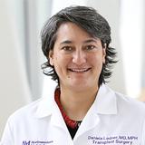 Daniela P Ladner合作论文数Division of Organ Transplantation, Department of Surgery, Feinberg School of Medicine, Northwestern University;Department of Medical Social Sciences, Feinberg School of Medicine, Northwestern University;National Institute of Diabetes and Digestive and Kidney Diseases, National Institutes of Health;Northwestern Medicine15
 Anna C. Pavlick合作论文数Weill Cornell Medical College, Cornell University;NewYork-Presbyterian Hospital15
 Grant Mcarthur合作论文数Peter MacCallum Cancer Centre;Victorian Comprehesive Cancer Centre;University of Melbourne15Ortiz-Urda Susana合作论文数15




