Abstract The purpose of this study is to investigate the formation and maintenance of germinal centers in TLS by defining mechanisms and spatial patterns of CXCL13 chemokine gradients in tumor tissue. In secondary lymphoid tissue, it is recognized that CXCL13 is immobilized by follicular dendritic cells (FDCs), but the mechanisms of immobilization remain unclear. Furthermore, spatial patterns and mechanisms of CXCL13 immobilization in TLS are unknown. We developed a CXCL13 Immunohistochemistry /in situ hybridization (ICH/ISH) dual stain to determine which cells are responsible for CXCL13 production and display. We stained 3 formalin-fixed paraffin-embedded tonsil specimens and found that CXCL13 protein and mRNA staining have distinct localizations. Specifically, spatial analyses were performed on 10 individual follicular structures from each tonsil to compare the percent surface area of either germinal centers or mantle zones (n=30) demonstrating staining by IHC or ISH. This analysis revealed that CXCL13 protein was highly expressed in germinal centers and expressed relatively lower in mantle zones (31% vs 12% surface area, p<0.0001). However, CXCL13 ISH staining was the inverse: mRNA expression was lower in germinal centers, and higher in mantle zones (20% vs 37%, p<0.0001). Flow cytometry analysis of fresh tonsil tissue revealed stromal cells displaying CXCL13, (PDPN+CD31-CXCL13+), supporting the CXCL13 IHC findings. Similarly, spatial transcriptomics showed similar findings to the CXCL13 ISH, providing additional validation of the spatial expression pattern. Together, these results support a model where CXCL13 is primarily secreted from mantle zones, then immobilized in germinal centers. To investigate if these findings extend to TLS in tumor tissues, we applied our CXCL13 IHC/ISH dual stain to lung cancer tissue from the definitive resection specimen from patients treated with neoadjuvant anti-PD-1 immunotherapy. Spatial analyses revealed that similar to what was observed in tonsil, the density of CXCL13 mRNA is highly correlated with CXCL13 protein staining (n=3, Spearman’s r: 0.9, p<9.311e-19), though these do not immediately overlap spatially, with an average distance of 30 μm between mRNA and protein expression, suggesting capture of the diffusing CXCL13. We also observed that stromal cells in mature TLS immobilize CXCL13, but stromal cells in immature TLS do not. In addition, we found CXCL13 immobilized on elastin fibers near mRNA staining. These findings show that CXCL13 immobilization may contribute to the formation and maintenance of TLS. New therapies enhancing the formation and maturation of TLS by modulating CXCL13 may warrant exploration. Ongoing single cell RNA sequencing and in vitro studies are focused on determining the mechanisms responsible for the immobilization of CXCL13 in lymphoid and tumor tissue. Citation Format: Jonathan Rex Skidmore, Aleksandra Ogurtsova, Grant Salvucci, Victoria Jacobs, Logan Engle, Robert A. Anders, Drew M. Pardoll, Janis Marie Taube. CXCL13 secretion and immobilization show distinct geographies within secondary and tertiary lymphoid structures (TLS) [abstract]. In: Proceedings of the American Association for Cancer Research Annual Meeting 2026; Part 1 (Regular Abstracts); 2026 Apr 17-22; San Diego, CA. Philadelphia (PA): AACR; Cancer Res 2026;86(7 Suppl):Abstract nr 2219.
This study investigates the spatial organization and the mechanisms responsible for forming soluble and insoluble CXCL13 chemokine gradients. CXCL13 attracts B-cells to follicles in secondary lymphoid tissue and tertiary lymphoid structures (TLS). Cosgrove’s et al. ex vivo tonsil tissue studies showed that CXCL13 forms a soluble and insoluble chemokine gradient via immobilization in the extra cellular matrix. Mechanisms of CXCL13 secretion, immobilization and spatial distribution are unknown. Quantitative CXCL13 IHC/ISH co-stain analysis in human tonsil demonstrates abundant CXCL13 mRNA in mantle zone compared to germinal center cells (p < 0.0001). However, more CXCL13 protein localizes in germinal center compared to mantle zone cells (p < 0.0001). Flow cytometry confirmed CXCL13 presence on PDPN+ CD31- stromal cells in tonsil. The ISH tissue staining pattern was validated and candidate genes responsible for CXCL13 immobilization were identified using public spatial and single-cell transcriptomics datasets. Lung cancer TLS showed a similar IHC/ISH tissue staining pattern. Additionally, we found that CXCL13 is immobilized by elastin fibers surrounding TLS. This study uncovers potential mechanisms regulating CXCL13 chemokine gradients. Enhancing intratumoral CXCL13 gradients may create a more favorable tumor microenvironment which could improve the effectiveness of immune based anti-cancer therapies. Supported by R01CA142779 from NCI & Mark Foundation for Cancer Research Cytokines and Chemokines and Their Receptors (CCR)
Pancreatic ductal adenocarcinoma (PDAC) is a rapidly progressing cancer that responds poorly to immunotherapies. Intratumoral tertiary lymphoid structures (TLS) have been associated with rare long-term PDAC survivors, but the role of TLS in PDAC and their spatial relationships within the context of the broader tumor microenvironment remain unknown. In this study, we report the generation of a spatial multiomic atlas of PDAC tumors and tumor-adjacent lymph nodes from patients treated with combination neoadjuvant immunotherapies. Using machine learning-enabled hematoxylin and eosin image classification models, imaging mass cytometry, and unsupervised gene expression matrix factorization methods for spatial transcriptomics, we characterized cellular states within and adjacent to TLS spanning distinct spatial niches and pathologic responses. Unsupervised learning identified TLS-specific spatial gene expression signatures that are significantly associated with improved survival in patients with PDAC. We identified spatial features of pathologic immune responses, including intratumoral TLS-associated B-cell maturation colocalizing with IgG dissemination and extracellular matrix remodeling. Our findings offer insights into the cellular and molecular landscape of TLS in PDACs during immunotherapy treatment.
Probabilistic spatial modelling techniques developed on large-scale tumor-immune Atlases (~35M individually mapped cells; 50,000 high power fields) were used to characterize predictive features of treatment-responsive lung cancer. We identified CD8+FoxP3+ cell density as a robust pre-treatment biomarker for outcomes across disease stages and therapy types. In parallel, single-cell RNAseq studies of CD8+FoxP3+ T-cells revealed an activated, early effector phenotype, substantiating an anti-tumor role, and contrasting with CD4+FoxP3+ T-regulatory cells. A spatial biomarker was developed using an empirical probabilistic model to define the immediate cell neighbors or niche surrounding CD8+FoxP3+ cells and proximity to the tumor-stromal boundary. The resultant 'Diversity of Niches Unlocking Treatment Sensitivity (DONUTS)' are more prevalent than the CD8+FoxP3+ cells themselves, mitigating sampling error in small biopsies. Further, the DONUTS only require four markers, are additive to PD-L1, and associate with tertiary lymphoid structure counts. Taken together, the DONUTS represent a next-generation predictive biomarker poised for clinical implementation.
Sialic acids are overexpressed in many cancers, and binding of sialic acid via sialic acid binding immunoglobulin-like lectins (Siglecs) may contribute significantly to immune evasion and cancer progression. This important resistance mechanism in the tumor immune microenvironment has been understudied, partially due to the lack of useful reagents. Here, we developed and optimized an immunohistochemistry staining protocol for novel reagents that detect 3 types of Siglec-engaging sialoglycans (HYDRA-3, -7, and -9, which detect sialoglycans recognized by Siglec-3, -7, and -9, respectively) in the tumor immune microenvironment. We evaluated HYDRA staining across 10 different cancer types across whole slides, finding that HYDRA-9 exhibited the highest overall staining range, with HYDRA-3 and -7 showing lower to moderate staining across all tested tumor types. To correlate HYDRA staining patterns and immune infiltration in melanoma, we stained melanoma tissue microarrays with the 3 HYDRA reagents and compared HYDRA staining profiles with a 6-plex multiplex immunofluorescence panel targeting CD8, CD163, FoxP3, PD1, PDL1, and Sox10/S100. Siglec-3 and -9 sialoglycan ligand expression negatively correlated with CD8 T cell infiltration (r = -0.28/P = .002 and r = -0.29/P = .001, respectively), particularly at the tumor-stromal interface (r = -0.37/P < .001 and r = -0.44/P < .001, respectively). Additionally, a high ratio of Siglec-3 and -9 ligand expression at the tumor-stromal interface versus the tumor core was associated with reduced overall survival (Hazard's ratio: 2.60 and 2.11, respectively), whereas CD8 infiltration was not associated with survival outcomes in our cohort. Taken together, the expression levels and spatial distribution of Siglec-engaging sialoglycans may play a role in patient prognosis, potentially representing a biomarker of survival that is independent of conventional metrics of an inflamed tumor microenvironment. This study highlights the need for further investigation of Siglec ligand expression as a predictive and prognostic biomarker of treatment response and resistance.
Merkel cell carcinoma (MCC) is an aggressive cutaneous malignancy with a poor prognosis. One of the major mechanisms of immune evasion in MCC involves downregulation of major histocompatibility complex class I (MHC-I). Anti-PD-1/programmed death ligand 1 checkpoint inhibitors have revolutionized treatment for MCC, producing objective responses in approximately 50% of patients, and are now the standard of care; however, a substantial proportion of patients either fail to respond or develop resistance to checkpoint inhibitors. Given these recent successes, identification of other targetable immune checkpoints in the MCC tumor microenvironment is of great interest. Additionally, γδ T cells may play critical roles in response to MHC-I-deficient cancers; therefore, evaluating γδ T cells as a prognostic biomarker is warranted. We characterized the expression of programmed death ligand 1, PD-1, CD3, CD8, lymphocyte activation gene 3 (LAG-3), MHC-I, and γδ T cells by immunohistochemistry in a preimmunotherapy retrospective cohort of 54 cases of MCC and quantified expression levels and marker density using HALO software. The increased density of LAG-3 and γδ T cells correlated with other markers of an inflamed tumor microenvironment, with significant positive associations across all 6 markers (P < .002). Reflective of their putative role in the response to MHC-I-suppressed cancers, cases with low human leukocyte antigen I density showed a trend toward a higher ratio of γδ T cells:CD3+ T cells (Spearman r = -0.1582; P = .21). Importantly, high CD3 density (hazard ratio [HR], 0.23; P = .002), LAG-3 density (HR, 0.47; P = .037), γδ T-cell density (HR, 0.26; P = .02), and CD8 density (HR, 0.27; P = .03) showed associations with improved progression-free survival. Conditional tree analysis demonstrated that high CD8 and TCRδ expression were nonsignificant predictors of improved progression-free survival and overall survival. Overall, LAG-3 is expressed in MCC infiltrates and is prognostic in preimmunotherapy MCC, suggesting a potential role for LAG-3 inhibition in MCC. Additionally, CD8 and γδ T cells may play a critical role in the response to MCC, and γδ T-cell density may represent a novel biomarker in MCC.
Abstract Background: In pancreatic ductal adenocarcinoma (PDAC), rare long-term survivors correlate with high intratumoral tertiary lymphoid structure (TLS) density. This finding prompted our investigation of clinically viable strategies to induce TLS in patients with immune-excluded tumors. We previously reported the induction of intratumoral TLS following administration of a neoadjuvant GM-CSF-secreting allogeneic vaccine (GVAX) to PDAC patients (NCT00727441). However, no clinical benefit was observed, likely owing to immune tolerance mechanisms governing the PDAC tumor microenvironment (TME). We previously observed upregulation of both the PD-1 and 4-1BB pathways with GVAX, and thus in a subsequent neoadjuvant trial combined GVAX with PD-1 blockade and 4-1BB agonism (NCT02451982) which was associated with pathologic responses. We hypothesized this combination strategy induced TLS of higher maturity and anti-tumor activity compared to GVAX alone. Methods: To explore how this therapeutic strategy affected TLS morphology and intercellular crosstalk, we leveraged the Visium spatial transcriptomics platform and a 35-marker customized TLS panel for imaging mass cytometry. We generated cellular and molecular maps of the TME after neoadjuvant treatment in 26 PDAC patients (GVAX n=19, GVAX+aPD1 n=2, GVAX+aPD1+a41BB n=5). To compare TLS maturation in parallel with secondary lymphoid organ-mediated tumor immunity, we also profiled tumor-adjacent lymph nodes. We applied unsupervised learning with non-negative matrix factorization (NMF) and trained AI-enabled image classification models to characterize cellular states within tissue structures of interest. Results: We identified spatial gene expression NMF patterns in PDAC TLS spanning across distinct morphologies and neoadjuvant treatment arms. Intratumoral TLS after GVAX were found to propagate activated B cells expressing immunoglobulins that infiltrated into malignant cellular niches. TLS NMF patterns were also associated with autoimmune disease signatures, such as diabetes, in a subset of patients. We scored TLS using tumor-draining lymph nodes as a reference and found increased maturation of TLS after PD-1 blockade, while addition of 4-1BB agonism significantly boosted the cytotoxic NK/T cell compartment compared to GVAX alone. Conclusions: We present machine learning approaches for spatial multi-omics analysis to characterize the TLS-enriched TME. We mined genome-wide spatial TLS gene expression patterns elucidating the spatial dynamics of humoral immunity of rare immunotherapy pathologic responders in PDAC. Altogether our findings shed light on the plasticity of TLS in neoadjuvant immunotherapy and suggest future immunotherapy approaches should target both humoral and cytotoxic NK/T cell compartments to augment responses in solid tumors. Citation Format: Dimitrios N. Sidiropoulos, Sarah M. Shin, Alexander Girgis, Daniel H. Shu, Janelle Montagne, Atul Deshpande, Jeanette A. Johnson, Lucie Dequiedt, Victoria Jacobs, Aleksandra Ogurtsova, Guanglan Mo, Xuan Yuan, Genevieve Stein-O’Brien, Mark Yarchoan, Qingfeng Zhu, Ashley Kiemen, Elizabeth M. Jaffee, Lei Zheng, Won Jin Ho, Robert Anders, Elana J. Fertig, Luciane T. Kagohara. Machine learning integrating spatial omics uncovers humoral immunity patterns in intratumoral tertiary lymphoid structures in pancreatic cancer pathologic responders [abstract]. In: Proceedings of the American Association for Cancer Research Annual Meeting 2024; Part 1 (Regular Abstracts); 2024 Apr 5-10; San Diego, CA. Philadelphia (PA): AACR; Cancer Res 2024;84(6_Suppl):Abstract nr 1159.
 Taube Janis M合作论文数27Haiying Xu合作论文数18
 Robert Albert Anders合作论文数Department of Pathology, Johns Hopkins University School of Medicine18
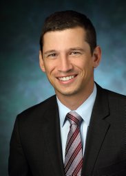 Joel C. Sunshine合作论文数Department of Biomedical Engineering, Johns Hopkins University School of Medicine13
 Ludmila Danilova合作论文数Sidney Kimmel Comprehensive Cancer Center, Johns Hopkins University;School of Medicine, Johns Hopkins University12
 Lipson Evan J合作论文数Sch Med, Johns Hopkins Univ11Berry Sneha合作论文数The Wilmer Eye Institute, Johns Hopkins University School of Medicine11
 Tricia Cottrell合作论文数Sinclair Cancer Research Institute, Faculty of Health Sciences, Queen's University11Peter Nguyen合作论文数8
 Elana J. Fertig合作论文数Institute for Genome Sciences, University of Maryland School of Medicine8Qingfeng Zhu合作论文数Pathology, and8Engle Elizabeth L合作论文数8
 Elizabeth M. Jaffee合作论文数Department of Pathology, School of Medicine, Johns Hopkins University;Institute for Clinical & Translational Research, Johns Hopkins University8Daniel Shu合作论文数7
 Xuan Yuan合作论文数7
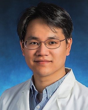 Won Jin Ho合作论文数Department of Otolaryngology-Head and Neck Surgery, School of Medicine, Johns Hopkins University7
 Benjamin Jonathan Green合作论文数Department of Animal Science, University of Vermont7
 Mark Yarchoan合作论文数Department of Oncology, Sidney Kimmel Comprehensive Cancer Center, Johns Hopkins University School of Medicine7Kagohara Luciane T合作论文数Sidney Kimmel Comprehensive Cancer Center, Johns Hopkins University School of Medicine7
 Drew Mark Pardoll合作论文数Department of Oncology, School of Medicine, Johns Hopkins University;Bloomberg~Kimmel Institute for Cancer Immunotherapy, School of Medicine, Johns Hopkins University;Sidney Kimmel Comprehensive Cancer Center, School of Medicine, Johns Hopkins University7Sarah Shin合作论文数6Jessica Esandrio合作论文数6Julie E Stein合作论文数6Janelle M Montagne合作论文数6
 Dimitrios N Sidiropoulos合作论文数Sidney Kimmel Comprehensive Cancer Center, Johns Hopkins University School of Medicine;Convergence Institute, Johns Hopkins University;Bloomberg-Kimmel Institute for Cancer Immunotherapy, Johns Hopkins University School of Medicine;Cellular and Molecular Medicine, Johns Hopkins University School of Medicine;Johns Hopkins University School of Medicine, Johns Hopkins University School of Medicine6
 Alexander Girgis合作论文数Biomedical Engineering, Johns Hopkins University6Mo Guanglan合作论文数6
 Giraldo Nicolas A合作论文数UPMC Univ Paris 06, Sorbonne Universités6Logan L. Engle合作论文数5Daphne Wang合作论文数5
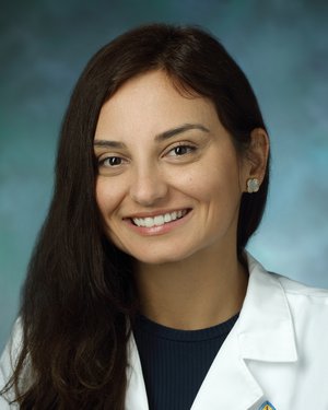 Farah Succaria合作论文数Department of Dermatology, School of Medicine, Johns Hopkins University5Kaunitz Genevieve合作论文数5Abha Soni合作论文数4Atul Deshpande合作论文数4
 Genevieve Stein-O'Brien合作论文数Johns Hopkins University;4
 Alexander Szalay合作论文数Department of Physics and Astronomy, Krieger School of Arts and Sciences, Johns Hopkins University;Department of Computer Science, Whiting School of Engineering, Johns Hopkins University;Institute for Data Intensive Engineering and Science, Johns Hopkins University;Eötvös University4
 Cornish Toby C合作论文数Dept Pathol, Johns Hopkins Univ4
 Alan Keith Meeker合作论文数Department of Pathology, School of Medicine, The Johns Hopkins University;Department of Oncology, School of Medicine, The Johns Hopkins University;Department of Urology, School of Medicine, The Johns Hopkins University4
 Lei Zheng合作论文数Department of Oncology and Surgery, School of Medicine, John Hopkins University;Pancreatic Cancer Precision Medicine Center of Excellence,;Sidney Kimmel Comprehensive Cancer Center,4Margaret Eminizer合作论文数3
 Jae W. Lee合作论文数3Hanfei Qi合作论文数3Lucie Dequiedt合作论文数3
 Erin Coyne合作论文数3Julie Stein Deutsch合作论文数3Kabeer Munjal合作论文数3Kathryn Howe合作论文数3Tamara Y Lopez-Vidal合作论文数3Jonathan Lai合作论文数3




