Abstract The purpose of this study is to investigate the formation and maintenance of germinal centers in TLS by defining mechanisms and spatial patterns of CXCL13 chemokine gradients in tumor tissue. In secondary lymphoid tissue, it is recognized that CXCL13 is immobilized by follicular dendritic cells (FDCs), but the mechanisms of immobilization remain unclear. Furthermore, spatial patterns and mechanisms of CXCL13 immobilization in TLS are unknown. We developed a CXCL13 Immunohistochemistry /in situ hybridization (ICH/ISH) dual stain to determine which cells are responsible for CXCL13 production and display. We stained 3 formalin-fixed paraffin-embedded tonsil specimens and found that CXCL13 protein and mRNA staining have distinct localizations. Specifically, spatial analyses were performed on 10 individual follicular structures from each tonsil to compare the percent surface area of either germinal centers or mantle zones (n=30) demonstrating staining by IHC or ISH. This analysis revealed that CXCL13 protein was highly expressed in germinal centers and expressed relatively lower in mantle zones (31% vs 12% surface area, p<0.0001). However, CXCL13 ISH staining was the inverse: mRNA expression was lower in germinal centers, and higher in mantle zones (20% vs 37%, p<0.0001). Flow cytometry analysis of fresh tonsil tissue revealed stromal cells displaying CXCL13, (PDPN+CD31-CXCL13+), supporting the CXCL13 IHC findings. Similarly, spatial transcriptomics showed similar findings to the CXCL13 ISH, providing additional validation of the spatial expression pattern. Together, these results support a model where CXCL13 is primarily secreted from mantle zones, then immobilized in germinal centers. To investigate if these findings extend to TLS in tumor tissues, we applied our CXCL13 IHC/ISH dual stain to lung cancer tissue from the definitive resection specimen from patients treated with neoadjuvant anti-PD-1 immunotherapy. Spatial analyses revealed that similar to what was observed in tonsil, the density of CXCL13 mRNA is highly correlated with CXCL13 protein staining (n=3, Spearman’s r: 0.9, p<9.311e-19), though these do not immediately overlap spatially, with an average distance of 30 μm between mRNA and protein expression, suggesting capture of the diffusing CXCL13. We also observed that stromal cells in mature TLS immobilize CXCL13, but stromal cells in immature TLS do not. In addition, we found CXCL13 immobilized on elastin fibers near mRNA staining. These findings show that CXCL13 immobilization may contribute to the formation and maintenance of TLS. New therapies enhancing the formation and maturation of TLS by modulating CXCL13 may warrant exploration. Ongoing single cell RNA sequencing and in vitro studies are focused on determining the mechanisms responsible for the immobilization of CXCL13 in lymphoid and tumor tissue. Citation Format: Jonathan Rex Skidmore, Aleksandra Ogurtsova, Grant Salvucci, Victoria Jacobs, Logan Engle, Robert A. Anders, Drew M. Pardoll, Janis Marie Taube. CXCL13 secretion and immobilization show distinct geographies within secondary and tertiary lymphoid structures (TLS) [abstract]. In: Proceedings of the American Association for Cancer Research Annual Meeting 2026; Part 1 (Regular Abstracts); 2026 Apr 17-22; San Diego, CA. Philadelphia (PA): AACR; Cancer Res 2026;86(7 Suppl):Abstract nr 2219.
TPS9596 Background: Prior studies demonstrated use of neoadjuvant plus adjuvant immune checkpoint inhibitors (ICIs) improves event-free survival (EFS) compared with upfront surgery and adjuvant ICI therapy alone, supporting that peri-operative (neoadjuvant plus adjuvant) ICI therapy improves survival outcomes in patients (pts) with clinical stage III and resectable IV melanoma (Mel). Initial efforts in Mel trials to explore combined blockade by anti-programmed cell death-1 (anti–PD-1) and anti-lymphocyte activation gene 3 (anti–LAG-3) antibodies produced incrementally better efficacy than blockade of the PD-1 pathway alone. However, these efforts may not have provided optimal blockade of the two pathways. We have utilized VelocImmune technology to create potentially best-in-class, high-affinity, fully human immunoglobulin G4-blocking antibodies, fianlimab (FIAN; anti–LAG-3) and cemiplimab (CEMI; anti–PD-1). In a multicohort study (NCT03005782), FIAN + CEMI demonstrated reproducibly high clinical activity (objective response rate [ORR]: 57%; median progression-free survival: 24 months; N=98) in three independent cohorts of pts who were naïve to anti–PD-1 treatment in the advanced Mel setting, with an acceptable safety profile. Thus, the combination of FIAN + CEMI warrants an investigation as a peri-operative regimen in resectable, clinically detectable, high-risk, stage III and IV cutaneous Mel. Methods: This is a randomized Phase 2 peri-operative study (NCT06190951) in pts with clinical stage III/IV Mel with resectable disease. Pts will receive 3 cycles of neoadjuvant therapy followed by complete surgical resection, and continue with an optional 15 cycles of adjuvant therapy, based on pathological response. The primary objective is to compare the effect of FIAN + CEMI versus CEMI alone as measured by the pathological complete response (pCR) rate. Approximately 150 pts will be randomized 1:1:1 to three arms (intravenously once every 3 weeks): Arm A, CEMI 350 mg + placebo; Arm B, High Dose FIAN + CEMI 350 mg; Arm C, Low Dose FIAN + CEMI 350 mg. Pts will be stratified based on tumor, node, metastasis (TNM) stage and geographical region. Key inclusion criteria: age ≥18 years; resectable clinical stage III/IV histologically confirmed Mel; pts with stage III Mel must have clinically detectable disease; Eastern Cooperative Oncology Group performance status 0 or 1; adequate bone marrow, hepatic, and kidney function. The primary endpoint is pCR rate by blinded independent pathological review performed centrally. The secondary endpoints are pCR rate (by local assessment), major pathological response (by local and central review), EFS, overall survival, distant metastasis-free survival, relapse-free survival, ORR, safety, pharmacokinetics, immunogenicity, and patient-reported outcomes. Clinical trial information: NCT06190951 .
Neoadjuvant immunotherapies have shown antitumor activity in melanoma. Substudy 02C of the global, rolling-arm, phase 1/2, adaptive-design KEYMAKER-U02 trial is evaluating neoadjuvant pembrolizumab (anti-PD-1) alone or in combination, followed by adjuvant pembrolizumab, for stage IIIB-D melanoma. Here we report results from the first three arms: pembrolizumab plus vibostolimab (anti-TIGIT), pembrolizumab plus gebasaxturev (coxsackievirus A21) and pembrolizumab monotherapy. Pathologic complete responses occurred in 10 of 26 patients (38%) with pembrolizumab plus vibostolimab, 7 of 25 (28%) with pembrolizumab plus gebasaxturev and 6 of 15 (40%) with pembrolizumab monotherapy. Major pathologic responses occurred in 13 (50%), 10 (40%) and 7 (47%) patients, respectively. Safety was manageable. Treatment-related adverse events occurred in 24 of 26 patients (92%) with pembrolizumab plus vibostolimab, 21 of 25 (84%) with pembrolizumab plus gebasaxturev and 12 of 15 (80%) with pembrolizumab monotherapy; grade 3 or 4 treatment-related adverse events occurred in 2 (8%), 7 (28%) and 1 (7%) patient in each arm, respectively. No deaths due to adverse events occurred. Exploratory objective responses per RECIST v1.1 were observed in 13 (50%), 8 (32%) and 4 (27%) patients, in each arm, respectively. In a post hoc analysis, scores for tumor mutational burden and an 18-gene T cell-inflamed gene expression profile were generally higher in patients with major pathologic response. Longer follow-up will provide insight into the incremental benefit of combining neoadjuvant pembrolizumab with other therapies in stage IIIB-D melanoma. ClinicalTrials.gov registration: NCT04303169 .
Objectives Multiplex immunohistochemistry and immunofluorescence (mIHC/IF) are emerging technologies that can be used to help define complex immunophenotypes in tissue, quantify immune cell subsets, and assess the spatial arrangement of marker expression. mIHC/IF assays require concerted efforts to optimize and validate the multiplex staining protocols prior to their application on slides. The best practice guidelines for staining and validation of mIHC/IF assays across platforms were previously published by this task force. The current effort represents a complementary manuscript for mIHC/IF analysis focused on the associated image analysis and data management. Methods The Society for Immunotherapy of Cancer convened a task force of pathologists and laboratory leaders from academic centers as well as experts from pharmaceutical and diagnostic companies to develop best practice guidelines for the quantitative image analysis of mIHC/IF output and data management considerations. Results Best-practice approaches for image acquisition, color deconvolution and spectral unmixing, tissue and cell segmentation, phenotyping, and algorithm verification are reviewed. Additional quality control (QC) measures such as batch-to-batch correction and QC for assembled images are also discussed. Recommendations for sharing raw outputs, processed results, key analysis programs and source code, and representative photomicrographs from mIHC/IF assays are included. Lastly, multi-institutional harmonization efforts are described. Conclusions mIHC/IF technologies are maturing and are routinely included in research studies and moving towards clinical use. Guidelines for how to perform and standardize image analysis on mIHC/IF-stained slides will likely contribute to more comparable results across laboratories and pave the way for clinical implementation. A checklist encompassing these two-part guidelines for the generation of robust data from quantitative mIHC/IF assays will be provided in a third publication from this task force. While the current effort is mainly focused on best practices for characterizing the tumor microenvironment, these principles are broadly applicable to any mIHC/IF assay and associated image analysis.
8009 Background: In AEGEAN, perioperative durvalumab (D) + neoadj CT significantly improved the primary endpoints of event-free survival (EFS) and pathological complete response (pCR) vs neoadj CT alone in pts with R-NSCLC. Prior analyses of AEGEAN suggest that pts without ctDNA clearance during neoadj Tx or with molecular residual disease (MRD; i.e. ctDNA detected) at a landmark timepoint after Sx (adj C1D1) had worse outcomes. Using data from all biomarker-evaluable pts, we report exploratory analyses for associations of post-Sx MRD status with pt characteristics, neoadj ctDNA dynamics, pathological response, genomic mutations and outcomes. Methods: AEGEAN is a double-blind PBO-controlled study (NCT03800134). Adults with Tx-naïve R-NSCLC (stage II–IIIB[N2]) and ECOG PS 0/1 were randomized 1:1 to receive neoadj platinum-based CT + D or PBO IV (Q3W, 4 cycles) before Sx followed by D or PBO IV (Q4W, 12 cycles) after Sx. Efficacy was assessed in the mITT population, which excluded pts with known EGFR / ALK aberrations. ctDNA analysis was performed on plasma collected before each neoadj Tx cycle, Sx, and adj C1, C3/4 and C10/11 using pt-specific tumor-informed assays. Whole exome sequencing analysis of diagnostic tumor biopsies was performed to identify mutations associated with MRD status at the post-Sx landmark. Results: Among MRD-evaluable pts, 10% (17/168) were MRD-positive (D, n=10; PBO, n=7) and 90% (151/168) were MRD-negative (D, n=78; PBO, n=73) at the landmark timepoint (median 6.9 wk post-Sx). 88% [15/17] of MRD-positive pts were initially diagnosed with stage III disease. In the D arm, the majority of MRD-positive pts (9/10) also had ctDNA detected at the pre-Sx visit. No MRD-positive pts in the D arm had pCR or major pathological response. As expected, overall disease-free survival (DFS) rates at 12 mo were worse in MRD-positive (14.3%; 95% CI, 2.4–36.3) vs MRD-negative pts (89.3%; 95% CI, 82.6–93.5). In both arms, MRD-positive pts had worse DFS outcomes vs MRD-negative pts (D: HR, 21.28; 95% CI, 7.70–58.83; PBO: HR, 14.29; 95% CI, 4.94–41.36) with DFS trends favoring the D vs PBO arm, particularly in pts with no ctDNA detected (MRD-negative: HR, 0.56; 95% CI, 0.26–1.20; MRD-positive: HR, 0.78; 95% CI, 0.26–2.36). Mutated genes associated with MRD-positive status in the D arm included KEAP1 and KMT2C ; despite small pt numbers, EFS benefit in the D vs PBO arm was not evident in pts with the mutations (m) (KEAP1 m: HR, 1.39; 95% CI, 0.29–6.77; KMT2C m : HR, 2.03; 95% CI, 0.70–5.91). In contrast, EFS benefit was evident in pts with wild type (wt) ( KEAP1 wt: HR, 0.54; 95% CI, 0.36–0.79; KMT2C wt: HR, 0.52; 95% CI, 0.35–0.78). Conclusions: Exploratory analyses based on post-Sx MRD status and genomic analysis identified a small high-risk subgroup of pts with markedly worse prognosis with potentially reduced benefit from the AEGEAN regimen. Clinical trial information: NCT03800134 .
In CheckMate 816, lower %RVT in primary tumor (PT) and lymph node (LN) after neoadjuvant (neoadj) NIVO + chemo correlated with improved EFS in patients (pts) with resectable NSCLC. To further evaluate %RVT as a surrogate for EFS, we report an exploratory analysis of efficacy with adjuvant (adj) NIVO after neoadj treatment (tx) by LN involvement, nodal (N) status, and %RVT in PT and LN in CheckMate 77T. Pts with resectable stage IIA-IIIB NSCLC were randomized to neoadj NIVO + chemo Q3W (up to 4 cycles [cyc]) followed by adj NIVO Q4W (up to 13 cyc) or neoadj placebo (PBO) + chemo Q3W (up to 4 cyc) followed by adj PBO Q4W (up to 13 cyc). Primary endpoint: EFS per BICR. This analysis, which included pts with pathologically evaluable samples who had definitive surgery and ≥ 1 adj tx dose, assessed EFS by LN involvement, N status, %RVT in PT and LN, and associations between %RVT and EFS per time-dependent ROC curve analysis. BL characteristics were similar between tx arms (NIVO, 123; PBO, 134; median f/u, 33.3 mo). NIVO improved EFS v PBO regardless of LN involvement or N status (Table). A higher proportion of pts treated with NIVO had 0% RVT in PT and/or LN v PBO (52% v 20%). In pts with LN involvement, 2-y EFS rates with NIVO were higher in pts with 0% RVT in both PT and LN (90%) or 0% RVT in PT or LN (85%) v > 0% RVT in both PT and LN (76%). Area under the ROC curve for %RVT-PT in pts with PT-only disease was 0.83. 2-y EFS rates with NIVO were 94%, 77%, and 50% in pts with 0-5%, > 5-80%, and > 80% RVT-PT, respectively; similar results were seen in all pts with pathologically evaluable samples whether they received adj tx or not. In this exploratory analysis, NIVO improved EFS v PBO, particularly in pts with LN involvement and regardless of N status. %RVT also associated with EFS in a continuous manner, supporting %RVT as a surrogate for EFS and highlighting its prognostic value in pts who receive perioperative NIVO. Julie Stein Deutsch, Ashley Cimino-Mathews, Elizabeth Thompson, Edward Gabrielson, Peter Illei, Jaroslaw Jedrych, Ezra Baraban, Alex S. Baras, Mariano Provencio Pulla, Tina Cascone, Jonathan D. Spicer, Mark M. Awad, Fumihiro Tanaka, Jie He, Shun Lu, Cinthya Coronado Erdmann, Vipul Devas, Sumeena Bhatia, Janis M. Taube. Associations between percent residual viable tumor (%RVT) and efficacy with perioperative nivolumab (NIVO) for resectable NSCLC in CheckMate 77T [abstract]. In: Proceedings of the American Association for Cancer Research Annual Meeting 2025; Part 2 (Late-Breaking, Clinical Trial, and Invited Abstracts); 2025 Apr 25-30; Chicago, IL. Philadelphia (PA): AACR; Cancer Res 2025;85(8_Suppl_2):Abstract nr CT097.
Purpose: Co-mutations of the Kirsten rat sarcoma virus (KRAS) and serine/threonine kinase 11 (STK11) genes in advanced non-small cell lung cancer (NSCLC) are associated with immune checkpoint blockade (ICB) resistance. Although neoadjuvant chemoimmunotherapy is now a standard-of-care treatment for resectable NSCLC, the clinical and immunologic impacts of KRAS and STK11 co-mutations in this setting are unknown.Experimental Design: We evaluated and compared recurrence-free survival of resectable KRAS-mutated NSCLC tumors, with or without co-occurring STK11 mutations, treated with neoadjuvant ICB. Single-cell transcriptomics was performed on tumor-infiltrating T cells from seven KRASmut/STK11wt tumors and six KRAS and STK11 co-mutated (KRASmut/STK11mut) tumors.Results: Relative to KRASmut/STK11wt tumors, KRASmut/STK11mut exhibited significantly higher recurrence risk. Single-cell transcriptomics showed enhanced oxidative phosphorylation with evidence of decreased prostaglandin E2 signaling and increased IL-2 signaling in CD8+ tumor-infiltrating lymphocytes (TIL) from KRASmut/STK11mut tumors, a finding that was mirrored in KRASwt tumors that relapsed. TILs from KRASmut/STK11mut tumors expressed high levels of molecules associated with tumor residence, including CD39 and ZNF683 (HOBIT).Conclusions: These divergent T-cell transcriptional fates suggest that T-cell maintenance and residence may be detrimental to antitumor immunity in the context of neoadjuvant ICB for resectable NSCLC, regardless of KRAS mutation status. Our work provides a basis for future investigations into the mechanisms underpinning prostaglandin E2 signaling and IL-2 signaling as they relate to T-cell immunity to cancer and to divergent clinical outcomes in KRASmut/STK11mut NSCLC treated with neoadjuvant ICB.
INTRODUCTION:In AEGEAN, perioperative durvalumab plus neoadjuvant chemotherapy, versus neoadjuvant chemotherapy alone, significantly improved event-free survival (p = 0.004) and pathologic complete response (p < 0.001; primary end points; modified intention-to-treat [mITT] population, which excluded patients with known EGFR or ALK aberrations) with a manageable safety profile in patients with resectable (R)-NSCLC. Here, we report surgical outcomes from AEGEAN. METHODS:Patients with treatment-naive R-NSCLC (stage II-IIIB [N2]) and Eastern Cooperative Oncology Group performance status 0 or 1 were randomized (1:1) to platinum-based chemotherapy plus durvalumab or placebo intravenously (every 3 wk, 4 cycles) before surgery, followed by durvalumab or placebo (every 4 wk, 12 cycles). Surgical outcomes were summarized for the mITT population using descriptive statistics. RESULTS:A total of 737 out of 740 mITT patients received treatment, 366 and 371 in the durvalumab and placebo arms, respectively; 80.6% and 80.7% underwent surgery, 77.6% and 76.7% completed surgery, and of the 295 and 302 patients who underwent surgery, 17.3% and 22.2% had delayed surgery. The median time from last neoadjuvant dose to surgery was 34.0 days in both arms. Of the patients who underwent surgery, similar proportions had open (49.2% versus 50.7%) and minimally invasive (49.2% versus 47.0%) procedures; lobectomy was the most common procedure (88.1% versus 85.4%). R0 resection rates were numerically higher in the durvalumab versus placebo arm (94.7% versus 91.3%). The median time from surgery to first adjuvant dose was 50.0 versus 52.0 days. In exploratory analyses, a numerically higher proportion of patients in the durvalumab versus placebo arm with baseline N2 nodal status had downstaging from N2 to N0 (47.3% versus 40.2%) or, with baseline N1 nodal status, from N1 to N0 (53.6% versus 46.2%) after surgery. Similar proportions had surgical complication(s) (59.1% versus 60.1%), primarily grade 1 or 2 (53.0% versus 51.8%, modified safety analysis set). CONCLUSION:The addition of durvalumab to neoadjuvant chemotherapy had no detrimental effect on the feasibility, approach, type, or timing of surgery and was associated with a tolerable surgical safety profile, versus neoadjuvant chemotherapy alone. CLINICAL TRIAL INFORMATION:ClinicalTrials.Gov Identifier: NCT03800134.
Probabilistic spatial modelling techniques developed on large-scale tumor-immune Atlases (~35M individually mapped cells; 50,000 high power fields) were used to characterize predictive features of treatment-responsive lung cancer. We identified CD8+FoxP3+ cell density as a robust pre-treatment biomarker for outcomes across disease stages and therapy types. In parallel, single-cell RNAseq studies of CD8+FoxP3+ T-cells revealed an activated, early effector phenotype, substantiating an anti-tumor role, and contrasting with CD4+FoxP3+ T-regulatory cells. A spatial biomarker was developed using an empirical probabilistic model to define the immediate cell neighbors or niche surrounding CD8+FoxP3+ cells and proximity to the tumor-stromal boundary. The resultant 'Diversity of Niches Unlocking Treatment Sensitivity (DONUTS)' are more prevalent than the CD8+FoxP3+ cells themselves, mitigating sampling error in small biopsies. Further, the DONUTS only require four markers, are additive to PD-L1, and associate with tertiary lymphoid structure counts. Taken together, the DONUTS represent a next-generation predictive biomarker poised for clinical implementation.
Sialic acids are overexpressed in many cancers, and binding of sialic acid via sialic acid binding immunoglobulin-like lectins (Siglecs) may contribute significantly to immune evasion and cancer progression. This important resistance mechanism in the tumor immune microenvironment has been understudied, partially due to the lack of useful reagents. Here, we developed and optimized an immunohistochemistry staining protocol for novel reagents that detect 3 types of Siglec-engaging sialoglycans (HYDRA-3, -7, and -9, which detect sialoglycans recognized by Siglec-3, -7, and -9, respectively) in the tumor immune microenvironment. We evaluated HYDRA staining across 10 different cancer types across whole slides, finding that HYDRA-9 exhibited the highest overall staining range, with HYDRA-3 and -7 showing lower to moderate staining across all tested tumor types. To correlate HYDRA staining patterns and immune infiltration in melanoma, we stained melanoma tissue microarrays with the 3 HYDRA reagents and compared HYDRA staining profiles with a 6-plex multiplex immunofluorescence panel targeting CD8, CD163, FoxP3, PD1, PDL1, and Sox10/S100. Siglec-3 and -9 sialoglycan ligand expression negatively correlated with CD8 T cell infiltration (r = -0.28/P = .002 and r = -0.29/P = .001, respectively), particularly at the tumor-stromal interface (r = -0.37/P < .001 and r = -0.44/P < .001, respectively). Additionally, a high ratio of Siglec-3 and -9 ligand expression at the tumor-stromal interface versus the tumor core was associated with reduced overall survival (Hazard's ratio: 2.60 and 2.11, respectively), whereas CD8 infiltration was not associated with survival outcomes in our cohort. Taken together, the expression levels and spatial distribution of Siglec-engaging sialoglycans may play a role in patient prognosis, potentially representing a biomarker of survival that is independent of conventional metrics of an inflamed tumor microenvironment. This study highlights the need for further investigation of Siglec ligand expression as a predictive and prognostic biomarker of treatment response and resistance.
BACKGROUND:Neoadjuvant nivolumab plus chemotherapy significantly improved pathological complete response and event-free survival in patients with resectable non-small-cell lung cancer (NSCLC) in a phase 3 trial. Data are needed on overall survival. METHODS:In this open-label, phase 3 trial, patients with stage IB to IIIA resectable NSCLC were randomly assigned to receive nivolumab plus chemotherapy or chemotherapy alone for three cycles, followed by surgery. The primary end points were event-free survival and pathological complete response. Here, we report the results of the planned analysis of overall survival. RESULTS:A total of 358 patients were concurrently assigned to receive nivolumab plus chemotherapy (179 patients) or chemotherapy alone (179 patients). The final analysis of overall survival significantly favored neoadjuvant nivolumab plus chemotherapy over chemotherapy (hazard ratio for death, 0.72; 95% confidence interval [CI], 0.523 to 0.998; P = 0.048). At a median follow-up of 68.4 months, the 5-year overall survival was 65.4% with nivolumab plus chemotherapy and 55.0% with chemotherapy alone, with consistency across most subgroups. In exploratory analyses, the 5-year overall survival in the nivolumab-plus-chemotherapy group was 95.3% (95% CI, 82.7 to 98.8) among the patients with a pathological complete response and 55.7% (95% CI, 46.9 to 63.7) among those without such a response; survival was 75.0% among the patients with presurgery clearance of circulating tumor DNA (ctDNA) and 52.6% among those without such clearance. No new safety signals were observed. CONCLUSIONS:Three cycles of neoadjuvant nivolumab plus chemotherapy significantly improved overall survival among patients with resectable NSCLC as compared with chemotherapy alone. (Funded by Bristol Myers Squibb; CheckMate 816 ClinicalTrials.gov number, NCT02998528.).
PURPOSE Cancer-related mortality rates among kidney transplant recipients (KTR) are high, but these patients have largely been excluded from trials of immune checkpoint inhibitors because of immunosuppression and risk of treatment-related allograft loss (TRAL). We conducted a prospective clinical trial testing nivolumab (NIVO) + tacrolimus (TACRO) + prednisone (PRED) ± ipilimumab (IPI) in KTR with advanced cutaneous cancers. METHODS Adult KTR with advanced melanoma or basal, cutaneous squamous, or Merkel cell carcinomas were eligible. Immunosuppression was standardized to TACRO (serum trough 2-5 ng/mL) + PRED 5 mg once daily. Patients then received NIVO 480 mg IV once every 4 weeks. The primary composite end point was partial or complete (tumor) response (CR) or stable disease per RECIST v1.1 without allograft loss at 16W. Patients with progressive disease (PD) could receive IPI 1 mg/kg IV + NIVO 3 mg/kg once every 3 weeks × 4 followed by NIVO. Donor-derived cell-free DNA (dd-cfDNA) levels were measured approximately once every 2 weeks as a potential predictor of allograft rejection. RESULTS Among eight evaluable patients, none met the trial's primary end point. All eight patients experienced PD on NIVO + TACRO + PRED; TRAL occurred in one patient. Six patients then received IPI + NIVO + TACRO + PRED. Best overall responses: two CR (one with TRAL) and four PD (one with TRAL). In total, 7 of 8 pre-NIVO tumor biopsies contained a paucity of infiltrating immune cells. In total, 2 of 5 on-NIVO biopsies demonstrated moderate immune infiltrates; both patients later experienced a CR to IPI + NIVO. In 2 of 3 patients with TRAL, dd-cfDNA elevations occurred 10 and 15 days before increases in serum creatinine. CONCLUSION In most KTR with advanced skin cancer, TACRO + PRED provides insufficient allograft protection and compromises immune-mediated tumor regression after administration of NIVO ± IPI. Elevated dd-cfDNA levels can signal treatment-related allograft rejection earlier than rises in serum creatinine.
BackgroundGastric carcinomas (GC) are aggressive malignancies, and only ~15% of patients respond to anti-programmed cell death (ligand) 1 (PD-(L)1) monotherapy. However, Epstein-Barr virus (EBV)-associated GCs (~5–10% of GCs) often harbor PD-L1 and PD-L2 chromosomal amplifications and robust CD8+ T cell infiltrates, and respond at a high rate to anti-PD-1. The current study compares the tumor immune microenvironments (TiMEs) of EBV+ versus EBV(−) GCs.MethodsOver 1000 cases of primary invasive GCs were screened to identify 25 treatment-naïve specimens for study (11 EBV+, 14 EBV(−)). Quantitative immunohistochemistry (IHC) was conducted for markers of immune cell subsets and co-regulatory molecules. Gene expression profiling (GEP) was performed on RNAs isolated from macrodissected areas of CD3+ T cell infiltrates abutting PD-L1+ stromal/tumor cells, using multiplex quantitative reverse transcriptase PCR for a panel of 122 candidate immune-related genes.ResultsIHC revealed that 17/25 GCs contained PD-L1+ stromal cells, with no significant difference between EBV+/- specimens; however, only 3/25 specimens (all EBV+) contained PD-L1+ tumor cells. CD8+ T cell densities were higher in EBV+ versus EBV(−) tumors (p=0.044). With GEP normalized to the pan-leukocyte markerPTPRC/CD45, EBV+ GCs overexpressedITGAE(CD103, marking intraepithelial T cells and a dendritic cell subset) and the interferon-inducible genesCXCL9andIDO1. In contrast, EBV(−) tumors overexpressed several functionally-related gene groups associated with myeloid cells (CD163,IL1A,NOS2, RIGI),immunosuppressive cytokines/chemokines (CXCL2,CXCR4,IL10, IL32),coinhibitory molecules (HAVCR2/TIM-3 andVSIR/VISTA), and adenosine pathway components (ENTPD1/CD39 andNT5E/CD73). Notably, compared with EBV+ GCs, EBV(−) GCs also overexpressed components of the cyclooxygenase 2 (COX-2)/prostaglandin E2 (PGE2) pathway associated with cancer-promoting inflammation, includingPTGS2/COX-2 (most highly upregulated gene, 32-fold, p=0.005); prostaglandin receptorsPTGER1(EP1; up 21-fold, p=0.015) andPTGER4(EP4; up twofold, p=0.022); and the major COX-2-inducing cytokineIL1B(up 11-fold, p=0.019). Consistent with these findings, COX-2 protein expression trended higher in EBV(−) versus EBV+ GCs (p=0.068).ConclusionsWhile certain markers of immunosuppression are found in the GC TiME regardless of EBV status, EBV(−) GCs, which are much more common than EBV+ GCs, overexpress components of the COX-2/PGE2 pathway. These findings provide novel insights into the immune microenvironments of EBV+ and EBV(−) GC, and offer potential targets to overcome resistance to anti-PD-(L)1 therapies.
8011 Background: In the phase 3 AEGEAN study (NCT03800134), perioperative D + neoadj chemotherapy (CT) vs neoadj CT alone significantly improved the primary EPs of event-free survival (EFS) and pathological complete response (pCR; absence of residual viable tumor [RVT] in resection specimen, incl. primary tumor and sampled lymph nodes) with manageable safety in pts with R-NSCLC (modified ITT [mITT] population). Here, we report exploratory analyses of pts in AEGEAN with baseline N2 nodal status. Methods: Pts with Tx-naïve R-NSCLC (stage II–IIIB[N2]; AJCC 8th ed) were randomized (1:1) to 4 cycles of platinum-based CT plus D 1500 mg IV or placebo (PBO) Q3W before surgery (Sx), followed by D or PBO (Q4W, 12 cycles) after Sx. The mITT population excluded pts with known EGFR/ ALK alterations. Efficacy was assessed in an mITT subpopulation with baseline N2 nodal status (per investigator). Safety was assessed in all N2 R-NSCLC pts who had ≥1 Tx dose. Results: Of 740 pts in the mITT population, 366 (49.5%) had baseline N2 nodal status (D, n = 181; PBO, n = 185). In the N2 subgroup, 83.4% in the D arm and 84.9% in the PBO arm completed 4 cycles of platinum-doublet CT; 77.9% and 77.8%, respectively, had Sx; and 73.5% and 71.9% completed Sx. In the N2 subgroup, similar to the overall mITT population, EFS was prolonged in the D vs PBO arm (HR, 0.63 [95% CI: 0.43–0.90]); rates of pCR (16.6% vs 4.9%; difference, 11.7% [95% CI: 5.6–18.4]) and major pathological response (MPR, ≤10% RVT in primary tumor; 32.6% vs 15.1%; difference, 17.5% [95% CI: 8.8–26.0]) were higher in the D vs PBO arm. While EFS benefit in the D vs PBO arm was similar among pts with single- (HR, 0.61 [95% CI: 0.39–0.94]) or multi-station N2 (HR, 0.69 [95% CI: 0.33–1.38]), pCR benefit was less pronounced in multi-station pts (difference in pCR rate, 13.9% [95% CI: 6.6–21.7] for single-station N2 vs 3.8% [95% CI: –9.2–18.8] for multi-station N2). Among pts in the N2 subgroup who had Sx, similar proportions in the D and PBO arms had open (47.5% vs 50.0%) and minimally invasive procedures (50.4% vs 46.5%); 9.2% vs 11.1% had pneumonectomy (9.2% vs 9.6% in the overall mITT population). Of pts in the N2 subgroup who completed Sx, the proportion with R0 resection in the D vs PBO arm (94.7% vs 91.7%) was similar to that in the overall mITT population. Sx was delayed in a similar proportion who had Sx in the D vs PBO arm (19.9% vs 23.6%, respectively), most commonly for logistical reasons (e.g., scheduling issues). Among 394 pts who received Tx (N2 safety analysis subset; D, n = 200; PBO, n = 194), max grade 3/4 any-cause AEs occurred in 38.5% vs 41.8% in the D and PBO arm, respectively, similar to rates in the overall safety analysis set. Conclusions: With clinically meaningful improvement in efficacy, no adverse impact on Sx outcomes and a manageable safety profile, the addition of perioperative D to neoadj CT remains a potential new Tx option for pts with N2 R-NSCLC. Clinical trial information: NCT03800134 .
 Drew Mark Pardoll合作论文数Department of Oncology, School of Medicine, Johns Hopkins University;Bloomberg~Kimmel Institute for Cancer Immunotherapy, School of Medicine, Johns Hopkins University;Sidney Kimmel Comprehensive Cancer Center, School of Medicine, Johns Hopkins University98
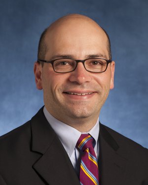 Robert Albert Anders合作论文数Department of Pathology, Johns Hopkins University School of Medicine69
 Tricia Cottrell合作论文数Sinclair Cancer Research Institute, Faculty of Health Sciences, Queen's University65
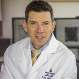 Lipson Evan J合作论文数Sch Med, Johns Hopkins Univ61
 Haiying Xu合作论文数56
 Charles Drake合作论文数Herbert Irving Comprehensive Cancer Center, Columbia University;The Janssen Pharmaceutical Companies of Johnson & Johnson49
 Julie Brahmer合作论文数School of Medicine, Johns Hopkins University;Sidney Kimmel Comprehensive Cancer Center, Johns Hopkins University;Bloomberg~Kimmel Institute for Cancer Immunotherapy, Johns Hopkins University;Johns Hopkins Sidney Kimmel Comprehensive Cancer Center46
 Suzanne Louise Topalian合作论文数Bloomberg-Kimmel Institute for Cancer Immunotherapy, Johns Hopkins University;School of Medicine, Johns Hopkins University43Patrick M Forde合作论文数39
 Kellie N Smith合作论文数Department of Oncology, The Johns Hopkins University School of Medicine35
 Jamie E. Chaft合作论文数Memorial Sloan Kettering Cancer Center32Tracee L. Mcmiller合作论文数30Julie E Stein合作论文数28
 Ludmila Danilova合作论文数Sidney Kimmel Comprehensive Cancer Center, Johns Hopkins University;School of Medicine, Johns Hopkins University28
 Hao Wang合作论文数Department of Oncology, Johns Hopkins University School of Medicine;Division of Biostatistics and Bioinformatics, Sidney Kimmel Comprehensive Cancer Center, Johns Hopkins University School of Medicine;Harvard T.H. Chan School of Public Health, Harvard University28Aleksandra Ogurtsova合作论文数27
 Valsamo Anagnostou合作论文数School of Medicine, Yale University27
 Peter B Illei合作论文数Department of Pathology, Memorial Sloan-Kettering Cancer Center27
 William H Sharfman合作论文数Department of Otolaryngology-Head and Neck Surgery, School of Medicine, Johns Hopkins University25Alan E. Berger合作论文数25
 Cimino-Mathews Ashley合作论文数Johns Hopkins Univ Hosp25
 Matthew Hellmann合作论文数Memorial Sloan Kettering Cancer Center25Gabrielson Edward合作论文数24
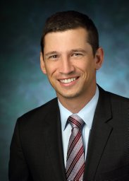 Joel C. Sunshine合作论文数Department of Biomedical Engineering, Johns Hopkins University School of Medicine24
 Victor E. Velculescu合作论文数Department of Genetic Medicine, School of Medicine, Johns Hopkins University;Delfi Diagnostics22
 William H. Westra合作论文数Icahn School of Medicine at Mount Sinai22
 Jarushka Naidoo合作论文数Royal College of Surgeons in Ireland21
 Benjamin Jonathan Green合作论文数Department of Animal Science, University of Vermont21
 Jiajia Zhang合作论文数19Engle Elizabeth L合作论文数19Kaunitz Genevieve合作论文数18
 Michael Lim合作论文数School of Medicine, Johns Hopkins University;School of Medicine, Stanford University18Julie Stein Deutsch合作论文数17Jinshui Fan合作论文数17
 Leslie Cope合作论文数Department of Otolaryngology-Head and Neck Surgery, School of Medicine, Johns Hopkins University17Berry Sneha合作论文数The Wilmer Eye Institute, Johns Hopkins University School of Medicine17
 Alexander Szalay合作论文数Department of Physics and Astronomy, Krieger School of Arts and Sciences, Johns Hopkins University;Department of Computer Science, Whiting School of Engineering, Johns Hopkins University;Institute for Data Intensive Engineering and Science, Johns Hopkins University;Eötvös University16Tam Ada合作论文数16
 Lieping Chen (陈列平)合作论文数School of Medicine, Yale University16
 Alexander Baras合作论文数Baltimore, MD16Kristen A Marrone合作论文数15
 Christina Jackson合作论文数Dept Neurosurg, Johns Hopkins Univ15
 Georgina Long合作论文数Faculty of Medicine and Health, The University of Sydney;Melanoma Institute Australia15
 Alan Keith Meeker合作论文数Department of Pathology, School of Medicine, The Johns Hopkins University;Department of Oncology, School of Medicine, The Johns Hopkins University;Department of Urology, School of Medicine, The Johns Hopkins University15
 David R. Jones合作论文数Department of Surgery, Memorial Sloan-Kettering Cancer Center15
 GEORGES J. NETTO合作论文数Department of Pathology, The University of Alabama at Birmingham15
 Igor Puzanov合作论文数Roswell Park Comprehensive Cancer Center15
 Maria Libera Ascierto合作论文数Department of Transfusion Medicine and Center for Human Immunology, National Institutes of Health15
 Taha Merghoub合作论文数Graduate School of Medical Sciences, Weill Cornell Medicine;Meyer Cancer Center, Weill Cornell Medicine15




