Myocardial infarction (MI) is a leading cause of morbidity and death worldwide. Endothelial cells (ECs) contribute to post-MI remodeling through angiogenesis, inflammation, and endothelial-to-mesenchymal transition (EndMT). ADAM17, a membrane-bound protease, is upregulated in ischemic heart disease, but its role in endothelial function post-MI is unknown. We investigated whether loss of endothelial ADAM17 could improve post-MI recovery using male and female mice with inducible endothelial-specific ADAM17 knockdown (Adam17f/f/Cdhr5-CreERT2; Adam17EC-KD). Surprisingly, male Adam17EC-KD mice exhibited compromised post-MI survival (42% death due to LV rupture vs. 13%), and progressive decline in cardiac function compared to controls (Adam17f/f-MI). Post-MI rupture was less drastic but detected in female Adam17EC-KD-MI mice. Adam17EC-KD hearts exhibited increased neutrophil infiltration, NETosis, and cytotoxic CD8+ T-cell accumulation post-MI; however, depletion of these immune cells did not improve post-MI survival. Single-nuclei RNA-seq analyses identified suppression of pro-angiogenic and EndMT markers, and emergence of an EC subpopulation enriched for necroptotic markers. Decreased vascularization was confirmed in the infarcted myocardium with reduced coronary density (CD31 staining; 3-D micro-CT) and pVEGFR2 signaling. Suppressed EndMT in Adam17EC-KD mice was linked to reduced collagen crosslinking, decreased activation of the SMAD pathway (pSMAD2/3), decreased expression of lysyl oxidase and Fibronectin in infarcted myocardium. In EC-fibroblast co-cultures in vitro, endothelial Adam17 knockdown suppressed tubular formation in hypoxic conditions and reduced EndMT. Conditioned media from hypoxic ECAd17-KD suppressed fibroblast activation. Increased necroptosis in vivo (Adam17EC-KD-MI), and in vitro (ECAd17-KD±hypoxia), was associated with increased TNFR1-RIPK3-RIPK1-MLKL signaling due to stabilization of TNFR1 in the absence of its ADAM17-mediated shedding. The critical role of necroptosis in impaired post-MI recovery was confirmed as inhibition of necroptosis (necrostatin-1) markedly improved post-MI survival and coronary vascularization in Adam17EC-KD-MI hearts. This study demonstrates that ADAM17 regulates post-MI endothelial functions, necroptosis, vascularization, and EndMT, with necroptosis as a critical factor in post-MI adverse myocardial remodeling and survival.
Chronic allograft vasculopathy limits graft and recipient survival after heart transplantation despite modern immune suppression. We examined the effect of sacubitril, an inhibitor of neprilysin neutral endopeptidase activity, on the progression of chronic allograft vasculopathy in HY-antigen-mismatched mouse heart transplantation. We found that sacubitril treatment of the recipient markedly blunted the progressive occlusion of the coronary arterial lumen versus the vehicle control. The proteome of the heart grafts was characterized, and notably identifies differential increased expression of several serine protease inhibitors, decreased transforming growth factor-beta superfamily pathway constituents, and matrix proteins among sacubitril-treated recipients. We observed reduced immune cell infiltration of the allograft, associated with suppression of graft vascular endothelial cell Cx3cl1 and Vcam1 expression among the sacubitril-treated recipients. Further, graft expression of proreparative apelin was increased, and endothelial cell-mesenchymal transdifferentiation was suppressed. In vitro, candidate signaling pathways via glucagon-like protein-1 and atrial natriuretic peptide receptor, but not apelin receptor, agonists phenocopied the effect of sacubitril in vivo. The results highlight direct and indirect proteinase inhibitory and favorable anti-inflammatory effects of sacubitril treatment that limit maladaptive repair of the graft vasculature.
Atherosclerosis is commonly known as an inflammatory disease that is characterized by lipid deposition in the arterial wall, causing gradual restriction or complete blockade of blood flow, which can cause complications such as myocardial infarction, stroke, or peripheral artery disease. Several factors contribute to initiation and progression of atherosclerotic plaque formation. The role of macrophages and leukocytes in atherosclerosis has been well explored. Here, we provide an overview of what has been reported on the role and impact of the arterial cells on plaque formation, and vice versa. The atherogenic environment can trigger transformation and dedifferentiation of the endothelial cells (ECs), smooth muscle cells, and fibroblasts (FBs) whereby they can either directly contribute to plaque formation or influence its composition. Recent studies have demonstrated the plasticity in the identity of the arterial cells, the formation of intermediate cell types that share the characteristics of multiple cell types, and have revealed novel roles and functions for these cells in atherosclerosis. The potential for all vascular cells to cross-transdifferentiate, and detection of cells with mosaic characteristics in the atherosclerotic plaques reveal that the plaque environment is a complex and dynamic environment that could regulate the disease progression independent from the circulating lipid levels. We will also provide an overview on the interplay between sex and atherosclerosis, which has remained an underexplored area.
Myristoylation is the modification of proteins by the 14-carbon fatty acid myristate. In humans, two N-myristoyltransferases (NMT1 and NMT2) catalyze myristate transfer onto >200 proteins to regulate membrane binding and signal transduction. The pan-NMT inhibitor PCLX-001 has been in phase I clinical trials over a year for the treatment of lymphoma and advanced solid malignancies. Lack of myristoylation promotes the degradation of numerous normally myristoylated proteins like the proto-oncogenic Src family kinases (SFKs). SFKs like Src and Lyn are essential for downstream signaling of Vascular Endothelial Growth Factor (VEGF) and Epidermal Growth Factor (EGF) receptor tyrosine kinases. Thus, SFKs are critical for angiogenesis and cell motility. Angiogenesis is essential for tumor growth and dissemination of cancer cells to distal locations. VEGF-induced angiogenesis requires Src activation, suggesting that PCLX-001 may have antiangiogenic properties. SFKs also promote cell migration/metastasis, the leading cause of cancer-associated mortality. Lyn SFK knockdown was shown to reduce breast cancer migration and neighbouring tissue invasion, suggesting that PCLX-001 may also inhibit cell migration/metastasis. We investigated the antiangiogenic properties of PCLX-001 in vitro using HUVEC umbilical cord cells. PCLX-001 treatment significantly reduced HUVEC sprouting and tube-formation to levels lower than two FDA-approved angiogenesis inhibitors Sorafenib and Sunitinib. PCLX-001 also significantly reduced neo-vascularisation in vivo at lower concentrations than these two drugs using a chicken chorioallantoic membrane model, demonstrating the high potential of myristoylation inhibitors to reduce angiogenesis. We then validated our observations in vivo using a HT-1376 bladder mouse xenograft model. Tumors from mice treated for 28 days at increasing PCLX-001 concentrations showed significant reduction of Src, Lyn, and VEGF receptor protein levels as measured by immunohistochemistry, further validating PCLX-001 effect in vivo. We hypothesize that PCLX-001 acts downstream of VEGF/EGF receptors by inducing degradation of un-myristoylated SFKs. We confirmed this by knocking down NMT1 or treating breast cancer cells with PCLX-001 and found that both significantly reduced SFK protein levels. In a wound-healing assay using PCLX-001, we observed significant dose-dependent cell migration inhibition. PCLX-001 also dose-dependently reduced migration and invasion in transwell migration/invasion assays, indicating that myristoylation is required for optimal migration and potentially cancer cell metastasis. Overall, by lowering SFK levels, PCLX-001 reduces angiogenesis, cell migration/invasion, and thus potentially cancer metastasis. Reducing these classical cancer hallmarks in malignant cells may benefit a wide array of cancer patients and ultimately improve cancer treatment outcomes. Citation Format: Rony Pain, Erwan Beauchamp, Katia Carmine-Simmen, Jay Gamma, Rebecca Reif, Abul Azad, Allan Murray, John Lewis, Luc Berthiaume. N-myristoylation inhibition reduces angiogenesis and cancer cell migration. [abstract]. In: Proceedings of the American Association for Cancer Research Annual Meeting 2023; Part 1 (Regular and Invited Abstracts); 2023 Apr 14-19; Orlando, FL. Philadelphia (PA): AACR; Cancer Res 2023;83(7_Suppl):Abstract nr 3620.
Heart transplant and recipient survival are limited by immune cell-mediated injury of the graft vasculature. We examined the role of the phosphoinositide 3-kinase-β (PI3Kβ) isoform in endothelial cells (EC) during coronary vascular immune injury and repair in mice. In minor histocompatibility-antigen mismatched allogeneic heart grafts, a robust immune response was mounted to each wild-type, PI3Kβ inhibitor-treated, or endothelial-selective PI3Kβ knockout (ECβKO) graft transplanted to wild-type recipients. However, microvascular EC loss and progressive occlusive vasculopathy only developed in control, but not PI3Kβ-inactivated hearts. We observed a delay in inflammatory cell infiltration of the ECβKO grafts, particularly in the coronary arteries. Surprisingly, this was accompanied by an impaired display of proinflammatory chemokine and adhesion molecules by the ECβKO ECs. In vitro, tumor necrosis factor α-stimulated endothelial ICAM1 and VCAM1 expression was blocked by PI3Kβ inhibition or RNA interference. Selective PI3Kβ inhibition also blocked tumor necrosis factor α-stimulated degradation of inhibitor of nuclear factor kappa Bα and nuclear translocation of nuclear factor kappa B p65 in EC. These data identify PI3Kβ as a therapeutic target to reduce vascular inflammation and injury.
Angiogenesis is required in embryonic development and tissue repair in the adult. Vascular endothelial growth factor (VEGF) initiates angiogenesis, and VEGF or its receptor is targeted therapeutically to block pathological angiogenesis. Additional pro-angiogenic cues, such as CXCL12 acting via the CXCR4 receptor, co-operate with VEGF/VEGFR2 to cue vascular patterning. We studied the role of FGD5, an endothelial Rho GTP/GDP exchange factor (RhoGEF), to regulate CXCR4-dependent signals in the endothelial cell (EC). Patient-derived renal cell carcinomas produce a complex milieu of growth factors that stimulated sprouting angiogenesis and endothelial tip cell differentiation ex vivo that was blocked by EC FGD5 loss. In a simplified model, CXCL12 augmented sprouting and tip gene expression under conditions where VEGF was limiting. CXCL12-stimulated tip cell differentiation was dependent on PI3 kinase (PI3K)-beta activity. Knockdown of EC FGD5 abolished CXCR4 signaling to PI3K-beta and Akt. Further, inhibition of Rac1, a Rho GTPase required for PI3K-beta activity, recapitulated the signaling defects of FGD5 deficiency, suggesting that FGD5 may regulate PI3K-beta activity through Rac1. Overexpression of a RhoGEF deficient, Dbl domain-deleted FGD5 mutant reduced CXCL12-stimulated Akt phosphorylation and failed to rescue PI3K signaling in native FGD5-deficient EC, indicating that FGD5 RhoGEF activity is required for FDG5 function. Endothelial expression of mutant PI3K-beta with an inactivated Rho binding domain confirmed that CXCL12-stimulated PI3K activity in EC requires Rac1-GTP co-regulation. Together, this data identify the role of FGD5 to generate Rac1-GTP to regulate pro-angiogenic CXCR4-dependent PI3K-beta signaling in EC. Inhibition of FGD5 activity may complement current angiogenesis inhibitor drugs.
Angiogenesis inhibitor drugs targeting vascular endothelial growth factor (VEGF) signaling to the endothelial cell (EC) are used to treat various cancer types. However, primary or secondary resistance to therapy is common. Clinical and pre-clinical studies suggest that alternative pro-angiogenic factors are upregulated after VEGF pathway inhibition. Therefore, identification of alternative pro-angiogenic pathway(s) is critical for the development of more effective anti-angiogenic therapy. Here we study the role of apelin as a pro-angiogenic G-protein-coupled receptor ligand in tumor growth and angiogenesis. We found that loss of apelin in mice delayed the primary tumor growth of Lewis lung carcinoma 1 and B16F10 melanoma when combined with the VEGF receptor tyrosine kinase inhibitor, sunitinib. Targeting apelin in combination with sunitinib markedly reduced the tumor vessel density, and decreased microvessel remodeling. Apelin loss reduced angiogenic sprouting and tip cell marker gene expression in comparison to the sunitinib-alone-treated mice. Single-cell RNA sequencing of tumor EC demonstrated that the loss of apelin prevented EC tip cell differentiation. Thus, apelin is a potent pro-angiogenic cue that supports initiation of tumor neovascularization. Together, our data suggest that targeting apelin may be useful as adjuvant therapy in combination with VEGF signaling inhibition to inhibit the growth of advanced tumors.
Background and objectives Immune checkpoint inhibitors are increasingly used to treat various malignancies, but their application in patients with kidney transplants is complicated by high allograft rejection rates. Immune checkpoint inhibitor–associated rejection is a novel, poorly understood entity demonstrating overlapping histopathologic features with immune checkpoint inhibitor–associated acute interstitial nephritis, which poses a challenge for diagnosis and clinical management. We sought to improve the understanding of these entities through biopsy-based gene expression analysis. Design, setting, participants, & measurements NanoString was used to measure and compare the expression of 725 immune-related genes in 75 archival kidney biopsies, including a 25-sample discovery cohort comprising pure T cell–mediated rejection and immune checkpoint inhibitor–associated acute interstitial nephritis and an independent 50-sample validation cohort comprising immune checkpoint inhibitor–associated acute interstitial nephritis, immune checkpoint inhibitor–associated T cell–mediated rejection, immune checkpoint inhibitor–associated crescentic GN, drug-induced acute interstitial nephritis, BK virus nephropathy, and normal biopsies. Results Significant molecular overlap was observed between immune checkpoint inhibitor–associated acute interstitial nephritis and T cell–mediated rejection. Nevertheless, IFI27 , an IFN- α– induced transcript, was identified and validated as a novel biomarker for differentiating immune checkpoint inhibitor–associated T cell–mediated rejection from immune checkpoint inhibitor–associated acute interstitial nephritis (validation cohort: P <0.001, area under the receiver operating characteristic curve =100%, accuracy =86%). Principal component analysis revealed heterogeneity in inflammatory gene expression patterns within sample groups; however, immune checkpoint inhibitor–associated T cell–mediated rejection and immune checkpoint inhibitor–associated acute interstitial nephritis both demonstrated relatively more molecular overlap with drug-induced acute interstitial nephritis than T cell–mediated rejection, suggesting potential dominance of hypersensitivity mechanisms in these entities. Conclusions These results indicate that, although there is significant molecular similarity between immune checkpoint inhibitor–associated rejection and acute interstitial nephritis, biopsy-based measurement of IFI27 gene expression represents a potential biomarker for differentiating these entities.
Background: PI3K alpha (Phosphoinositide 3-kinase alpha) regulates multiple downstream signaling pathways controlling cell survival, growth, and proliferation and is an attractive therapeutic target in cancer and obesity. The clinically-approved PI3K alpha inhibitor, BYL719, is in further clinical trials for cancer and overgrowth syndrome. However, the potential impact of PI3K alpha inhibition on the heart and following myocardial infarction (MI) is unclear. We aim to determine whether PI3K alpha inhibition affects cardiac physiology and post-MI remodeling and to elucidate the underlying molecular mechanisms. Methods and results: Wildtype (WT) 12-wk old male mice receiving BYL719 (daily, p.o.) for 10 days showed reduction in left ventricular longitudinal strain with normal ejection fraction, weight loss, mild cardiac atrophy, body composition alteration, and prolonged QT(C) interval. RNASeq analysis showed gene expression changes in multiple pathways including extracellular matrix remodeling and signaling complexes. After MI, both p110 alpha and phospho-Akt protein levels were increased in human and mouse hearts. Pharmacological PI3K alpha inhibition aggravated cardiac dysfunction and resulted in adverse post-MI remodeling, with increased apoptosis, elevated inflammation, suppressed hypertrophy, decreased coronary blood vessel density, and inhibited Akt/GSK3 beta/eNOS signaling. Selective genetic ablation of PI3K alpha in endothelial cells was associated with worsened post-MI cardiac function and reduced coronary blood vessel density. In vitro, BYL719 suppressed Akt/eNOS activation, cell viability, proliferation, and angiogenic sprouting in coronary and human umbilical vein endothelial cells. Cardiomyocyte-specific genetic PI3K alpha ablation resulted in mild cardiac systolic dysfunction at baseline. After MI, cardiac function markedly deteriorated with increased mortality concordant with greater apoptosis and reduced hypertrophy. In isolated adult mouse cardiomyocytes, BYL719 decreased hypoxia-associated activation of Akt/GSK3 beta signaling and cell survival. Conclusions: PI3K alpha is required for cell survival (endothelial cells and cardiomyocytes) hypertrophic response, and angiogenesis to maintain cardiac function after MI. Therefore, PI3K alpha inhibition that is used as anti-cancer treatment, can be cardiotoxic, especially after MI.
Sustained, indolent immune injury of the vasculature of a heart transplant limits long-term graft and recipient survival. This injury is mitigated by a poorly characterized, maladaptive repair response. Vascular endothelial cells respond to proangiogenic cues in the embryo by differentiation to specialized phenotypes, associated with expression of apelin. In the adult, the role of developmental proangiogenic cues in repair of the established vasculature is largely unknown. We found that human and minor histocompatibility-mismatched donor mouse heart allografts with alloimmune-mediated vasculopathy upregulated expression of apelin in arteries and myocardial microvessels. In vivo, loss of donor heart expression of apelin facilitated graft immune cell infiltration, blunted vascular repair, and worsened occlusive vasculopathy in mice. In vitro, an apelin receptor agonist analog elicited endothelial nitric oxide synthase activation to promote endothelial monolayer wound repair and reduce immune cell adhesion. Thus, apelin acted as an autocrine growth cue to sustain vascular repair and mitigate the effects of immune injury. Treatment with an apelin receptor agonist after vasculopathy was established markedly reduced progression of arterial occlusion in mice. Together, these initial data identify proangiogenic apelin as a key mediator of coronary vascular repair and a pharmacotherapeutic target for immune-mediated injury of the coronary vasculature.
Angiogenesis inhibitors, such as the receptor tyrosine kinase (RTK) inhibitor sunitinib, target vascular endothelial growth factor (VEGF) signaling in cancers. However, only a fraction of patients respond, and most ultimately develop resistance to current angiogenesis inhibitor therapies. Activity of alternative pro-angiogenic growth factors, acting via RTK or G-protein coupled receptors (GPCR), may mediate VEGF inhibitor resistance. The phosphoinositide 3-kinase (PI3K)β isoform is uniquely coupled to both RTK and GPCRs. We investigated the role of endothelial cell (EC) PI3Kβ in tumor angiogenesis. Pro-angiogenic GPCR ligands were expressed by patient-derived renal cell carcinomas (PD-RCC), and selective inactivation of PI3Kβ reduced PD-RCC-stimulated EC spheroid sprouting. EC-specific PI3Kβ knockout (ΕC-βKO) in mice potentiated the sunitinib-induced reduction in subcutaneous growth of LLC1 and B16F10, and lung metastasis of B16F10 tumors. Compared to single-agent sunitinib treatment, tumors in sunitinib-treated ΕC-βKO mice showed a marked decrease in microvessel density, and reduced new vessel formation. The fraction of perfused mature tumor microvessels was increased in ΕC-βKO mice suggesting immature microvessels were most sensitive to combined sunitinib and PI3Kβ inactivation. Taken together, EC PI3Kβ inactivation with sunitinib inhibition reduces microvessel turnover and decreases heterogeneity of the tumor microenvironment, hence PI3Kβ inhibition may be a useful adjuvant antiangiogenesis therapy with sunitinib.
IntroductionDrugs targeting the VEGF‐pathway have been approved to treat various cancers. However, tumors become resistant to this therapy due to the recruitment of alternative growth factors, acting via cognate endothelial cells (EC) receptor tyrosine kinases (RTK) or g‐protein coupled receptors (GPCR), to cue neo‐angiogenesis. Facio‐genital dysplasia 5 (FGD5) regulates vascular endothelial growth factor (VEGF)‐mediated angiogenesis, and VEGF receptor‐2 trafficking at the endosome. However, the role of FGD5 to regulate other pro‐angiogenic signaling pathways that co‐operate with VEGF has not been explored. Here, we identify a novel role of FGD5 to control G protein‐coupled receptor (GPCR) signaling.MethodsHuman umbilical vein endothelial cells (HUVEC) were mounted on microcarrier beads, suspended in fibrin gels, then co‐cultured with freshly harvested patient‐derived renal cell carcinoma (PD‐RCC) samples. The effect of FGD5 loss on sprouting angiogenesis was evaluated using RNAi against FGD5 in HUVEC. Stromal cell derived factor (SDF)‐1 was used as a model GPCR ligand to study the role of FGD5 in endothelial angiogenesis and PI3K signaling. Further, FDG5 domain‐deletant mutants were transiently overexpressed to rescue the effect of FGD5 knockdown on endothelial PI3K signaling. Western blot and confocal imaging were performed to readout the role of FGD5 in the GPCR/PI3K signaling pathway.ResultsLoss of FGD5 in HUVEC decreased sprouting angiogenesis stimulated by growth factors released from PD‐RCCs. The defect in angiogenesis was accompanied by downregulation of the expression of endothelial tip cell specific marker genes. Knockdown of endothelial FGD5 abolished the SDF‐1 angiogenic effect and SDF‐1 signaling to PI3K‐β and Akt. Inhibition of Rac1, a Rho GTP‐binding protein required for PI3K‐β activity, recapitulated the signaling defects of FGD5 deficiency, suggesting that FGD5 may control PI3K‐β activity through Rac1‐GTP. Overexpression of Dbl (Rho guanine nucleotide exchange factor) domain‐deleted FGD5 (FGD5ΔDbl) reduced SDF‐1‐stimulated Akt phosphorylation, and failed to rescue defective PI3K signalling in FGD5‐deficient cells, indicating that the Dbl domain of FGD5 regulates the activity of PI3K‐β. PI3K‐β and phosphorylated Akt localized to early endosomes after SDF‐1 stimulation, but loss of FGD5 decreased endosomal phosphorylated‐Akt. Failure of Akt activation at PI3K‐β‐positive endosomes suggests a defect in PI3K‐β activity after FGD5 loss.ConclusionsWe identify a novel role of FGD5 to regulate GPCR signaling to PI3K‐β. FGD5 acts at an endosomal convergence node regulating RTK and GPCR angiogenic pathways that may serve as a target for anti‐angiogenic therapy.Support or Funding InformationUniversity of Alberta Doctoral Recruitment Scholarship, Translational Medicine Research Award, Alberta Graduate Excellence Scholarship, Canadian Cancer Society, Division of Nephrology
FYVE domains are highly conserved protein modules that typically bind phosphatidylinositol 3-phosphate (PI3P) on the surface of early endosomes. Along with pleckstrin homology (PH) and phox homology (PX) domains, FYVE domains are the principal readers of the phosphoinositide (PI) code that mediate specific recognition of eukaryotic organelles. Of all the human FYVE domain containing proteins, those within the faciogenital dysplasia (Fgd) subfamily are particularly divergent and couple with GTPases to exert unique cellular functions. The subcellular distributions and functions of these evolutionarily conserved signal transducers, which also include Dbl homology (DH) and two PH domains, are discussed here to better understand the biological range of processes that such multidomain proteins engage in. Determinants of their various functions include specific multidomain architectures, posttranslational modifications including PIP stops that have been discovered in sorting nexins, PI recognition motifs, and phospholipid-binding surfaces as defined by the Membrane Optimal Docking Area (MODA) program. How these orchestrate Fgd function remains unclear but has implications for developmental diseases including Aarskog-Scott syndrome, which is also known as faciogenital dysplasia, and forms of cancer that are associated with mutations and amplifications of Fgd genes.
Endothelial cells play a central role in physiological function and pathophysiology of blood vessels in health and disease. However, the molecular mechanism that establishes the endothelial phenotype, and contributes to its signature cell type-specific gene expression, is not yet understood. We studied the regulation of a highly endothelial-specific gene, von Willebrand factor (VWF), in induced pluripotent stem cells generated from primary endothelial cells (human umbilical vein endothelial cells [HUVEC] into a pluripotent state [HiPS]) and subsequently differentiated back into endothelial cells. This allowed us to explore how VWF expression is regulated when the endothelial phenotype is revoked (endothelial cells to HiPS), and re-established (HiPS back to endothelial cells [EC-Diff]). HiPS were generated from HUVECs, their pluripotency established, and then differentiated back to endothelial cells. We established phenotypic characteristics and robust angiogenic function of EC-Diff. Gene array analyses, VWF chromatin modifications, and transacting factors binding assays were performed on the three cell types (HUVEC, HiPS, and EC-Diff). The results demonstrated that generally cohorts of transacting factors that function as transcriptional activators, and those that contribute to histone acetylation and DNA demethylation, were significantly decreased in HiPS compared with HUVECs and EC-Diff. In contrast, there were significant increases in the gene expression levels of epigenetic modifiers that function as methyl transferases in HiPS compared with endothelial cells. The results demonstrated that alterations in chromatin modifications of the VWF gene, in addition to expression and binding of transacting factors that specifically function as activators, are responsible for establishing endothelial specific regulation of the VWF gene. Stem Cells 2019;37:542-554.
INTRODUCTION:Anastomotic pseudoaneurysm is one of the rarest vascular complications after renal transplant surgery. Therapeutic options include open surgical repair or endovascular stenting.CASE PRESENTATION:Case 1 had pseudoaneurysm involving external iliac artery and was managed by jump graft to allograft using cadaveric donor iliac arteries and patch angioplasty repair of external iliac artery after excising pseudoaneurysm. Case 2 had undergone orthotopic renal transplant with spleno-renal arterial anastomosis and developed a massive pseudoaneurysm proximal to spleno-renal arterial anastomosis. This patient underwent endovascular stenting preserving allograft vascularity and graft function. Outcome in both patients was successful with normalization of renal function to baseline levels.CONCLUSION:Treatment of renal transplant anastomotic pseudoaneurysms is difficult and associated with high rates of graft loss. Open surgery is the gold standard providing several possibilities for arterial reconstruction preserving graft and limb circulation. Endovascular treatment should be considered in high-risk surgical patients with favorable anatomy.
Rho proteins are signalling molecules that control cellular dynamics, movement and morphological changes. They are activated by Rho guanine-nucleotide exchange factors (Rho GEFs) that transduce upstream signals into Rho-mediated activation of downstream processes. Fgd5 is a Rho GEF involved in angiogenesis and its target Rho protein for this process has been linked to Cdc42 activation. Here, we examined the function of purified Fgd5, specifically, which Rho proteins it activates and pinpoint the structural domains required for enzymatic activity. Using a GEF enzyme assay, we found that purified Fgd5 showed preferential activation of Rac1 and direct binding of Rac1 in pull-down and co-immunoprecipitation assays. Structural comparisons showed that the Fgd5 DH domain is highly similar to the Rac1 GEF, TrioN, supporting a role for Fgd5 as a Rac1 GEF. Compounds that bind to purified Fgd5 DH-PH protein were identified by screening a small molecule library via surface plasmon resonance. The effects of eleven ligands were further examined for their ability to inhibit the Fgd5 GEF enzymatic activity and Rac1 interaction. From these studies, we found that the compound aurintricarboxylic acid, and to a lesser extent mitoxantrone dihydrochloride, inhibited both Fgd5 GEF activation of Rac1 and their interaction. Aurintricarboxylic acid had no effect on the activity or binding of the Rac1 GEF, TrioN, thus demonstrating the feasibility of selectively disrupting Rho GEF activators. Abbreviations: a.a.: amino acid; ATA: aurintricarboxylic acid; DH: Dbl homology; DOCK: dictator of cytokinesis; Fgd: faciogenital dysplasia; GEF: guanine-nucleotide exchange factor; GST: glutathione S-transferase; LOPAC: library of pharmacologically active compounds; PH: pleckstrin homology; PDB: protein data bank; s.e.m.: standard error of the mean; SPR: surface plasmon resonance.
AIMS Cardiac remodelling in the ischaemic heart determines prognosis in patients with ischaemic heart disease (IHD), while enhancement of angiogenesis and cell survival has shown great potential for IHD despite translational challenges. Phosphoinositide 3-kinase (PI3K)/Akt signalling pathways play a critical role in promoting angiogenesis and cell survival. However, the effect of PI3Kβ in the ischaemic heart is poorly understood. This study investigates the role of endothelial and cardiomyocyte (CM) PI3Kβ in post-infarct cardiac remodelling. METHODS AND RESULTS PI3Kβ catalytic subunit-p110β level was increased in infarcted murine and human hearts. Using cell type-specific loss-of-function approaches, we reported novel and distinct actions of p110β in endothelial cells (ECs) vs. CMs in response to myocardial ischaemic injury. Inactivation of endothelial p110β resulted in marked resistance to infarction and adverse cardiac remodelling with decreased mortality, improved systolic function, preserved microvasculature, and enhanced Akt activation. Cultured ECs with p110β knockout or inhibition displayed preferential PI3Kα/Akt/endothelial nitric oxide synthase signalling that consequently promoted protective signalling and angiogenesis. In contrast, mice with CM p110β-deficiency exhibited adverse post-infarct ventricular remodelling with larger infarct size and deteriorated cardiac function, which was due to enhanced susceptibility of CMs to ischaemia-mediated cell death. Disruption of CM p110β signalling compromised nuclear p110β and phospho-Akt levels leading to perturbed gene expression and elevated pro-cell death protein levels, increasing the susceptibility to CM death. A similar divergent response of PI3Kβ endothelial and CM mutant mice was seen using a model of myocardial ischaemia-reperfusion injury. CONCLUSION These data demonstrate novel, differential, and cell-specific functions of PI3Kβ in the ischaemic heart. While the loss of endothelial PI3Kβ activity produces cardioprotective effects, CM PI3Kβ is protective against myocardial ischaemic injury.
IntroductionAngiogenesis‐inhibitor drugs targeting Vascular Endothelial Growth Factor (VEGF) signalling to the endothelial cell (EC) are used to treat various cancers. However, tumors become resistant to this therapy due to the recruitment of alternative growth factors, acting via cognate EC receptor tyrosine kinases (RTK) or g‐protein coupled receptors (GPCR), to cue neo‐angiogenesis. In ECs, the PI3 kinase p110β isoform is uniquely coupled to both RTKs and GPCRs. Endothelial‐specific p110β inactivation impairs angiogenic sprouting and tip cell marker gene expression in vitro. These data indicate that p110β mediates pro‐angiogenic signals. We hypothesize that EC PI3 kinase‐β activity mediates tumor angiogenesis escape from sunitinib therapy.MethodsMouse Lewis lung carcinoma (LLC1) or B16F10 melanoma cells were implanted subcutaneously in EC‐specific p110β knockout (ECβKO) or control mice. Sunitinib (40mg/kg/day) treatment was initiated when the tumors reached an average volume of 200 mm3, then tumor growth was monitored, and all mice were euthanized when the average tumor volume reached 1500 mm3. Second, to model metastasis, B16F10 cells were injected intravenously in ECβKO or control mice, then treated with sunitinib for 20 days. Pimonidazole was administrated 1 hour before euthanasia. Immunohistochemical analyses were performed for CD31‐positive vessels and pimonidazole‐positive hypoxic areas.ResultsEC‐specific p110β loss with sunitinib treatment decreases the growth of subcutaneous LLC1 and B16F10 melanoma cells among syngeneic mice vs the sunitinib alone‐treated control mice. Similarly, EC p110β loss with sunitinib decreases B16F10 metastases in the lung, and the overall tumor area in lung cross‐section, vs sunitinib‐treated control mice. Further, subcutaneous primary and metastatic tumors had a marked decrease in CD31‐positive microvessels in ECβKO vs control mice, accompanied by a significant reduction in tip cell marker gene expression. Surprisingly, pimonidazole‐positive hypoxic area in the tumors was normalized in ECβKO vs control mice. We found that inactivation of EC‐p110β increased NG2‐positive pericyte coverage of tumor microvessels and arterioles.ConclusionsThese findings demonstrate that EC‐specific inactivation of p110β in combination with sunitinib decreases primary tumor growth and tumor metastasis vs sunitinib treatment alone. The density of the tumor vasculature and tip cell gene expression is reduced, but tumor oxygen delivery is normalized. Inhibition of endothelial p110β may be useful as adjuvant therapy with sunitinib, and may facilitate delivery and/or response of the tumor to conventional chemotherapy agents.Support or Funding InformationCanadian Cancer SocietyThis abstract is from the Experimental Biology 2019 Meeting. There is no full text article associated with this abstract published in The FASEB Journal.
450 Background: mTOR (mammalian target of rapamycin) and autophagy are increasingly recognized as being a central cellular and pathological process for numerous human diseases, including renal cell carcinoma (RCC). Depending on the cellular context, autophagy may promote cancer cell survival or cell death. However, little is known about the mechanisms of regulating mTOR activity and autophagic function in RCC. We hypothesize that autophagy promotes cell survival via mTOR mediated-phosphatidylinositol 3-kinase (PI3K)/AKT pathway and is regulated by the von Hippel-Lindau (VHL) tumor suppressor. Methods: RCC cells were stably lentiviral transduced with expression of VHL or mCherry-EGFP tandemfluorescent-tagged LC3B for studying autophagic flux. Cell viability was evaluated by cytotoxic XTT and clonogenic assays and flow cytometry. The efficacy of PI3K/AKT/mTOR pathway inhibition by RAD001, PI-103, MK2206, AZD8055, and/or lysosomotropic inhibitors were evaluated by immunoblots and immunofluorescence for autophagy process of autophagosome and lysosome. Results: We show that mTOR is hyperactive in VHL-deficient cells compared to cells with wild-type VHL or VHL-expressing cells. AZD8055-induced toxicity occurs in a VHL-independent manner via cell cycle arrest and clonogenic senescent cell death, but results in significantly increased expression of autophagic marker LC3-II and the formation of autophagic vacuoles. Pharmacologic inhibition or siRNA silencing of autophagy pathway components promotes AZD8055-induced cell death in VHL-deficient cells. Interestingly, defective autophagy marked by the presence of sustained p62 expression in VHL-deficient cells appears to contribute to cell survival via mTOR signaling, which in turn influences autophagosome-lysosome fusion, and thus controls autophagic flux by acting at the termination stage of the process. Conclusions: These results support mTOR and autophagy pathways as potential targets of anticancer drugs and reveal VHL in control of the autophagic program in RCC. Further, this work suggests that combined inhibition of autophagy along with mTOR inhibitors could be a novel therapeutic strategy for the treatment of RCC.
Objective— VEGF (vascular endothelial growth factor-A) signaling to the endothelial cell (EC) through VEGFR2 (VEGF receptor-2) is the principal cue driving new blood vessel formation. FGD5 (faciogenital dysplasia-5)—a Rho-family guanine nucleotide exchange factor—is selectively expressed in EC. Deficiency of FGD5 is embryonically lethal in mice and perturbs angiogenesis and VEGF signal transduction. However, the mechanism of FGD5 regulation of VEGF signaling is poorly understood. Approach and Results— Angiogenic sprouting and EC cytoskeletal remodeling were evaluated in a 3-dimensional in vitro model. We examined the subcellular localization of FGD5 and VEGFR2 in EC by immunofluorescent staining and studied the association by immunoprecipitation. FGD5 deficiency reduced the number of angiogenic sprouts and tip cell filopodia by ≈80% and ≈70%, respectively. These defects were accompanied by downregulation of the expression of tip cell-specific markers. FGD5 inactivation led to a decrease in EC migration and early protrusion (lamellipodia) formation. In resting and VEGF-stimulated EC, FGD5 forms a complex with VEGFR2 and was enriched at the leading edge of the cell and among endosomes. FGD5 loss reduced mTORC2 (mammalian target of rapamycin complex-2)/Akt-dependent cortactin activation downstream of VEGFR2 but did not alter VEGFR2 plasma membrane expression, Y1175 phosphorylation, or endocytosis. However, FGD5 loss decreased endosomal VEGFR2 coupling to phosphoinositide-3 kinase and diverted VEGFR2 to lysosomal degradation. Conclusions— FGD5 regulates VEGFR2 retention in recycling endosomes and coupling to PI3 (phosphoinositide-3) kinase/mTORC2-dependent cytoskeletal remodeling.
 Gavin Oudit合作论文数Department of Medicine, Faculty of Medicine & Dentistry, University of Alberta;PEARKO Therapeutics Inc.16
 Maryam Nakhaei-Nejad合作论文数13
 Zamaneh Kassiri合作论文数Department of Physiology, Faculty of Medicine & Dentistry, University of Alberta13
 Zhabyeyev Pavel合作论文数Department of Medicine, University of Alberta10
 Maikel A Farhan合作论文数10
 Azad Abul Kalam合作论文数Dept Med, Univ Alberta9Qiu-Xia Zhang合作论文数9
 Bart Vanhaesebroeck合作论文数Research Department of Oncology, University College London;Cancer Institute, University College London8
 Jordan Pober合作论文数Department of Immunobiology for Section of Human and Translational Immunology, Yale University School of Medicine;Department of Medicine, Cambridge University8George Haddad合作论文数6
 Ron Moore合作论文数Pacific Northwest National Laboratory6
 Brent Mclean合作论文数5
 Philip Askenase合作论文数Section of Rheumatology, Allergy and Immunology, School of Medicine, Yale University;Yale New Haven Hospital5Rendon C Nelson合作论文数5Gary Eitzen合作论文数5
 Md. Abul Kalam Azad合作论文数4
 J F Elliott合作论文数Div Dermatol, Univ Alberta4Teresa Zhang合作论文数4
 Ratnadeep Basu合作论文数Dept Physiol, Univ Alberta4Desaulniers Jessica合作论文数4
 John C. Vederas合作论文数Department of Chemistry, University of Alberta4Amer M. Hussain合作论文数4ATL Young合作论文数3
 Vaibhav B Patel合作论文数3MI LORBER合作论文数3JRT LAKEY合作论文数3
 Jennifer Madison McNiff合作论文数Department of Dermatology, Yale School of Medicine, Yale University/Department of Pathology, Yale School of Medicine, Yale University3
 McKinnie Shaun M K合作论文数3Chen Xueyi合作论文数3
 Paul Kubes合作论文数Snyder Institute for Chronic Diseases, Department of Physiology and Pharmacology, Cumming School of Medicine, University of Calgary3David Rayner合作论文数3Parvez Sultan合作论文数3C. Phan合作论文数3Nadia Jahroudi合作论文数3May Ho合作论文数3Ann W. McMahon合作论文数3Alm Bothwell合作论文数2Katia Carmine-Simmen合作论文数2Tyler McDonald合作论文数2Js Schechner合作论文数2Lin-Fu Zhu合作论文数2
 John D Lewis合作论文数2Cameron Carlos Smithers合作论文数2Faqi Wang合作论文数Univ Alberta, Dept Pediat, Edmonton, AB T6R 2G3, Canada2Pu Zhang合作论文数2
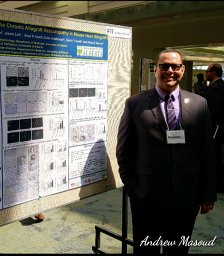 Andrew G Masoud合作论文数University of Alberta2Jiaxin Lin合作论文数University of Manitoba2John Gibson合作论文数School of Medicine, Faculty of Medicine and Health, The University of Sydney2
 Michael J. Hickey合作论文数Leukocyte Trafficking Group, Centre for Inflammatory Disease Monash Health, Monash University2




