A stem cell niche is a microenvironment where stem cells reside in a quiescent state, until activated. In a previous rat model, we combined 5-bromo-2-deoxy-uridine labeling with activation of endogenous stem cells by physical exercise and revealed a distinct region, in the atrioventricular junction (AVj), with features of a stem cell niche. In this study, we aim to investigate whether a similar niche exists in the human heart. Paired biopsies from AVj and left ventricle (LV) were collected both from explanted hearts of organ donors, not used for transplantation (N = 7) and from severely failing hearts from patients undergoing heart transplantation (N = 7). Using antibodies, we investigated the expression of stem cell, hypoxia, proliferation and migration biomarkers. In the collagen-dense region of the AVj in donor hearts, progenitor markers, MDR1, SSEA4, ISL1, WT1, and hypoxia marker, HIF1-α, were clearly detected. The expression gradually decreased with distance from the valve. At the myocardium border in the AVj costaining of the proliferation marker Ki67 with cardiomyocyte nuclei marker PCM1 and cardiac Troponin-T (cTnT) indicated proliferation of small cardiomyocytes. In the same site we also detected ISL1+/WT1+/cTnT cells. In addition, heterogeneity in cardiomyocyte sizes was noted. Altogether, these findings indicate different developmental stages of cardiomyocytes below the region dense in stem cell marker expression. In patients suffering from heart failure the AVj region showed signs of impairment generally displaying much weaker or no expression of progenitor markers. We describe an anatomic structure in the human hearts, with features of a progenitor niche that coincided with the same region previously identified in rats with densely packed cells expressing progenitor and hypoxia markers. The data provided in this study indicate that the adult heart contains progenitor cells and that AVj might be a specific niche region from which the progenitors migrate at the time of regeneration.
Low back pain is a major health issue and one main cause to this condition is believed to be intervertebral disc (IVD) degeneration. Stem cell therapy for degenerated discs using mesenchymal stromal cells (MSCs) has been suggested. The aim of the study was to investigate the presence and distribution pattern of autologous MSCs transplanted into degenerated IVDs in patients and explanted posttransplantation. IVD tissues from four patients (41, 45, 47, and 47 years of age) participating in a clinical feasibility study on MSC transplantation to degenerative discs were investigated. Three patients decided to undergo fusion surgery at time points 8 months and one patient at 28 months posttransplantation. Pretransplantation, MSCs from bone marrow aspirate were isolated by centrifugation in FICOLL (R) test tubes and cultured (passage 1). Before transplantation, MSCs were labeled with 1 mg/mL iron sucrose (Venofer (R)) and 1 x 10(6) MSCs were transplanted into degenerated IVDs. At the time point of surgery, IVD tissues were collected. IVD tissue samples were fixated, embedded in paraffin, and sections prepared. IVD samples were stained with Prussian Blue, by which iron deposits are visualized and examined (light microscopy). Immunohistochemistry (IHC), including SOX9 (sex determining region Y box 9), Coll2A1 (collagen 2A1), and cell viability (TUNEL) were performed. Cells positive for iron deposits were observed in IVD tissues (3/4 patients). The cells/iron deposits were observed in clusters and/or as solitary cells in regions in IVD tissue samples [regions of interest (ROIs)]. By IHC, SOX9- and Coll2A1-positive cells were detected in the same regions as the detected cells/iron deposits. A few nonviable cells were detected by TUNEL assay in ROIs. Results demonstrated that MSCs, labeled with iron sucrose, transplanted into degenerated IVDs were detectable 8 months posttransplantation. The detected cellular activity indicates that MSCs have differentiated into chondrocyte-like cells and that the injected MSCs and/or their progeny have survived since the cells were found in large cluster and as solitary cells which were distributed at different parts of the IVD.
Background:Despite recent advances in culture techniques for undifferentiated human Embryonic Stem Cells (hESCs), further improvements are required to facilitate research and translation of these cells in clinical settings. We have previously derived hESC lines that can be cultured in their undifferentiated state on regular plastic culture dishes, without the need for feeder cells or other coating supports, denoted Matrix-Free Growth hESCs (MFG-hESCs).Objective:In this study, we further characterize and compare MFG-hESCs to hESCs in order to understand the molecular differences responsible for the unique ability of MFG-hESCs.Results:Microarray analysis demonstrated that MFG-hESCs highly resemble feeder-cultured hESCs in global gene expression profile. Two identified groups of genes with differential expression were those encoding for ribosomal proteins and attachment proteins, such as the RGD (Arg-Gly-Asp)-associated proteins. Real-time PCR and flow cytometry corroborated the microarray results. Culture of MFG-hESCs in the presence of RGD peptides resulted in decreased attachment ability compared to cells cultured in the presence of RGES (Arg-Gly-Asp-Ser) peptides.Conclusion:This study demonstrates that MFG-hESC lines overexpress cell attachment proteins but retain the typical characteristics of undifferentiated feeder-cultured hESCs. The ability to culture high-quality pluripotent stem cells in feeder- and matrix-free conditions creates a new opportunities for their large-scale manufacturing for experimental research and translational applications.
Chondrocytes are effectively involved in the pathophysiological processes of inflammation in joints. They form cellular processes in the superficial layer of the articular cartilage and form gap junction coupled syncytium to facilitate cell-to-cell communication. However, very little is known about their physiological cellular identity and communication. The aim with the present work is to evaluate the physiological behavior after stimulation with the inflammatory inducers interleukin-1β and lipopolysaccharide. The cytoskeleton integrity and intracellular Ca2+ release were assessed as indicators of inflammatory state. Cytoskeleton integrity was analyzed through cartilage oligomeric matrix protein and actin labeling with an Alexa 488-conjugated phalloidin probe. Ca2+ responses were assessed through the Ca2+ sensitive fluorophore Fura-2/AM. Western blot analyses of several inflammatory markers were performed. The results show reorganization of the actin filaments. Glutamate, 5-hydoxytryptamine, and ATP evoked intracellular Ca2+ release changed from single peaks to oscillations after inflammatory induction in the chondrocytes. The expression of toll-like receptor 4, the glutamate transporters GLAST and GLT-1, and the matrix metalloproteinase-13 increased. This work demonstrates that chondrocytes are a key part in conditions that lead to inflammation in the cartilage. The inflammatory inducers modulate the cytoskeleton, the Ca2+ signaling, and several inflammatory parameters. In conclusion, our data show that the cellular responses to inflammatory insults from healthy and inflammatory chondrocytes resemble those previously observed in astrocyte and cardiac fibroblasts networks.
Anthracyclines, such as doxorubicin, are highly efficient chemotherapeutic agents against a variety of cancers. However, anthracyclines are also among the most cardiotoxic therapeutic drugs presently on the market. Chemotherapeutic-induced cardiomyopathy is one of the leading causes of disease and mortality in cancer survivors. The exact mechanisms responsible for doxorubicin-induced cardiomyopathy are not completely known, but the fact that the cardiotoxicity is dose-dependent and that there is a variation in time-to-onset of toxicity, and gender- and age differences suggests that several mechanisms may be involved. In this study, we investigated doxorubicin-induced cardiotoxicity in human pluripotent stem cell-derived cardiomyocytes using proteomics. In addition, different sources of omics data (protein, mRNA, and microRNA) from the same experimental setup were further combined and analyzed using newly developed methods to identify differential expression in data of various origin and types. Subsequently, the results were integrated in order to generate a combined visualization of the findings. In our experimental model system, we exposed cardiomyocytes derived from human pluripotent stem cells to doxorubicin for up to 2 days, followed by a wash-out period of additionally 12 days. Besides an effect on the cell morphology and cardiomyocyte functionality, the data show a strong effect of doxorubicin on all molecular levels investigated. Differential expression patterns that show a linkage between the proteome, transcriptome, and the regulatory microRNA network, were identified. These findings help to increase the understanding of the mechanisms behind anthracycline-induced cardiotoxicity and suggest putative biomarkers for this condition.
Visual integration of multiple omics data from human pluripotent stem cell-derived cardiomyocytes
We report the first detailed chemical, microstructural and thermal analyses of a growing corpus of metallurgical tuyeres from the wider archaeological landscape of Great Zimbabwe in southern Africa ...
This review addresses the progress in cartilage repair technology over the decades with an emphasis on cartilage regeneration with cell therapy. The most abundant cartilage is the hyaline cartilage that covers the surface of our joints and, due to avascularity, this tissue is unable to repair itself. The cartilage degeneration seen in osteoarthritis causes patient suffering and is a huge burden to society. The surgical approach to cartilage repair was non-existing until the 1950s when new surgical techniques emerged. The use of cultured cells for cell therapy started as experimental studies in the 1970s that developed over the years to a clinical application in 1994 with the introduction of the autologous chondrocyte transplantation technique (ACT). The technology is now spread worldwide and has been further refined by combining arthroscopic techniques with cells cultured on matrix (MACI technology). The non-regenerating hypothesis of cartilage has been revisited and we are now able to demonstrate cell divisions and presence of stem-cell niches in the joint. Furthermore, cartilage derived from human embryonic stem cells and induced pluripotent stem cells could be the base for new broader cell treatments for cartilage injuries and the future technology base for prevention and cure of osteoarthritis.
This chapter deals with the tissue engineering aspects of the mesenchymal tissues—cartilage and bone. It includes a brief description of the tissue types and their embryonal origin. Tissue structures including cell and extracellular matrix components are described in detail. The disease aspect of hyaline cartilage with emphasis on cartilage injuries and the tissue engineering approach to cartilage regeneration with the autologous chondrocyte implantation technique is described in depth. Bone fracture healing and biomaterials for bone repair is also described in depth. The future aspects of tissue regeneration techniques with potential cell types including stem cells as well as new promising scaffold techniques are described.
For everyday use and special occasions : A multi-analytical study of pottery from two Early Neolithic Funnel Beaker (TRB) sites on the island of Oland, SE Sweden
During the last 35 years almost 5000 ceramic thin-sections have been analysed at the Laboratory for Ceramic Research, Lund University and of those have 1650 analyses been performed on material from the Iron Age in Northern Europe (here c. 500 BC-1100 AD). Currently there is a project to register these analyses in a database and evaluate the results. We can for the first time get an overall picture of the various handicraft traditions that have existed at different periods in Northern Europe and in particular Scandinavia. The study presented here aims primarily to identify regional diversity during the Iron Age. The focus will be on analyses performed on pottery from Eastern Denmark, Scania, Gotland and Malardalen with some examples from other parts of Northern Europe. A further aim of this survey, which is based on a large statistical material, will also investigate how cultural choices were reflected in material culture. Interactions between different geological conditions and various cultural choices played a decisive role in how the pottery craft developed in different regions. The different geological conditions, with different types of clays and temper, can partly explain the differences in ceramic handicrafts. These factors may also have affected e.g. regional differences in firing methods. Various vessel types, styles, and ultimately table manners have also influenced the craft. Regional differences in usage of various types of vessels also affected how the pottery was manufactured. The method of studying thin-sections can reveal hidden differences in the handicraft and modify or even overturn groupings based on style and decoration. This study will set focus on that. (Less)
The brick kiln at Saltsjo Boo is one of very few such structures known from medieval Sweden. Approx ¾ of the kiln has been excavated in successive short excavations from 2006 to 2011. Carbon 14 and TL (Thermoluminiscence) analyses indicate that the kiln has been in operation from c. 1250 to c.1450. This paper presents the results of the excavations and discusses the production of bricks and industrial logistics of the site. To study brick manufacture and changes that occurred in composition of the raw material during over a period of 200 years we have used a handheld XRF, which offers great possibilities for chemical analyses on ceramics. A clear advantage of the method is that a large amount of samples may be analysed at low cost and during a short period of time. The data acquired at the analysis have been processed by means of various statistical methods from interpretations of scatter plots of two elements at time to multivariate tests. Of special interest has been to determine what may be considered as a normal variation in a sample from this type of context and explain anomalies in the material. (Less)
An abstract is not available for this content so a preview has been provided. Please use the Get access link above for information on how to access this content.
By using premixed calcium phosphate cement (CPC) the handling properties of the cement are drastically improved, which is a challenge for traditional injectable CPC. In this article, a premixed acidic CPC has been compared to a conventional water mixed brushite cement to evaluate whether the premixed concept affects the biological response. The cements were evaluated regarding the pH-variation in simulated body fluid (SBF). Further, the biocompatibility of the cement with human mesenchymal stem cells (MSC) was studied in vitro and eventual inflammation properties studied in vivo, after subcutaneous material injections in rats. A larger pH decrease was seen for the conventional cement than the premixed cement in SBF. For both materials, > 90% of the MSC remained alive in vitro. No systemic or macroscopic inflammation was detected, only a mild microscopic inflammation was detected around both materials. In addition to the handling benefit of premixed cements compared to conventional water mixed CPC, the premixed CPC in the present study demonstrated high and in comparison to conventional CPC comparable biocompability, both in vitro and in vivo.
Since 1987, autologous chondrocyte implantation (ACI) has been performed in Gothenburg, Sweden in more than 1600 patients. Out of the first 442 patients operated with ACI, 153 (35%) had patella lesions and 91 (21%) had trochlea lesions. Forty two patients (9.5%) had kissing lesions of the patellofemoral joint. The aim of the study was to evaluate the current clinical status of operated patients. Lysholm and Tegner-Wallgren self-assessment questionnaires were used. The patients were requested to compare their current status to previous states and to report whether they would do the operation again. Concomitant realignment procedures of the patellofemoral joint were also recorded and preoperative scores were also assessed from the medical files. Patients were divided into groups according to the location of lesion. All the groups showed a significant improvement compared with the preoperative assessment. Over 90% of the treated patients were satisfied with the ACI and would have undergone the procedure again. It seems that correcting the coexisting background factors with realignment, stabilizing or unloading procedures is improving the results over time. Despite the initial controversy about the results and indication for ACI in patellofemoral lesions, it is clear that ACI provides a satisfactory result even for the difficult cases with concomitant patellar instability. Our study reveals preservation of the good results and of high level of patients’ activities, even 10 to 20 years after the implantation.
We assessed 224 patients treated with Autologous Chondrocyte Implantation performed 10–20 years ago (average 12.8 years). Average age at the time of the implantation was 33.3 years. Average size of lesion was 5.3 cm2 (range 0.6–16), while 55 patients sustained multiple lesions. The participants filled out five questionnaires. Lysholm score, Tegner-Walgren, modified Cincinnati (Noyes), Brittberg score, and KOOS were assessed. In addition, the patients were asked to grade their current situation compared to their previous follow up as better, worse of unchanged. Finally, they were asked if they would do the operation again, answering with yes or no. The patients were divided into groups according to the location and characteristics of the cartilage lesions, or concomitant surgeries during the ACI. Assessment of the outcomes reveals a significant improvement in all groups, compared with the preoperative values. There is no other study assessing a cartilage treatment with such a long follow up. According to the results of that study, autologous chondrocyte implantation seems to be an effective and durable solution for the treatment of large full thickness cartilage and osteochondral lesions of the knee joint
 Brittberg Mats合作论文数14Lars Peterson合作论文数11Camilla Karlsson合作论文数9
 Minghao Zheng合作论文数School of Biomedical Sciences, The University of Western Australia;Oceans Institute, The University of Western Australia;Perron Institute for Neurological and Translational Science5
 Innocent Pikirayi合作论文数Department of Anthropology and Archaeology, University of Pretoria4
 Ilkka Kiviranta合作论文数4Tommi Tallheden合作论文数4
 Camilla Brantsing合作论文数3Anna I. Vasara合作论文数3
 Helena Brisby合作论文数Sahlgrenska University Hospital, Göteborg University3
 Julia Asp合作论文数3Joakim Sandstedt合作论文数3
 Sandro Palla合作论文数2J. A. M. Korfage合作论文数2
 Helena Barreto Henriksson合作论文数2
 W. Robb MacLellan合作论文数Medicine Heart Institute, University of Washington;University of Washington Medical Center2Heydarkhan-Hagvall Sepideh合作论文数2
 Vera Colombo合作论文数2
 Gustav Holmgren合作论文数2T.M.G.J. van Eijden合作论文数2Wickelgren Ruth合作论文数2Robert Henry合作论文数2
 Katrin Engelmann合作论文数2
 Francisco José Barcellos Sampaio合作论文数Departamento De Anatomia, Universidade Do Estado Do Rio De Janeiro2
 Jukka Jurvelin合作论文数Department of Technical Physics, Faculty of Science, Forestry and Technology, University of Eastern Finland2
 David Wood合作论文数Divisions of Cardiology & Cardiovascular Surgery, Department of Medicine, Faculty of Medicine, The University of British Columbia2
 Peter Thomsen合作论文数Department of Biomaterials, University of Gothenburg2Marianne Jonsson合作论文数2
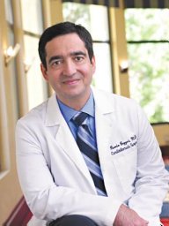 Ramin E. Beygui合作论文数Stanford University, Stanford University2
 Jane Synnergren合作论文数University of Skövde2Monika Valtink合作论文数2Hanna Stenhamre合作论文数2T. Van Wessel合作论文数2Elisabeth Hansson合作论文数2
 Peter Sartipy合作论文数Cellartis AB2Marco A. Pereira-Sampaio合作论文数2Sanaz Heydarkhan合作论文数2
 Luigi Gallo合作论文数Disorders andComplete Dentures;Dental andMaxillo-Facial Surgery;University ofZurich;Dental andMaxillo-Facial Surgery, University ofZurich2Sebastian Concaro合作论文数2
 Patricia Zuk合作论文数2
 Hans-Werner Denker合作论文数Abteilung Anatomie2Eva Sjögren-Jansson合作论文数2Richard H. W. Funk合作论文数2Luciano A. Favorito合作论文数2Katja Schenke-Layland合作论文数2P. Brugman合作论文数2Christian X. Andersson合作论文数2
 Geerling E.J. Langenbach合作论文数Department of Functional Anatomy;Academic Center for Dentistry Amsterdam2Eva Skiöldebrand合作论文数2




