Image-based profiling captures rich phenotypic signatures for drug discovery and functional genomics. Large public datasets like JUMP Cell Painting now provide millions of images for systematic study. However, the scale of these resources, 115 TB for JUMP alone, and fragmented evaluation practices make systematic comparison of representation methods intractable for many researchers. Here we present Nahual, an open-source framework for reproducible model deployment, and JUMP-lite, a curated 116 GB subset of JUMP that is 1000 times smaller while preserving phenotypic diversity through careful selection of perturbations with high-confidence annotations and a storage reduction via lossy JPEG XL compression. With these, we benchmark five representation methods, including classical features (CellProfiler) and deep learning models (MorphEM, OpenPhenom, SubCell, DINOv2), and demonstrate that compression preserves downstream signal while standardized phenotypic activity and consistency metrics reveal meaningful performance differences across methods. Together, JUMP-lite and Nahual provide a foundation for accessible, reproducible benchmarking of image-based cell representations.
Therapeutics working by novel mechanisms are needed for patients with psychiatric conditions. Cell-based assays to identify candidates that reverse observed abnormalities could accelerate the process. Here, we imaged peripheral cells (skin fibroblasts) of 168 patients, stained for DNA, actin, and mitochondria. We found mitochondria tend to be farther from the cell border for patients who experience psychosis (including subsets of individuals with bipolar disorder, schizophrenia, and schizoaffective disorder). We observed a reverse trend, albeit not statistically significant, for patients diagnosed with major depression. Because the phenotype could be identified by a single metric, we could query existing databases of cells stained for their mitochondria and treated with various chemical or genetic perturbations. We identified compounds and genes both negatively and positively affecting the psychosis-associated phenotype, including some known to impact psychiatric conditions. Developing therapeutics with novel mechanisms is a complex multi-step challenge. This cell-based assay holds promise for virtual and physical screening to identify candidates for treating psychiatric conditions.
Over 90% of missense variants across ~4,000 disease-associated genes are variants of uncertain significance (VUS). Experimental variant effect measurements provide critical evidence about pathogenicity and inform disease biology, but most variants lack data and clinical translation has been limited. The Impact of Genomic Variation on Function Consortium generated experimental data for 62,215 variants across ten genes using multiplexed assays and 1,407 variants across 163 genes using arrayed assays, curated 193,139 additional community-generated variant effect measurements across 30 additional genes, and developed automated calibration methods for translating experimental data and variant effect predictions into clinical evidence. To reduce current VUS, we developed a scalable workflow using only experimental and predictive evidence, enabling reclassification of 75% of the 16,115 VUS in these genes as pathogenic or benign with <1% error. To minimize future VUS, we analyzed >90,000 unobserved variants; 62% had enough evidence to be "preclassified" as pathogenic or benign. We validated our data, evidence and classifications using All of Us and created interactive resources to enable clinical use of the calibrated data. Thus, for 40 genes, representing 1% of the clinical genome, we resolve most existing VUS and future variants, illustrating how systematic use of scalable evidence can empower genomic medicine.
Accurately predicting the activity of a chemical in each bioactivity assay based on its already known properties is extremely useful in drug development. Unfortunately, we discovered that many assays in widely used assay-activity benchmark datasets directly relate to cell health and cytotoxicity. Many other assays intend to capture a more specific phenotype, but their active compounds impact cell count, while inactives do not. In both cases, counting cells achieves unexpectedly high performance in these benchmarks, making them less useful for discerning whether additional properties, such as phenotypic profiles (mRNA or Cell Painting), provide additional useful information on bioactivity. To accomplish this goal, we recommend filtering benchmarks to exclude such assays and including a cell-count baseline. Using a benchmark with 24 protein-target assays, we confirm that models leveraging Cell Painting image-based profiles outperformed the baseline cell count model. We propose several other practical recommendations for benchmarking machine learning models for predicting bioactivity and assessing the added value of mRNA, protein, or image-based profiles.
Scalable, human-relevant approaches for detecting compound modes of action are needed to improve chemical safety evaluation. Here, we apply image-based profiling (Cell Painting) alongside two cytotoxicity assays in primary human hepatocytes exposed to eight concentrations of 1,085 compounds spanning pharmaceuticals, pesticides, and industrial chemicals. We compared three computational approaches (CellProfiler, a Cell Painting-specific convolutional neural network, and a pretrained vision transformer) to extract morphological features from single cells or whole images. These features were used to predict activity in the measured cytotoxicity assays and in ToxCast assays covering cytotoxicity, cell-based, and cell-free assay endpoints. Morphological profiles detected bioactivity at lower concentrations than standard cytotoxicity assays and provided mode-of-action insights. In supervised analyses, they predicted cytotoxicity and targeted cell-based assays but not cell-free assays. Feature extraction methods performed similarly, and filtering concentrations did not improve performance. We envision that image-based profiling could be a key component of modern safety assessment.
AI agents are emerging as transformative tools in drug discovery, with the ability to autonomously reason, act and learn through complicated research workflows. Building on large language models and specialized tools, these systems can integrate biomedical data, execute tasks, conduct experiments and iteratively refine hypotheses. We provide a conceptual overview of agentic AI architectures and illustrate their applications across key stages of drug discovery, including literature synthesis, automated protocol generation, toxicity prediction, small-molecule synthesis, drug repurposing and end-to-end decision-making. Early implementations demonstrate substantial gains in speed, reproducibility and scalability. We discuss the challenges related to data heterogeneity, system reliability, privacy, benchmarking and outline future directions toward technology in support of science and translation.
Understanding cellular responses to external stimuli is critical for parsing biological mechanisms and advancing therapeutic development. High-content image-based assays provide a cost-effective approach to examine cellular phenotypes induced by diverse interventions, which offers valuable insights into biological processes and cellular states. We introduce MorphoDiff, a generative pipeline to predict high-resolution cell morphological responses under different conditions based on perturbation encoding. To the best of our knowledge, MorphoDiff is the first framework capable of producing guided, high-resolution predictions of cell morphology that generalize across both chemical and genetic interventions. The model integrates perturbation embeddings as guiding signals within a 2D latent diffusion model. The comprehensive computational, biological, and visual validations across three open-source Cell Painting datasets show that MorphoDiff can generate high-fidelity images and produce meaningful biology signals under various interventions. We envision the model will facilitate efficient in silico exploration of perturbational landscapes towards more effective drug discovery studies.
Advances in high-throughput microscopy have enabled the rapid acquisition of large numbers of high-content microscopy images. Whether by deep learning or classical algorithms, image analysis pipelines then produce single-cell features. To process these single-cells for downstream applications, we present Pycytominer, a user-friendly, open-source python package that implements the bioinformatics steps, known as image-based profiling. We demonstrate Pycytominers usefulness in a machine learning project to predict nuisance compounds that cause undesirable cell injuries.
Phenotypic profiling methods, such as Cell Painting and gene expression, have been widely used to predict compound bioactivity, often showing improvement over predictive models based on chemical structures alone. We discovered that a large subset of assays in widely-used benchmark datasets either directly relate to cell health and cytotoxicity or are assays intending to capture a more specific phenotype but whose active compounds impact cell count, while inactives do not. As a result, counting cells can achieve similar predictive performance as Cell Painting or gene expression data. Filtering benchmarks to include only assays relating to protein targets reveals that Cell Painting can capture information that cannot be predicted by mere cell counting. We re-evaluated three benchmark datasets used with Cell Painting data and observed that, in many cases, cell count models produced an AUC comparable to models using the full Cell Painting profiles. However, in protein-target-specific benchmarks across 17 distinct protein targets, Cell Painting features demonstrated unique predictive power, outperforming mean balanced accuracy from cell count models with a relative improvement of 19.6%. We propose five practical recommendations for benchmarking machine learning models for predicting bioactivity, including using cell count as a baseline feature. Although multi-class classification applications (such as matching samples based on their morphological profile) are less likely to be predictable by cell count than bioactivity benchmarks, these recommendations are broadly applicable to machine learning for drug discovery. ### Competing Interest Statement S. Singh and A.E.C. serve as scientific advisors for companies that use image-based profiling and Cell Painting (A.E.C.: Recursion, SyzOnc, Quiver Bioscience; S. Singh: Waypoint Bio, Dewpoint Therapeutics, DeepCell) and receive honoraria for occasional talks at pharmaceutical and biotechnology companies. J.C.P. and O.S. declare ownership in Phenaros Pharmaceuticals. National Institutes of HealthNational Institutes of Health, , NIH MIRA R35 GM122547 Massachusetts Life Sciences Center Bits to Bytes Capital CallMassachusetts Life Sciences Center Bits to Bytes Capital Call, , Swedish Research CouncilSwedish Research Council, , 2020-03731, 2020-01865, 2024-03566, 2024-04576 FORMASFORMAS, , 2022-00940 Swedish Cancer FoundationSwedish Cancer Foundation, , 22 2412 Pj 03 H Horizon Europe grant agreementHorizon Europe grant agreement, , #101057014 (PARC), #101057442 (REMEDI4ALL). Biotechnology and Biological Sciences Research CouncilBiotechnology and Biological Sciences Research Council, , BB/X511791/1
Large-scale profiling assays capture a cell population's state by measuring thousands of biological properties per cell or sample. However, evaluating profile strength and similarity remains challenging due to the high dimensionality and non-linear, heterogeneous nature of measurements. Here, we develop a statistical framework using mean average precision (mAP) as a single, data-driven metric to address this challenge. We validate the mAP framework against established metrics through simulations and real-world data, revealing its ability to capture subtle and meaningful biological differences in cell state. Specifically, we use mAP to assess a sample's phenotypic activity relative to controls, as well as the phenotypic consistency of groups of perturbations (or samples). We evaluate the framework across diverse datasets and on different profile types (image, protein, mRNA), perturbations (CRISPR, gene overexpression, small molecules), and resolutions (single-cell, bulk). The mAP framework, together with our open-source software package copairs, is useful for evaluating high-dimensional profiling data in biological research and drug discovery.
Modern quantitative image analysis techniques have enabled high-throughput, high-content imaging experiments. Image-based profiling leverages the rich information in images to identify similarities or differences among biological samples, rather than measuring a few features, as in high-content screening. Here, we review a decade of advancements and applications of Cell Painting, a microscopy-based cell-labeling assay aiming to capture a cell's state, introduced in 2013 to optimize and standardize image-based profiling. Cell Painting's ability to capture cellular responses to various perturbations has expanded owing to improvements in the protocol, adaptations for different perturbations, and enhanced methodologies for feature extraction, quality control, and batch-effect correction. Cell Painting is a versatile tool that has been used in various applications, alone or with other -omics data, to decipher the mechanism of action of a compound, its toxicity profile, and other biological effects. Future advances will likely involve computational and experimental techniques, new publicly available datasets, and integration with other high-content data types.
A key challenge of the modern genomics era is developing data-driven representations of gene function. Here, we present the first unbiased morphology-based genome-wide perturbation atlas in human cells, containing three genome-scale genotype-phenotype maps comprising >20,000 single-gene CRISPR-Cas9-based knockout experiments in >30 million cells. Our optical pooled cell profiling approach (PERISCOPE) combines a de-stainable high-dimensional phenotyping panel (based on Cell Painting1,2) with optical sequencing of molecular barcodes and a scalable open-source analysis pipeline to facilitate massively parallel screening of pooled perturbation libraries. This approach provides high-dimensional phenotypic profiles of individual cells, while simultaneously enabling interrogation of subcellular processes. Our atlas reconstructs known pathways and protein-protein interaction networks, identifies culture media-specific responses to gene knockout, and clusters thousands of human genes by phenotypic similarity. Using this atlas, we identify the poorly-characterized disease-associated transmembrane protein TMEM251/LYSET as a Golgi-resident protein essential for mannose-6-phosphate-dependent trafficking of lysosomal enzymes, showing the power of these representations. In sum, our atlas and screening technology represent a rich and accessible resource for connecting genes to cellular functions at scale.
Cell Painting images offer valuable insights into a cell's state and enable many biological applications, but publicly available arrayed datasets only include hundreds of genes perturbed. The JUMP Cell Painting Consortium perturbed roughly 75% of the protein-coding genome in human U-2 OS cells, generating a rich resource of single-cell images and extracted features. These profiles capture the phenotypic impacts of perturbing 15,243 human genes, including overexpressing 12,609 genes (using open reading frames) and knocking out 7,975 genes (using CRISPR-Cas9). Here we mitigated technical artifacts by rigorously evaluating data processing options and validated the dataset's robustness and biological relevance. Analysis of phenotypic profiles revealed previously undiscovered gene clusters and functional relationships, including those associated with mitochondrial function, cancer and neural processes. The JUMP Cell Painting genetic dataset is a valuable resource for exploring gene relationships and uncovering previously unknown functions.
Quantitative analysis of multidimensional biological images is useful for understanding complex cellular phenotypes and accelerating advances in biomedical research. As modern microscopy generates ever-larger 2D and 3D datasets, existing computational approaches are increasingly limited by their scalability, efficiency, and integration with modern scientific computing workflows. Existing bioimage analysis tools often lack application programmable interfaces (APIs), do not support graphics processing unit (GPU) acceleration, lack broad 3D image processing capabilities, and/or have poor interoperability for compute-heavy workflows. Here, we introduce cubic, an open-source Python library that addresses these challenges by augmenting widely used SciPy and scikit-image APIs with GPU-accelerated alternatives from CuPy and RAPIDS cuCIM. cubic's API is device-agnostic and dispatches operations to GPU when data reside on the device and otherwise executes on CPU, seamlessly accelerating a broad range of image processing routines. This approach enables GPU acceleration of existing bioimage analysis workflows, from preprocessing to segmentation and feature extraction for 2D and 3D data. We evaluate cubic both by benchmarking individual operations and by reproducing existing deconvolution and segmentation pipelines, achieving substantial speedups while maintaining algorithmic fidelity. These advances establish a robust foundation for scalable, reproducible bioimage analysis that integrates with the broader Python scientific computing ecosystem, including other GPU-accelerated methods, enabling both interactive exploration and automated high-throughput analysis workflows. cubic is openly available at https://github.com/alxndrkalinin/cubic
Microscopy enables direct observation of cellular morphology in 3D, with transmitted-light methods offering low-cost, minimally invasive imaging and fluorescence microscopy providing specificity and contrast. Virtual staining combines these strengths by using machine learning to predict fluorescence images from label-free inputs. However, training of existing methods typically relies on loss functions that treat all pixels equally, thus reproducing background noise and artifacts instead of focusing on biologically meaningful signals. We introduce Spotlight, a simple yet powerful virtual staining approach that guides the model to focus on relevant cellular structures. Spotlight uses histogram-based foreground estimation to mask pixel-wise loss and to calculate a Dice loss on soft-thresholded predictions for shape-aware learning. Applied to a 3D benchmark dataset, Spotlight improves morphological representation while preserving pixel-level accuracy, resulting in virtual stains better suited for downstream tasks such as segmentation and profiling.
Image-based profiling has been used to analyze cell health, drug mechanism of action, CRISPR-edited cells, and overall cytotoxicity. Cell Painting is a broadly used imagebased assay that uses morphological features to capture how cells respond to treatments. However, this method requires cell fixation for staining, which prevents examining live cells. To address this limitation, here we present Live-Cell Painting (LCP), a high-content method based on acridine orange, a metachromatic dye that labels different organelles and cellular structures. We began by showing that LCP can be applied to follow acidic vesicle redistribution of cells exposed to acidic vesicles inhibitors. Next, we show that LCP can identify subtle changes in cells exposed to silver nanoparticles that are not detected by techniques such as MTT assay. In drug treatments, LCP was helpful in assessing the dose-response relationship and creating profiles that allow clustering of drugs that cause liver injury. Here, we present an affordable and easy-to-use image-based assay capable of assessing overall cell health and showing promise for use in various applications such as assessing drugs and nanoparticles. We envisage the use of LCP as an initial screening of overall cell health while providing insights into new biological questions.
Neuropsychiatric disorders remain difficult to treat due to complex and poorly understood mechanisms. NeuroPainting is a high-content morphological profiling assay based on Cell Painting and optimized for human stem cell-derived neural cell types, including neurons, progenitors, and astrocytes. The assay quantifies over 4000 features of cell structure and organelle organization, generating a dataset suitable for phenotypic screening in neural models. Here, we show that, in studies of the 22q11.2 deletion-a strong genetic risk factor for schizophrenia-we observe cell-type-specific effects, particularly in astrocytes, including mitochondrial disruption, altered endoplasmic reticulum organization, and cytoskeletal changes. Transcriptomic analysis shows reduced expression of cell adhesion genes in deletion astrocytes, consistent with post-mortem brain data. Integration of RNA and morphology data suggests a link between adhesion gene dysregulation and mitochondrial abnormalities. These results illustrate how combining image-based profiling with gene expression analysis can reveal cellular mechanisms associated with genetic risk in neuropsychiatric disease.
Cell images contain a vast amount of quantifiable information about the status of the cell: for example, whether it is diseased, whether it is responding to a drug treatment, or whether a pathway has been disrupted by a genetic mutation. We aim to go beyond measuring individual cell phenotypes that biologists already know are relevant to a particular disease. Instead, in a strategy called image-based profiling, often using the Cell Painting assay, we extract hundreds of features of cells from microscopy images. Just like transcriptional or proteomic profiling, the similarities and differences in the patterns of extracted features reveal connections among diseases, drugs, and genes, with many applications in cancer research. In fact, these strategies underpin drug discovery platform companies such as Recursion and SyzOnc. Because images are inexpensive and high-throughput, we can carry out experiments at very large scale, yielding single-cell profiles for hundreds of thousands of samples through public-private consortia (JUMP, OASIS, VISTA, NIH IGVF) and pooled barcode-based optical screens. Cell morphology is therefore a powerful data source for cancer systems biology, alongside molecular omics. Anne E. Carpenter. Accelerating oncology drug discovery with the power of microscopy & AI [abstract]. In: Proceedings of the AACR Special Conference in Cancer Research: Artificial Intelligence and Machine Learning; 2025 Jul 10-12; Montreal, QC, Canada. Philadelphia (PA): AACR; Clin Cancer Res 2025;31(13_Suppl):Abstract nr IA03.
We present the nELISA, a high-throughput, high-fidelity, and high-plex protein profiling platform. DNA oligonucleotides are used to pre-assemble antibody pairs on spectrally encoded microparticles and perform displacement-mediated detection. Spatial separation between non-cognate antibodies prevents the rise of reagent-driven cross-reactivity, while read-out is performed cost-efficiently and at high-throughput using flow cytometry. We assembled an inflammatory panel of 191 targets that were multiplexed without cross-reactivity or impact on performance vs 1-plex signals, with sensitivities as low as 0.1pg/mL and measurements spanning 7 orders of magnitude. We then performed a large-scale secretome perturbation screen of peripheral blood mononuclear cells (PBMCs), with cytokines as both perturbagens and read-outs, measuring 7,392 samples and generating ∼1.5M protein datapoints in under a week, a significant advance in throughput compared to other highly multiplexed immunoassays. We uncovered 447 significant cytokine responses, including multiple putatively novel ones, that were conserved across donors and stimulation conditions. We also validated the nELISA’s use in phenotypic screening, and propose its application to drug discovery.
Machine learning (ML) is increasingly valuable for predicting molecular properties and toxicity in drug discovery. However, toxicity-related end points have always been challenging to evaluate experimentally with respect to in vivo translation due to the required resources for human and animal studies; this has impacted data availability in the field. ML can augment or even potentially replace traditional experimental processes depending on the project phase and specific goals of the prediction. For instance, models can be used to select promising compounds for on-target effects or to deselect those with undesirable characteristics (e.g., off-target or ineffective due to unfavorable pharmacokinetics). However, reliance on ML is not without risks, due to biases stemming from nonrepresentative training data, incompatible choice of algorithm to represent the underlying data, or poor model building and validation approaches. This might lead to inaccurate predictions, misinterpretation of the confidence in ML predictions, and ultimately suboptimal decision-making. Hence, understanding the predictive validity of ML models is of utmost importance to enable faster drug development timelines while improving the quality of decisions. This perspective emphasizes the need to enhance the understanding and application of machine learning models in drug discovery, focusing on well-defined data sets for toxicity prediction based on small molecule structures. We focus on five crucial pillars for success with ML-driven molecular property and toxicity prediction: (1) data set selection, (2) structural representations, (3) model algorithm, (4) model validation, and (5) translation of predictions to decision-making. Understanding these key pillars will foster collaboration and coordination between ML researchers and toxicologists, which will help to advance drug discovery and development.
 Shantanu Singh合作论文数Dept. of Computer Science and Engg., The Ohio State University97
 Beth A Cimini合作论文数Broad Inst MIT & Harvard, Imaging Platform, Cambridge, MA 02142 USA50
 Mark-Anthony Bray合作论文数Disease Biophysics Group, Harvard University39
 Juan Caicedo合作论文数Department of Biomedical Engineering, College of Engineering, University of Wisconsin-Madison25
 Allen Goodman合作论文数Broad Inst MIT & Harvard, Imaging Platform, Cambridge, MA 02142 USA18
 Way Gregory P合作论文数Department of Biology, Saint Joseph's University18
 Vebjorn Ljosa合作论文数University of California, Santa Barbara17
 David Sabatini合作论文数Institute of Organic Chemistry and Biochemistry, Czech Academy of Sciences16
 Maria Kost-Alimova合作论文数Broad Inst MIT & Harvard, Cambridge, MA 02142 USA15
 David C Logan合作论文数Institut de Recherche en Horticulture et Semences, University of Angers15Thouis R. Jones合作论文数Insitut Curie in the BioPhenics platform15
 Marzieh Haghighi合作论文数Broad Institute of MIT and Harvard14
 Minh Doan合作论文数Broad Inst MIT & Harvard, Imaging Platform, 415 Main St, Cambridge, MA 02142 USA14
 Paul Rees合作论文数14
 Kevin Eliceiri合作论文数Department of Medical Physics, School of Medicine and Public Health, University of Wisconsin–Madison;Center for Quantitative Cell Imaging, University of Wisconsin–Madison;Carbone Cancer Center, University of Wisconsin–Madison14Kyle W. Karhohs合作论文数13Kamentsky Lee合作论文数13
 Andreas Bender合作论文数Department of Chemistry, University of Cambridge13
 John E. Arevalo合作论文数Bioingenium Research Group, Universidad Nacional de Colombia13Erin Weisbart合作论文数11
 Tim Becker合作论文数IAV GmbH, Gifhorn, Germany.11Yu Han合作论文数11Claire Mcquin合作论文数Broad Institute11
 Holger Hennig合作论文数Max Planck Institute for Dynamics and Self-Organization, Göttingen, Germany. holgerh@nld.ds.mpg.de11
 David Root合作论文数Broad Institute11Paul A. Clemons合作论文数Broad Institute of Harvard and MIT11
 Ola Spjuth合作论文数Dept Pharmaceut Biosci, Uppsala Univ11
 Mohammad Hossein Rohban合作论文数Advanced Information & Communications Research Center, Sharif University of Technology11Katherine L Sokolnicki合作论文数10
 Srijit Seal合作论文数Yusuf Hamied Dept Chem, Univ Cambridge9
 Srinivas N. Chandrasekaran合作论文数Department of Biochemistry and Biophysics,;Program in Molecular and Cellular Biophysics, and;Department of Biochemistry and Biophysics,;Program in Molecular and Cellular Biophysics, and9
 Andrew Filby合作论文数Institute of Cellular Medicine, Newcastle University;Institute of Cellular Medicine, Newcastle University9
 Alexandr A. Kalinin合作论文数Chan Zuckerberg Biohub San Francisco9
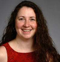 Polina Golland合作论文数Department of Electrical Engineering and Computer Science, Massachusetts Institute of Technology;Computer Science & Artificial Intelligence Laboratory, Massachusetts Institute of Technology;Institute for Medical Engineering & Science, Massachusetts Institute of Technology8
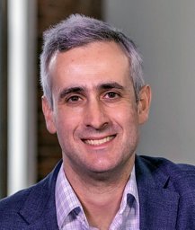 Jesse S. Boehm合作论文数The Broad Institute of MIT and Harvard, 7 Cambridge Center, Cambridge, MA 02142, USA8
 Huw D. Summers合作论文数8Hamdah Shafqat Abbasi合作论文数7
 Hanchuan Peng (彭汉川)合作论文数Institute for Brain and Intelligence, Fudan University7David R Stirling合作论文数UCL, Div Infect & Immun, London, England7
 Molnar, C.合作论文数Image Process. & Comput. Graphics Dept., Univ. of Szeged;c;Image Process. & Comput. Graphics Dept., Univ. of Szeged7Dančík Vlado合作论文数7Jamali Nasim合作论文数7
 Stuart L. Schreiber合作论文数Department of Chemistry and Chemical Biology, Faculty of Arts and Sciences, Harvard University;Broad Institute7
 Pavel Tomancak合作论文数Max Planck Institute of Molecular Cell Biology and Genetics;Central European Institute of Technology7
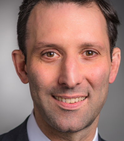 Benjamin Levine Ebert合作论文数Harvard Medical School;Dana-Farber Cancer Institute;Harvard Stem Cell Institute;Broad Institute7
 Jane Hung合作论文数Uber6Shamji Alykhan F合作论文数6
 Conery Annie L合作论文数Massachusetts Gen Hosp, Dept Mol Biol, Boston, MA 02114 USA6
 Puigvert Jordi C合作论文数6




