Advances in high-throughput microscopy have enabled the rapid acquisition of large numbers of high-content microscopy images. Whether by deep learning or classical algorithms, image analysis pipelines then produce single-cell features. To process these single-cells for downstream applications, we present Pycytominer, a user-friendly, open-source python package that implements the bioinformatics steps, known as image-based profiling. We demonstrate Pycytominers usefulness in a machine learning project to predict nuisance compounds that cause undesirable cell injuries.
The identification of genetic and chemical perturbations with similar impacts on cell morphology can elucidate compounds' mechanisms of action or novel regulators of genetic pathways. Research on methods for identifying such similarities has lagged due to a lack of carefully designed and well-annotated image sets of cells treated with chemical and genetic perturbations. Here we create such a Resource dataset, CPJUMP1, in which each perturbed gene's product is a known target of at least two chemical compounds in the dataset. We systematically explore the directionality of correlations among perturbations that target the same protein encoded by a given gene, and we find that identifying matches between chemical and genetic perturbations is a challenging task. Our dataset and baseline analyses provide a benchmark for evaluating methods that measure perturbation similarities and impact, and more generally, learn effective representations of cellular state from microscopy images. Such advancements would accelerate the applications of image-based profiling of cellular states, such as uncovering drug mode of action or probing functional genomics. The CPJUMP1 Resource comprises Cell Painting images and profiles of 75 million cells treated with hundreds of chemical and genetic perturbations. The dataset enables exploration of their relationships and lays the foundation for the development of advanced methods to match perturbations.
Cellular exposure to free fatty acids (FFAs) is implicated in the pathogenesis of obesity-associated diseases. However, there are no scalable approaches to comprehensively assess the diverse FFAs circulating in human plasma. Furthermore, assessing how FFA-mediated processes interact with genetic risk for disease remains elusive. Here, we report the design and implementation of fatty acid library for comprehensive ontologies (FALCON), an unbiased, scalable, and multimodal interrogation of 61 structurally diverse FFAs. We identified a subset of lipotoxic monounsaturated fatty acids associated with decreased membrane fluidity. Furthermore, we prioritized genes that reflect the combined effects of harmful FFA exposure and genetic risk for type 2 diabetes (T2D). We found that c-MAF-inducing protein (CMIP) protects cells from FFA exposure by modulating Akt signaling. In sum, FALCON empowers the study of fundamental FFA biology and offers an integrative approach to identify much needed targets for diverse diseases associated with disordered FFA metabolism.
In image-based profiling, software extracts thousands of morphological features of cells from multi-channel fluorescence microscopy images, yielding single-cell profiles that can be used for basic research and drug discovery. Powerful applications have been proven, including clustering chemical and genetic perturbations on the basis of their similar morphological impact, identifying disease phenotypes by observing differences in profiles between healthy and diseased cells and predicting assay outcomes by using machine learning, among many others. Here, we provide an updated protocol for the most popular assay for image-based profiling, Cell Painting. Introduced in 2013, it uses six stains imaged in five channels and labels eight diverse components of the cell: DNA, cytoplasmic RNA, nucleoli, actin, Golgi apparatus, plasma membrane, endoplasmic reticulum and mitochondria. The original protocol was updated in 2016 on the basis of several years’ experience running it at two sites, after optimizing it by visual stain quality. Here, we describe the work of the Joint Undertaking for Morphological Profiling Cell Painting Consortium, to improve upon the assay via quantitative optimization by measuring the assay’s ability to detect morphological phenotypes and group similar perturbations together. The assay gives very robust outputs despite various changes to the protocol, and two vendors’ dyes work equivalently well. We present Cell Painting version 3, in which some steps are simplified and several stain concentrations can be reduced, saving costs. Cell culture and image acquisition take 1–2 weeks for typically sized batches of ≤20 plates; feature extraction and data analysis take an additional 1–2 weeks. This protocol is an update to Nat. Protoc. 11, 1757–1774 (2016): https://doi.org/10.1038/nprot.2016.105 We provide an updated protocol for image-based profiling with Cell Painting. A detailed procedure, with standardized conditions for the assay, is presented, along with a comprehensive description of parameters to be considered when optimizing the assay.
Quantitative microscopy is a powerful method for performing phenotypic screens from which image-based profiling can extract a wealth of information, termed profiles. These profiles can be used to elucidate the changes in cellular phenotypes across cell populations from different patient samples or following genetic or chemical perturbations. One such image-based profiling method is the Cell Painting assay, which provides morphological insight through the imaging of eight cellular compartments. Here, we examine the performance of the Cell Painting assay across multiple high-throughput microscope systems and find that all are compatible with this assay. Furthermore, we determine independently for each microscope system the best performing settings, providing those who wish to adopt this assay an ideal starting point for their own assays. We also explore the impact of microscopy setting changes in the Cell Painting assay and find that few dramatically reduce the quality of a Cell Painting profile, regardless of the microscope used.
Widespread sequencing has yielded thousands of missense variants predicted or confirmed as disease-causing. This creates a new bottleneck: determining the functional impact of each variant - largely a painstaking, customized process undertaken one or a few genes or variants at a time. Here, we established a high-throughput imaging platform to assay the impact of coding variation on protein localization, evaluating 3,547 missense variants of over 1,000 genes and phenotypes. We discovered that mislocalization is a common consequence of coding variation, affecting about one-sixth of all pathogenic missense variants, all cellular compartments, and recessive and dominant disorders alike. Mislocalization is primarily driven by effects on protein stability and membrane insertion rather than disruptions of trafficking signals or specific interactions. Furthermore, mislocalization patterns help explain pleiotropy and disease severity and provide insights on variants of unknown significance. Our publicly available resource will likely accelerate the understanding of coding variation in human diseases.
Genetic and chemical perturbations impact diverse cellular phenotypes, including multiple indicators of cell health. These readouts reveal toxicity and antitumorigenic effects relevant to drug discovery and personalized medicine. We developed two customized microscopy assays, one using four targeted reagents and the other three targeted reagents, to collectively measure 70 specific cell health phenotypes including proliferation, apoptosis, reactive oxygen species, DNA damage, and cell cycle stage. We then tested an approach to predict multiple cell health phenotypes using Cell Painting, an inexpensive and scalable image-based morphology assay. In matched CRISPR perturbations of three cancer cell lines, we collected both Cell Painting and cell health data. We found that simple machine learning algorithms can predict many cell health readouts directly from Cell Painting images, at less than half the cost. We hypothesized that these models can be applied to accurately predict cell health assay outcomes for any future or existing Cell Painting dataset. For Cell Painting images from a set of 1500+ compound perturbations across multiple doses, we validated predictions by orthogonal assay readouts. We provide a web app to browse predictions: http://broad.io/cell-health-app. Our approach can be used to add cell health annotations to Cell Painting datasets.
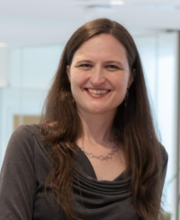 Anne E. Carpenter合作论文数Broad Institute7
 Shantanu Singh合作论文数Dept. of Computer Science and Engg., The Ohio State University6
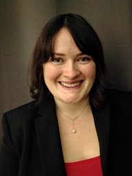 Beth A Cimini合作论文数Broad Inst MIT & Harvard, Imaging Platform, Cambridge, MA 02142 USA5
 Maria Kost-Alimova合作论文数Broad Inst MIT & Harvard, Cambridge, MA 02142 USA4Jamali Nasim合作论文数4
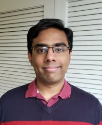 Srinivas N. Chandrasekaran合作论文数Department of Biochemistry and Biophysics,;Program in Molecular and Cellular Biophysics, and;Department of Biochemistry and Biophysics,;Program in Molecular and Cellular Biophysics, and3Desiree Hernandez合作论文数2Briana Fritchman合作论文数2Adam Skepner合作论文数2Lisa Miller合作论文数2
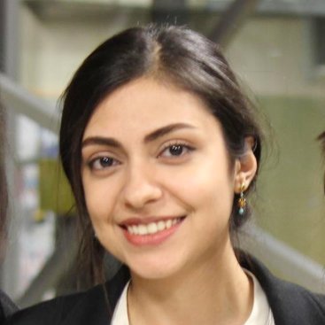 Marzieh Haghighi合作论文数Broad Institute of MIT and Harvard2Erin Weisbart合作论文数2Callum Tromans-Coia合作论文数Broad Institute, MIT2Amy Goodale合作论文数Broad Inst MIT & Harvard, Cambridge, MA 02142 USA2
 Tim Becker合作论文数IAV GmbH, Gifhorn, Germany.2Michelle Melanson合作论文数2
 Rebecca A. Senft合作论文数Ctr Brain Sci, Harvard Univ2Garg Sakshi合作论文数2
 Way Gregory P合作论文数Department of Biology, Saint Joseph's University2
 Susanne E. Swalley合作论文数2
 Juan Caicedo合作论文数Department of Biomedical Engineering, College of Engineering, University of Wisconsin-Madison2
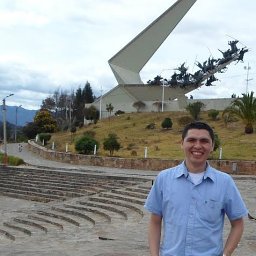 John E. Arevalo合作论文数Bioingenium Research Group, Universidad Nacional de Colombia2
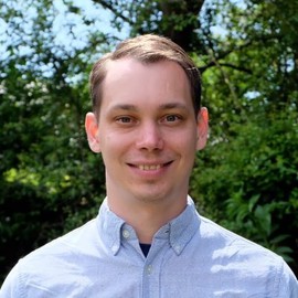 Alexandr A. Kalinin合作论文数Chan Zuckerberg Biohub San Francisco2Dave Bunten合作论文数1Jenna Tomkinson合作论文数1Julian A. Pacheco合作论文数1Dmitri Segal合作论文数1Tamara Gilbert合作论文数1Michael Yang合作论文数1Mitsunari Yamaguchi合作论文数1Stefan Letzsch合作论文数1Erika Kaneko合作论文数1Mai Hagimoto合作论文数1Kenneth A Giuliano合作论文数1Aaron Baublis合作论文数1John Concannon合作论文数1Alexander Schreiner合作论文数1Elizabeth Mouchet合作论文数1
 Jessica Lacoste合作论文数1James Berstler合作论文数1Mahomi Suzuki合作论文数1Fumiki Yanagawa合作论文数1Kevin Jan合作论文数1Gisela Hormel合作论文数1Shahan Haider合作论文数1Ruifan Pei合作论文数1Xueting Xiong合作论文数1Vincent Rubinetti合作论文数1Patrick Byrne合作论文数1




