For over two decades, image-based profiling has revolutionized cellular phenotype analysis. Image-based profiling processes rich, high-throughput, microscopy data into unbiased measurements that reveal phenotypic patterns powerful for drug discovery, functional genomics, and cell state classification. Here, we review the evolving computational landscape of image-based profiling, detailing current procedures, discussing limitations, and highlighting future development directions. Deep learning has fundamentally reshaped image-based profiling, improving feature extraction, scalability, and multimodal data integration. Methodological advancements such as single-cell analysis and batch effect correction, drawing inspiration from single-cell transcriptomics, have enhanced analytical precision. The growth of open-source software ecosystems and the development of community-driven standards have further democratized access to image-based profiling, fostering reproducibility and collaboration across research groups. Despite these advancements, the field still faces significant challenges requiring innovative solutions. By focusing on the technical evolution of image-based profiling rather than the wide-ranging biological applications, our aim with this review is to provide researchers with a roadmap for navigating the progress and new challenges in this rapidly advancing domain.
Quantifying cell morphology using images and machine learning has proven to be a powerful tool to study the response of cells to treatments. However, the models used to quantify cellular morphology are typically trained with a single microscopy imaging type and under controlled experimental conditions. This results in specialized models that cannot be reused across biological studies because the technical specifications do not match (e.g., different number of channels), or because the target experimental conditions are out of distribution. Here, we present CHAMMI-75, a dataset of heterogeneous, multi-channel microscopy images with 2.8M multi-channel images from 75 diverse biological studies. We curated this resource from publicly available sources to investigate cellular morphology models that are channel-adaptive and can process any microscopy image type. Our experiments show that training with CHAMMI-75 can improve performance in multi-channel bioimaging tasks, opening the way to create the next generation of cellular morphology models for biological studies.
Prior work using Masked Autoencoders (MAEs) typically relies on random patch masking based on the assumption that images have significant redundancies across different channels, allowing for the reconstruction of masked content using cross-channel correlations. However, this assumption does not hold in Multi-Channel Imaging (MCI), where channels may provide complementary information with minimal feature overlap. Thus, these MAEs primarily learn local structures within individual channels from patch reconstruction, failing to fully leverage cross-channel interactions and limiting their MCI effectiveness. In this paper, we present ChA-MAEViT, an MAE-based method that enhances feature learning across MCI channels via four key strategies: (1) dynamic channel-patch masking, which compels the model to reconstruct missing channels in addition to masked patches, thereby enhancing cross-channel dependencies and improving robustness to varying channel configurations; (2) memory tokens, which serve as long-term memory aids to promote information sharing across channels, addressing the challenges of reconstructing structurally diverse channels; (3) hybrid token fusion module, which merges fine-grained patch tokens with a global class token to capture richer representations; and (4) Channel-Aware Decoder, a lightweight decoder utilizes channel tokens to effectively reconstruct image patches. Experiments on satellite and microscopy datasets, CHAMMI, JUMP-CP, and So2Sat, show that ChA-MAEViT significantly outperforms state-of-the-art MCI-ViTs by 3.0-21.5%, highlighting the importance of cross-channel interactions in MCI. Our code is publicly available at https://github.com/chaudatascience/cha_mae_vit.
Advances in high-throughput microscopy have enabled the rapid acquisition of large numbers of high-content microscopy images. Whether by deep learning or classical algorithms, image analysis pipelines then produce single-cell features. To process these single-cells for downstream applications, we present Pycytominer, a user-friendly, open-source python package that implements the bioinformatics steps, known as image-based profiling. We demonstrate Pycytominers usefulness in a machine learning project to predict nuisance compounds that cause undesirable cell injuries.
Accurately quantifying cellular morphology at scale could substantially empower existing single-cell approaches. However, measuring cell morphology remains an active field of research, which has inspired multiple computer vision algorithms over the years. Here, we show that DINOv2, a vision-transformer based, self-supervised algorithm, has a remarkable ability for learning rich representations of cellular morphology without manual annotations or any other type of supervision. We apply DINOv2 to cell phenotyping problems, and compare the performance of resulting models, called Cell-DINO models, on a wide variety of tasks across two publicly available imaging datasets of diverse specifications and biological focus. Compared to supervised and other self-supervised baselines, Cell-DINO models demonstrate improved performance, especially in low annotation regimes. For instance, to classify protein localization using only 1% of annotations on a challenging single-cell dataset, Cell-DINO performs 70% better than a supervised strategy, and 24% better than another self-supervised alternative. The results show that Cell-DINO can support the study of unknown biological variation, including single-cell heterogeneity and relationships between experimental conditions, making it an excellent tool for image-based biological discovery.
Dynamic contrast-enhanced magnetic resonance imaging (DCE-MRI) is essential for breast cancer diagnosis because it can characterize tissue based on contrast agent kinetics. Conventional DCE-MRI protocols require multiple imaging phases, including both early and late post-contrast acquisitions, leading to prolonged scanning times that can cause patient discomfort, motion artifacts, and contribute to higher costs and limited availability in clinical settings. To address these limitations, this paper presents a comprehensive pipeline for synthesizing long-term (late-phase) contrast-enhanced breast MRI images from short-term (early-phase) counterparts, aiming to replicate the behavior of the time-intensity (TI) curve in enhanced regions while maintaining visual properties across the entire image. The proposed approach introduces a new loss function called the Time Intensity Loss (TI-loss), which leverages the temporal behavior of the contrast agent to guide the training of a generative model. Furthermore, as established normalization strategies show undesirable effects on the enhancement behavior, a novel normalization strategy (TI-norm) is also proposed, which preserves the contrast enhancement pattern across multiple image sequences at various timestamps. Additionally, two new metrics are proposed to evaluate the synthesized image quality, i.e., the Contrast agent Pattern score (CPs), which determines the validity of annotated regions according to their enhancement patterns (plateau, persistent, washout), and the average difference in enhancement (ED), which quantifies the difference between the real and generated enhancement in selected regions. Evaluation was performed using a public DCE-MRI dataset that includes studies from 3T and 1.5T scanners with different imaging techniques. Experimental results demonstrate that our method accurately synthesizes the contrast enhancement response in terms of the TI curve in regions of interest and significantly outperforms other models, while maintaining visual properties comparable to real late-phase contrast-enhanced images. By enabling accurate synthesis of late-phase contrast-enhanced images from early-phase data, our method has the potential to optimize DCE-MRI protocols, reducing scanning time without compromising diagnostic accuracy. This advancement brings generative models closer to practical implementation in clinical scenarios, enhancing efficiency in breast cancer imaging.
The identification of genetic and chemical perturbations with similar impacts on cell morphology can elucidate compounds' mechanisms of action or novel regulators of genetic pathways. Research on methods for identifying such similarities has lagged due to a lack of carefully designed and well-annotated image sets of cells treated with chemical and genetic perturbations. Here we create such a Resource dataset, CPJUMP1, in which each perturbed gene's product is a known target of at least two chemical compounds in the dataset. We systematically explore the directionality of correlations among perturbations that target the same protein encoded by a given gene, and we find that identifying matches between chemical and genetic perturbations is a challenging task. Our dataset and baseline analyses provide a benchmark for evaluating methods that measure perturbation similarities and impact, and more generally, learn effective representations of cellular state from microscopy images. Such advancements would accelerate the applications of image-based profiling of cellular states, such as uncovering drug mode of action or probing functional genomics. The CPJUMP1 Resource comprises Cell Painting images and profiles of 75 million cells treated with hundreds of chemical and genetic perturbations. The dataset enables exploration of their relationships and lays the foundation for the development of advanced methods to match perturbations.
Measuring the phenotypic effect of treatments on cells through imaging assays is an efficient and powerful way of studying cell biology, and requires computational methods for transforming images into quantitative data. Here, we present an improved strategy for learning representations of treatment effects from high-throughput imaging, following a causal interpretation. We use weakly supervised learning for modeling associations between images and treatments, and show that it encodes both confounding factors and phenotypic features in the learned representation. To facilitate their separation, we constructed a large training dataset with images from five different studies to maximize experimental diversity, following insights from our causal analysis. Training a model with this dataset successfully improves downstream performance, and produces a reusable convolutional network for image-based profiling, which we call Cell Painting CNN. We evaluated our strategy on three publicly available Cell Painting datasets, and observed that the Cell Painting CNN improves performance in downstream analysis up to 30% with respect to classical features, while also being more computationally efficient.
Through digital imaging, microscopy has evolved from primarily being a means for visual observation of life at the micro- and nano-scale, to a quantitative tool with ever-increasing resolution and throughput. Artificial intelligence, deep neural networks, and machine learning are all niche terms describing computational methods that have gained a pivotal role in microscopy-based research over the past decade. This Roadmap is written collectively by prominent researchers and encompasses selected aspects of how machine learning is applied to microscopy image data, with the aim of gaining scientific knowledge by improved image quality, automated detection, segmentation, classification and tracking of objects, and efficient merging of information from multiple imaging modalities. We aim to give the reader an overview of the key developments and an understanding of possibilities and limitations of machine learning for microscopy. It will be of interest to a wide cross-disciplinary audience in the physical sciences and life sciences.
Predicting assay results for compounds virtually using chemical structures and phenotypic profiles has the potential to reduce the time and resources of screens for drug discovery. Here, we evaluate the relative strength of three high-throughput data sources—chemical structures, imaging (Cell Painting), and gene-expression profiles (L1000)—to predict compound bioactivity using a historical collection of 16,170 compounds tested in 270 assays for a total of 585,439 readouts. All three data modalities can predict compound activity for 6–10% of assays, and in combination they predict 21% of assays with high accuracy, which is a 2 to 3 times higher success rate than using a single modality alone. In practice, the accuracy of predictors could be lower and still be useful, increasing the assays that can be predicted from 37% with chemical structures alone up to 64% when combined with phenotypic data. Our study shows that unbiased phenotypic profiling can be leveraged to enhance compound bioactivity prediction to accelerate the early stages of the drug-discovery process.
Most neural networks assume that input images have a fixed number of channels (three for RGB images). However, there are many settings where the number of channels may vary, such as microscopy images where the number of channels changes depending on instruments and experimental goals. Yet, there has not been a systemic attempt to create and evaluate neural networks that are invariant to the number and type of channels. As a result, trained models remain specific to individual studies and are hardly reusable for other microscopy settings. In this paper, we present a benchmark for investigating channel-adaptive models in microscopy imaging, which consists of 1) a dataset of varied-channel single-cell images, and 2) a biologically relevant evaluation framework. In addition, we adapted several existing techniques to create channel-adaptive models and compared their performance on this benchmark to fixed-channel, baseline models. We find that channel-adaptive models can generalize better to out-of-domain tasks and can be computationally efficient. We contribute a curated dataset (https://doi.org/10.5281/zenodo.7988357) and an evaluation API (https://github.com/broadinstitute/MorphEm.git) to facilitate objective comparisons in future research and applications.
Accurately quantifying cellular morphology at scale could substantially empower existing single-cell approaches. However, measuring cell morphology remains an active field of research, which has inspired multiple computer vision algorithms over the years. Here, we show that DINO, a vision-transformer based, self-supervised algorithm, has a remarkable ability for learning rich representations of cellular morphology without manual annotations or any other type of supervision. We evaluate DINO on a wide variety of tasks across three publicly available imaging datasets of diverse specifications and biological focus. We find that DINO encodes meaningful features of cellular morphology at multiple scales, from subcellular and single-cell resolution, to multi-cellular and aggregated experimental groups. Importantly, DINO successfully uncovers a hierarchy of biological and technical factors of variation in imaging datasets. The results show that DINO can support the study of unknown biological variation, including single-cell heterogeneity and relationships between samples, making it an excellent tool for image-based biological discovery.
We consider the problem of predicting early-to-late Dynamic Contrast Enhanced Magnetic Resonance Imaging (DCE-MRI) in breast cancer sequences. This is approached with conditional generative adversarial networks that synthesize the late response image given the early response. We propose a novel loss function to improve the ability of GAN models to learn the relevant temporal tissue dynamics under this setting, as well as a clinically relevant metric to assess performance. Our experiments show that the proposed strategy predicts accurate responses and could serve as a solution to implement fast diagnostic protocols.
Real-world deployment of computer vision systems, including in the discovery processes of biomedical research, requires causal representations that are invariant to contextual nuisances and generalize to new data. Leveraging the internal replicate structure of two novel single-cell fluorescent microscopy datasets, we propose generally applicable tests to assess the extent to which models learn causal representations across increasingly challenging levels of OOD-generalization. We show that despite seemingly strong performance, as assessed by other established metrics, both naive and contemporary baselines designed to ward against confounding, collapse on these tests. We introduce a new method, Interventional Style Transfer (IST), that substantially improves OOD generalization by generating interventional training distributions in which spurious correlations between biological causes and nuisances are mitigated. We publish our code and datasets.
Quantitative evaluation of image segmentation algorithms is crucial in the field of bioimage analysis. The most common assessment scores, however, are often misinterpreted and multiple definitions coexist with the same name. Here we present the ambiguities of evaluation metrics for segmentation algorithms and show how these misinterpretations can alter leaderboards of influential competitions. We also propose guidelines for how the currently existing problems could be tackled.
Cells can be perturbed by various chemical and genetic treatments and the impact on gene expression and morphology can be measured via transcriptomic profiling and image-based assays, respectively. The patterns observed in these high-dimensional profile data can power a dozen applications in drug discovery and basic biology research, but both types of profiles are rarely available for large-scale experiments. Here, we provide a collection of four datasets with both gene expression and morphological profile data useful for developing and testing multimodal methodologies. Roughly a thousand features are measured for each of the two data types, across more than 28,000 chemical and genetic perturbations. We define biological problems that use the shared and complementary information in these two data modalities, provide baseline analysis and evaluation metrics for multi-omic applications, and make the data resource publicly available ( https://broad.io/rosetta/ ).
Background: Different asthma phenotypes are driven by molecular endotypes. A Th1-high phenotype is linked to severe, therapy-refractory asthma, subclinical infections and neutrophil inflammation. Previously, we found neutrophil granulocytes (NGs) from asthmatics exhibit decreased chemotaxis towards leukotriene B-4 (LTB4), a chemoattractant involved in inflammation response. We hypothesized that this pattern is driven by asthma in general and aggravated in a Th1-high phenotype. Methods: NGs from asthmatic nd healthy children were stimulated with 10 nM LTB4/100 nM N-formylmethionine-leucyl-phenylalanine and neutrophil migration was documented following our prior SiMA (simplified migration assay) workflow, capturing morphologic and dynamic parameters from single-cell tracking in the images. Demographic, clinical and serum cytokine data were determined in the ALLIANCE cohort. Results: A reduced chemotactic response towards LTB4 was confirmed in asthmatic donors regardless of inhaled corticosteroid (ICS) treatment. By contrast, only NGs from ICS-treated asthmatic children migrate similarly to controls with the exception of Th1-high donors, whose NGs presented a reduced and less directed migration towards the chemokines. ICS-treated and Th1-high asthmatic donors present an altered surface receptor profile, which partly correlates with migration. Conclusions: Neutrophil migration in vitro may be affected by ICS-therapy or a Th1-high phenotype. This may be explained by alteration of receptor expression and could be used as a tool to monitor asthma treatment.
In recent years, artificial intelligence (AI) has played an ever-increasing role in research across the physical sciences and beyond. In this Voices piece, published as part of the Cell Reports portfolio joint special issue on AI in healthcare, we ask a panel of experts: How do you think AI will change drug discovery?
Morphological and gene expression profiling can cost-effectively capture thousands of features in thousands of samples across perturbations by disease, mutation, or drug treatments, but it is unclear to what extent the two modalities capture overlapping versus complementary information. Here, using both the L1000 and Cell Painting assays to profile gene expression and cell morphology, respectively, we perturb human A549 lung cancer cells with 1,327 small molecules from the Drug Repurposing Hub across six doses, providing a data resource including dose-response data from both assays. The two assays capture both shared and complementary information for mapping cell state. Cell Painting profiles from compound perturbations are more reproducible and show more diversity but measure fewer distinct groups of features. Applying unsupervised and supervised methods to predict compound mechanisms of action (MOAs) and gene targets, we find that the two assays not only provide a partially shared but also a complementary view of drug mechanisms. Given the numerous applications of profiling in biology, our analyses provide guidance for planning experiments that profile cells for detecting distinct cell types, disease phenotypes, and response to chemical or genetic perturbations.
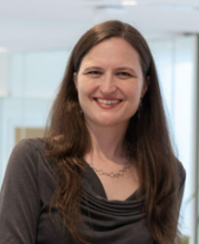 Anne E. Carpenter合作论文数Broad Institute25
 Shantanu Singh合作论文数Dept. of Computer Science and Engg., The Ohio State University23
 Fabio A. Gonzalez合作论文数Systems and Computer Engineering Department, National University of Colombia15
 Allen Goodman合作论文数Broad Inst MIT & Harvard, Imaging Platform, Cambridge, MA 02142 USA12
 Beth A Cimini合作论文数Broad Inst MIT & Harvard, Imaging Platform, Cambridge, MA 02142 USA11
 Jorge E. Camargo合作论文数Departamento de Ingeniería de Sistemas e Industrial, Universidad Nacional de Colombia11Claire Mcquin合作论文数Broad Institute10
 Tim Becker合作论文数IAV GmbH, Gifhorn, Germany.9
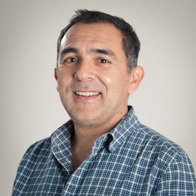 Fabio A. González O.合作论文数Department of Computing Systems and Industrial Engineering, National University of Colombia9
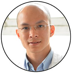 Minh Doan合作论文数Broad Inst MIT & Harvard, Imaging Platform, 415 Main St, Cambridge, MA 02142 USA7
 Michael Doron合作论文数Q.ai7
 Eduardo Romero合作论文数Universidad Nacional de Colombia6
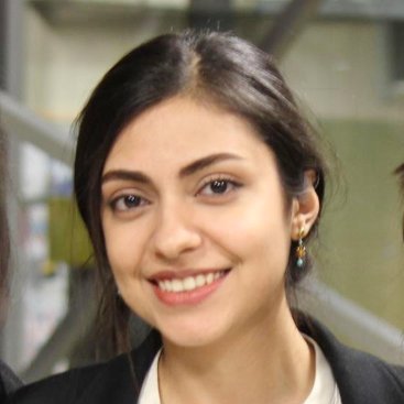 Marzieh Haghighi合作论文数Broad Institute of MIT and Harvard5
 Nikita Moshkov合作论文数These authors contributed equally5Kyle W. Karhohs合作论文数5
 Peter Horvath合作论文数Institute of Biochemistry, Biological Research Centre;Helmholtz Munich5
 Paul Rees合作论文数5
 Angel Cruz-Roa合作论文数Bioingenium Research Group, Universidad Nacional de Colombia5
 Bryan A. Plummer合作论文数Image and Video Computing Group, Department of Computer Science, Boston University5
 Holger Hennig合作论文数Max Planck Institute for Dynamics and Self-Organization, Göttingen, Germany. holgerh@nld.ds.mpg.de4
 Jorge A. Vanegas合作论文数4
 John E. Arevalo合作论文数Bioingenium Research Group, Universidad Nacional de Colombia4
 Fabio Gonzalez合作论文数Systems and Computer Engineering,Department National University of Colombia,Bogotá4
 Mohammad Hossein Rohban合作论文数Advanced Information & Communications Research Center, Sharif University of Technology4
 Chau Pham合作论文数Department of Computer Science, College of Arts and Sciences, Boston University3Michael Bornholdt合作论文数3
 Aditya Pratapa合作论文数Broad Inst3
 Srinivas N. Chandrasekaran合作论文数Department of Biochemistry and Biophysics,;Program in Molecular and Cellular Biophysics, and;Department of Biochemistry and Biophysics,;Program in Molecular and Cellular Biophysics, and3
 Wolfgang M. Pernice合作论文数3
 Way Gregory P合作论文数Department of Biology, Saint Joseph's University3
 Antoine Doucet合作论文数University of Caen3Edwin A. Niño合作论文数Universidad Nacional de Colombia3
 Olfa Nasraoui合作论文数Department of Computer Engineering, J.B. Speed School of Engineering, University of Louisville3
 Mickael Coustaty合作论文数L3I, University of La Rochelle3
 Olaf Wolkenhauer合作论文数Department of Systems Biology and Bioinformatics, Faculty of Computer Science and Electrical Engineering, University of Rostock;Department of Electrical Engineering and Computer Science, Case Western Reserve University;Chhattisgarh Swami Vivekanand Technical University3
 Nicholas Journet合作论文数L3I, La Rochelle, France3
 Alexandr A. Kalinin合作论文数Chan Zuckerberg Biohub San Francisco3
 Théo Moutakanni合作论文数CentraleSupélec;Meta - FAIR2Zitong S. Chen合作论文数2Liliana Hernández合作论文数2
 Mathilde Caron合作论文数Google2Alex Quach合作论文数2Hamdah Shafqat Abbasi合作论文数2Sultan Kenjeyev合作论文数2Erin Weisbart合作论文数2
 Kevin Yang合作论文数UC Berkeley;Meta AI2Maria Kost-Alimova合作论文数Broad Inst MIT & Harvard, Cambridge, MA 02142 USA2
 Jeanelle Ackerman合作论文数2
 Rebecca A. Senft合作论文数Ctr Brain Sci, Harvard Univ2




