Sperm in most mammalian species including rat, mice and human are kept completely quiescent (motionless) and viable for up to a few weeks in the cauda epididymis before ejaculation. Vigorous motility is initiated almost instantly upon sperm release from cauda during ejaculation. The molecular mechanisms that suppress sperm motility but increase cell survival during storage in cauda epididymis are not known. Intracellular signaling via phosphorylation cascades is quick events that may regulate motility and survival of transcriptionally inactive sperm. Pathscan intracellular signaling array provided the preliminary picture of cell signaling in quiescent and motile rat sperm, indicating upregulation of cell-survival pathways in quiescent sperm, which were downregulated during motility activation. Interactome of signaling proteins involved in motility activation was constructed by Search Tool for the Retrieval of Interacting Genes (STRING) software, which identified mitogen activated protein kinase-p38 (MAPK-p38), AKT, mTOR and their downstream target p70S6K as the key kinases regulating sperm function. Further validation was achieved by western blotting and pathway activators/inhibitors. Immunofluorescence localized the kinase proteins in the sperm mid-piece region (mitochondria), a known extra-nuclear target for these signaling pathways. Activators of these kinases inhibited sperm motility but increased viability, and vice versa was true for inhibitors, in most of the cases. Activators and inhibitors also affected sperm mitochondrial membrane potential, ATP content and reactive oxygen species (ROS) levels. Data suggest that sperm motility and survival are inversely complementary and critically regulated by intracellular cell signaling. Aberrant cell signaling in caudal sperm may affect cell survival (sperm concentration) and motility of ejaculated sperm.
Objectives To identify the sequence of inflammation‐driven signaling cascades and other molecular events that might cause tumor‐like transformation of prostatic cells. Methods Cytokine array analysis, Reactome and STRING analysis, immunoblotting, and immunocytochemistry were used to investigate the molecular mechanisms governing inflammation‐driven adverse changes in human prostatic cells caused by the sexually transmitted infection, Trichomonas vaginalis , resulting in prostatitis, benign prostatic hyperplasia and prostate cancer. Results Array analysis showed upregulation of 23 cytokines within 24 h of infection of human prostatic epithelial RWPE‐1 cells with the parasite, in vitro . Reactome and STRING analysis of array data identified interleukin‐6, interleukin‐8, nuclear factor kappa B, signal transducer and activator of transcription 3 and cyclooxygenase 2 as chief instigators of prostatic anomaly, which were found to be significantly upregulated by immunofluorescence and western blotting analyses. STRING further connected these instigators with macrophage migration inhibitory factor, PIM‐1 and prostate‐specific antigen; which was confirmed by their marked stimulation in infected prostatic cells by immunoblotting and immunocytochemistry. Upregulated proliferation markers, such as Ki67, proliferating cell nuclear antigen and B‐cell lymphoma 2, suggested tumor‐like signaling in infected RWPE‐1 cells, which was further supported by downregulation of E‐cadherin, upregulation of vimentin and activation of focal adhesion kinase. Prostate tumor DU145 cells were more sensitive to parasite invasion, and showed rapid upregulation with nuclear translocation of sensitive parameters, such as nuclear factor kappa B, signal transducer and activator of transcription 3, and macrophage migration inhibitory factor. The migration of DU145 cells augmented when incubated in spent media from parasite‐infected RWPE‐1 cells. Conclusion The initiation of inflammation driven tumor‐like cell signaling in parasite‐infected human prostatic epithelial cells is apparent, with the prostate tumor (DU145) cells being more sensitive to T. vaginalis than normal (RWPE‐1) prostatic cells.
Spermatogenesis is a critical and tightly regulated process that ultimately generates mature haploid sperm from diploid spermatogonia. The mammalian target of rapamycin (mTOR) signaling regulates cell growth, metabolism, proliferation and survival by several extracellular and intracellular signals and is considered as one of the key pathways in the regulation of spermatogenesis. This kinase assembles into two, evolutionarily conserved multiprotein complexes, the mTORC1 and mTORC2, which can be differentiated from each other by their unique binding partners and substrates. During spermatogenesis, a fine balance between the spermatogonial stem cell renewal and differentiation is crucial for ensuring continual sperm production, and mTOR signaling regulates this. Further, the polarity of Sertoli cells that helps in providing the essential structural framework and microenvironment for the development of germ cells also requires precise mTOR signaling. The junctional restructuring of the blood-testes barrier (BTB) is synchronized with the timing of the translocation of germ cells into the adluminal compartment by mTOR signaling in the seminiferous tubules. The current chapter presents a brief account of the role of mTOR activity in regulating the crucial balance between spermatogonial stem cells' (SSCs) self-renewal and differentiation as well as the functionality of the BTB and the Sertoli cell polarity to ensure incessant spermatogenesis. Further, the antagonistic effects of two mTOR complexes in maintaining a functional BTB are also summarized.
Spermatogenesis in most mammals (including human and rat) occurs at ~ 3 °C lower than body temperature in a scrotum and fails rapidly at 37 °C inside the abdomen. The present study investigates the heat-sensitive transcriptome and miRNAs in the most vulnerable germ cells (spermatocytes and round spermatids) that are primarily targeted at elevated temperature in a bid to identify novel targets for contraception and/or infertility treatment.
 Maikhuri Jagdamba P合作论文数3
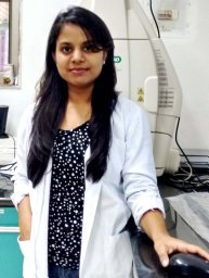 Bhavana Kushwaha合作论文数Endocrinology Division, CSIR-Central Drug Research Institute3
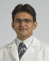 Singh Rajender合作论文数Centre for Cellular and Molecular Biology, Hyderabad, India3
 Gopal Gupta合作论文数Department of Urology, Stritch School of Medicine, Loyola University Chicago3
 Santosh Kumar Yadav合作论文数1
 Rahul Vishvkarma合作论文数1
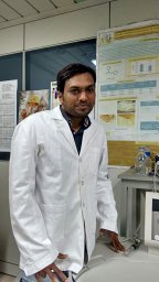 Lokesh Kumar合作论文数Medicinal &Process Chemistry Division, Endocrinology Division, and Molecular &Structural Biology Division, CSIR-Central Drug Research Institute;and Molecular & Structural Biology Division, CSIR-Central Drug Research Institute1
 Pandey Aastha合作论文数1




