BACKGROUND. Coronary artery calcium (CAC) scoring is used to stratify acute coronary syndrome (ACS) risk. Nonetheless, patients with a CAC score of zero (CAC0) remain at risk from noncalcified plaque components. OBJECTIVE. The purpose of this study was to explore CTA-derived coronary artery plaque characteristics in symptomatic patients with CAC0 who subsequently have ACS through comparisons with patients with a CAC score greater than 0 (CAC> 0) who subsequently have ACS as well as with patients with CAC0 who do not subsequently have ACS. METHODS. This study entailed a secondary retrospective analysis of prior prospective registry data. The international multicenter CONFIRM (Coronary CT Angiography Evaluation for Clinical Outcomes: An International Multicenter) registry collected longitudinal observational data on symptomatic patients who underwent clinically indicated coronary CTA from January 2004 to May 2010. ICONIC (Incident Coronary Syndromes Identified by CT) was a nested cohort study conducted within CONFIRM that identified patients without known coronary artery disease (CAD) at the time of CTA who did and did not subsequently have ACS (i.e., the ACS and control groups, respectively) and who were propensity matched in a 1:1 ratio on the basis of CAD risk factors and CAD severity on CTA. The present ICONIC substudy selected matched patients in the ACS and control groups who both had documented CAC scores. CTA examinations were analyzed using artificial intelligence software for automated quantitative plaque assessment. In the ACS group, invasive angiography findings were used to identify culprit lesions. RESULTS. The present study included 216 patients (mean age, 55.6 years; 91 women and 125 men), with 108 patients in each of the ACS and control groups. In the ACS group, 23% (n = 25) of patients had CAC0. In the ACS group, culprit lesions in the subsets of patients with CAC0 and CAC> 0 showed no significant differences in fibrous, fibrofatty, or necrotic-core plaque volumes (p > .05). In the CAC0 subset, patients with ACS, compared with control patients, had greater mean (± SD) fibrous plaque volume (29.4 ± 42.0 vs 5.5 ± 15.2 mm3, p < .001), fibrofatty plaque volume (27.3 ± 52.2 vs 1.3 ± 3.7 mm3, p < .001), and necrotic-core plaque volume (2.8 ± 6.4 vs 0.0 ± 0.1 mm3, p < .001). CONCLUSION. After propensity-score matching, 23% of patients with ACS had CAC0. Patients with CAC0 in the ACS and control groups showed significant differences in volumes of noncalcified plaque components. CLINICAL IMPACT. Methods that identify and quantify noncalcified plaque forms may help characterize ACS risk in symptomatic patients with CAC0.
Pre-procedural imaging is critical for transcatheter mitral valve repair planning in patients with mitral valve disease. As differences among various measurement techniques for valve evaluation are still poorly understood, we sought to assess the intra- and interobserver agreement of complex measurements derived from a prototype mitral evaluation tool (Siemens) and a commercially available tool (CVI42) using both saddle- and D-shaped mitral annulus techniques. Multiphasic cardiac computed tomography angiography data were loaded into each software. Three expert readers independently measured the annuli on systolic- and diastolic-phase images using both tools. Measurement agreement between the tools was assessed with t tests, with p ≤ 0.05 considered statistically significant. Intraclass correlation coefficient (ICC) was used for interobserver agreement. Bland–Altman plots were used to assess for systematic differences. Ten patients (mean age: 61.9 ± 9.9 years, 70
INTRODUCTION: Coronary CT angiography (CCTA) is a powerful noninvasive tool for identifying high-risk plaque, such as low-density non-calcified plaque (LD-NCP). Though, the optimal treatment of patients with LD-NCP remains unclear. This study explored the association of revascularization in the setting of LD-NCP with the occurrence of acute coronary syndrome (ACS). Methods: This was a post-hoc analysis of the ICONIC study. A subset of 234 patients that underwent CCTA with subsequent ACS were matched to 234 control patients who also underwent CCTA but did not have ACS during follow-up. Patients were also followed for occurrence of revascularization, either coronary artery bypass graft or percutaneous coronary intervention. Atherosclerosis imaging-enabled quantitative CT (AI-QCT) was used to measure diameter stenosis, and LD-NCP, non-calcified plaque, and calcified plaque volumes from each CCTA. LD-NCP was defined as plaque with -190 to 30 Hounsfield Units. Patients were stratified based on the presence of LD-NCP. Subgroup analysis was conducted to compare the occurrence of ACS with the rate of revascularization. Kaplan-Meier survival curves and extended Cox regression analysis were used to evaluate the effect size of revascularization and LD-NCP on occurrence of ACS. Results: AI-QCT was completed in 448/468 subjects (follow-up time [MEAN±SD] 2.44±2.48 years). The median of LD-NCP was 1.2 mm 3 for patients with >0 mm 3 LD-NCP. There were 85 patients with LD-NCP >1.2 mm 3 and 363 patients with LD-NCP ≤1.2 mm 3 . In patients with LD-NCP >1.2 mm 3 , the rate of revascularization in patients with and without ACS was 3/52 (5.8%) versus 14/33 (42.4%) (p<0.001). In patients with LD-NCP ≤1.2 mm 3 , the rate of revascularization in patients with and without ACS was 36/170 (21.2%) versus 39/193 (20.2%) (p=0.897). In comparison to patients without revascularization and LD-NCP ≤1.2 mm 3 , patients with LD-NCP >1.2 mm 3 and revascularization were less likely to have ACS during follow-up (adjusted HR: 0.20 [0.07, 0.61]; p=0.005). Additionally, patients with LD-NCP >1.2 mm 3 who did not undergo revascularization were more likely to have ACS (adjusted HR: 1.47 [1.03, 2.12]; p=0.036). Hazard ratios were adjusted for diameter stenosis, and non-calcified and calcified plaque volume. Time-dependent coefficients were included for diameter stenosis. Conclusion: Revascularization of patients with LD-NCP >1.2 mm 3 identified on CCTA with AI-QCT was associated with less risk for ACS.
Donor-derived cell-free DNA (dd-cfDNA) may safely assess kidney allograft rejection. Molecular Microscope (MMDx®) gene expression may offer increased precision to histology. This single-center retrospective study monitored kidney transplant recipients for rejection at specified time intervals by utilizing creatinine (SCr), proteinuria, donor-specific antibodies (DSAs), and dd-cfDNA. A clinically indicated biopsy sample was sent for histopathology and MMDx®. Patients were categorized into rejection (Rej) and non-rejection (NRej) groups, and further grouped according to antibody-mediated rejection (ABMR) subtypes. Rej and NRej groups included 52 and 37 biopsies, respectively. Median follow-up duration was 506 days. DSAs were positive in 53% and 22% of patients in both groups, respectively (p = 0.01). Among these groups, pre- and post-intervention median SCr, proteinuria, and dd-cfDNA at 1 month, 2 months, and at the last follow-up revealed significant difference for dd-cfDNA (all p = 0.01), however, no difference was found for SCr and proteinuria (p > 0.05). The AUC was 0.80 (95% CI: 0.69–0.91), with an optimal dd-cfDNA criterion of 2.2%. Compared to histology, MMDx® was more likely to diagnose ABMR (79% vs. 100%) with either C4d positivity or negativity and/or DSA positivity or negativity. Hence, a pre- and post-intervention allograft monitoring protocol in combination with dd-cfDNA, MMDx®, and histology has aided in early diagnosis and timely individualized intervention.
Rizvi, Asim; Hussain, Syed A.; Mujtaba, Muhammad A.; Lea, Alfred S.; Kueht, Michael; Gamilla-Crudo, Ann Kathleen N. Author Information
Rizvi, Asim; Hussain, Syed A.; Gamilla-Crudo, Ann Kathleen N.; Kueht, Michael; Mujtaba, Muhammad A. Author Information
 James K. Min合作论文数Cleerly;Weill Cornell Medical College49
 Fay Y. Lin合作论文数Department of Medicine, Weill Cornell Medical College—New York Presbyterian Hospital40
 Donghee Han合作论文数Departments of Medicine (Division of Artificial Intelligence in Medicine),, Cedars-Sinai Medical Center37
 Ji-Hyun Lee合作论文数37
 Bríain Ó Hartaigh合作论文数Novo Nordisk33
 Hyuk-Jae Chang合作论文数Department of Internal Medicine, College of Medicine, Yonsei University;Division of Cardiology, Severance Cardiovascular Hospital, College of Medicine, Yonsei University;Division of Medical Information and Technology, Severance Hospital;Ontact Healthcare28
 Heidi Gransar合作论文数24Gamilla-Crudo Ann K合作论文数University of Texas Medical Branch22
 Hadi Mirhedayati Roudsari合作论文数Internal Med Residency Program, Medstar Hlth18
 Syed Ameenuddin Hussain合作论文数Department of Zoology, Government College University18
 Matthew Budoff合作论文数The Lundquist Institute;Division of Cardiology, Harbor-UCLA Medical Center;David Geffen School of Medicine, University of California, Los Angeles18
 Martin Hadamitzky合作论文数Deutsches Herzzentrum München;German Heart Center Munich18
 Waleed Brinjikji合作论文数Division of Neuroradiology, Department of Radiology, Mayo Clinic18Mahn-Won Park合作论文数17
 Jonathon A. Leipsic合作论文数Department of Radiology, University of British Columbia17Daniele Andreini合作论文数17Yong-Jin Kim合作论文数16
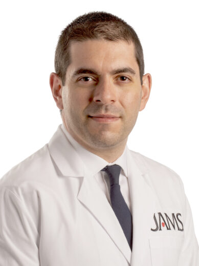 Subhi J. Al'Aref合作论文数University of Arkansas for Medical Sciences16
 Gianluca Pontone合作论文数16
 Erica Maffei合作论文数15
 Jessica M. Pena合作论文数Joan and Sanford I. Weill Department of Medicine, Weill Cornell Medicine, Cornell University15
 Filippo Cademartiri合作论文数Department of Radiology, SDN IRCCS, Naples, Italy15Kavitha Chinnaiyan合作论文数15Leslee Shaw合作论文数15Michael Kueht合作论文数14
 Daniel Berman合作论文数Cedars-Sinai;Medicine School, University of California, Los Angeles14
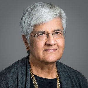 Renu Virmani合作论文数CVPath Institute Inc.14
 Hugo Marques合作论文数13M. Mujtaba合作论文数Univ Texas Med Branch13
 David F. Kallmes合作论文数Department of Radiology, Mayo Clinic;Comprehensive Cancer Center, Mayo Clinic13
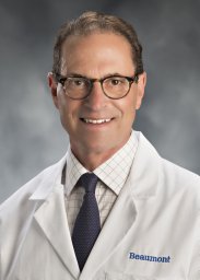 Gilbert L. Raff合作论文数Cardiology Division, William Beaumont Hospital13Lohendran Baskaran合作论文数12
 Muhammad Mujtaba合作论文数VTT Technical Research Centre of Finland Ltd11
 Todd Villines合作论文数11
 Yao Lu (陆瑶)合作论文数Colgate-Palmolive;Vaccine and Infectious Disease Organization - International Vaccine Centre11
 Jeroen Bax合作论文数Echo Laboratory, Department of Cardiology, Leiden University Medical Centre11
 Sean Fitzgerald合作论文数Biomedical Diagnostics Institute, Dublin City University;School of Biotechnology, Dublin City University;Dublin City University, Dublin City University10Khalil Anchouche合作论文数Weill Cornell Med, New York, NY 10065 USA9
 Sang-Eun Lee合作论文数Department of Life Sciences, CHA University;College of Medicine, Seoul National University;Dementia Research Center, University College London9
 Edoardo Conte合作论文数9
 Ran Heo合作论文数9
 Ashley N. Beecy合作论文数Sutter Health9Stuijfzand Wijnand J合作论文数9
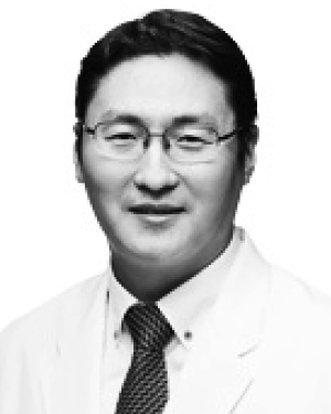 Park Hyung-Bok合作论文数9
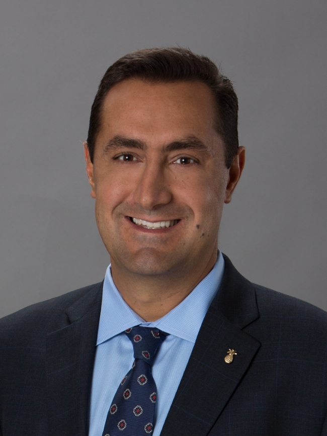 Ricardo Cury合作论文数Department of Radiology, Herbert Wertheim College of Medicine, Florida International University9
 Eun Ju Chun合作论文数9
 Benjamin Chow合作论文数The University of Ottawa Heart Institute;University of Ottawa9
 Seyed Mohammad Seyedsaadat合作论文数8
 Ramanathan Kadirvel合作论文数8




