Purpose To develop and evaluate a Deep learnIng-bAsed MONoenergetic imaging at Different energies (DIAMOND) framework for generating virtual monoenergetic images (VMIs) from conventional energy-integrating detector (EID) CT, aiming to reduce blooming artifacts and improve stenosis assessment in coronary CT angiography (CCTA) with heavily calcified plaques. Materials and Methods This study (August 2022-September 2023) used a combination of retrospective and prospective imaging data. DIAMOND was trained using a simplified U-Net architecture on a retrospective dataset of 10 CCTA examinations performed with ultrahigh-resolution (UHR) photon-counting detector (PCD) CT; 70-keV PCD VMIs (energy equivalent to 120-kV single-energy EID CT) served as inputs and 100-keV PCD VMIs as targets. The trained model was then applied to a prospective dataset of participants with heavily calcified plaques who underwent EID CT at 120 kV followed by same-day PCD CT. Percent diameter stenosis (PDS) was quantified for a phantom and participants by using commercial software and compared across EID CT, DIAMOND, and PCD CT using Bland-Altman analysis. Changes in stenosis severity categorization based on PDS were evaluated. Results DIAMOND reduced blooming artifacts and improved lumen visualization, with image quality resembling PCD CT. In 23 participants (mean age, 69 years ± 8 [SD]; 18 male), average PDS decreased from 35.65% (EID CT) to 25.19% (DIAMOND, P < .05), approaching 24.27% with UHR PCD CT (P < .05). Relative to EID CT, DIAMOND led to Coronary Artery Disease Reporting and Data System reclassification in 11 of 26 (42%) lesions, mainly in mild to moderate stenosis ranges. Processing time was approximately 0.21 second per axial section on a standard graphics processing unit. Conclusion This study demonstrated the feasibility of using DIAMOND to generate high-kiloelectron volt VMIs from single-energy EID CT, providing artifact-reduced coronary imaging and improved stenosis quantification for heavily calcified plaques comparable to PCD CT without hardware upgrades. Keywords: Coronary CT Angiography, Coronary Artery Stenosis, Energy-Integrating Detector CT, Photon-Counting Detector CT, Deep Learning, CT-Photon Counting, Angiography, Coronary Arteries Supplemental material is available for this article. © RSNA, 2026.
Purpose To evaluate the preliminary technical development, safety, and feasibility of dynamic contrast-enhanced MR lymphangiography (DCE-MRL) performed with nodal and/or dermal injection of gadopiclenol. Materials and Methods This institutional review board-approved retrospective study included consecutive patients referred to the complex lymphatic disorders clinic who underwent DCE-MRL at 1.5 T with gadopiclenol administered via inguinal nodal and/or interstitial transpedal injection. Technical performance, safety, and feasibility for imaging peripheral and central conducting lymphatic anatomy, flow, and drainage were assessed. Results A total of 23 patients (12 female) underwent DCE-MRL with gadopiclenol. Contrast material was administered via inguinal lymph nodes in all patients (23 of 23, 100%) and via interstitial transpedal injection in one patient (one of 23, 4.3%). No serious or nonserious adverse events were observed (0 of 23). Evaluation of central conducting lymphatic anatomy, flow, and drainage into the central venous system was feasible in all patients (23 of 23), and evaluation of lower extremity superficial lymphatic anatomy was feasible in one patient. Signal contrast increased with flip angle up to approximately 35° at 1.5 T, supporting protocol optimization. Preliminary results suggest a near-optimal flip angle of 35° at 1.5 T, a higher flip angle than when using other gadolinium-based contrast agents. Conclusion Dynamic gadopiclenol-enhanced nodal and pedal MR lymphangiography was safe and feasible for imaging peripheral and central conducting lymphatic anatomy. Keywords: Lymphangiography, MR-Angiography, Lymphatic, MR-Dynamic Contrast Enhanced, MR-Contrast Agent Supplemental material is available for this article. © RSNA, 2026.
BACKGROUND:Radiomics extracts quantitative imaging features from computed tomography (CT) data for clinical decision-making. However, variations in acquisition parameters-particularly x-ray tube voltage (kV)-introduce non-biological variability in attenuation values, limiting the reproducibility of radiomic features across scanners, protocols, and institutions. PURPOSE:To develop and evaluate a CT dAta harmoNiZAtion framework based on deep learNed vIrTual monoEnergetic imaging (TANZANITE), which leverages the keV flexibility of virtual monoenergetic images (VMIs) to enable cross-kV scan translation and radiomics harmonization. METHODS:TANZANITE is a model hub consisting of multiple pre-trained convolutional neural networks (CNNs), each designed to translate VMIs from 1 keV level to another. Phantom-based calibration was first used to determine energy-equivalent (Eff_E) keV levels corresponding to each tube potential (e.g., Eff_E(A) keV for source kV and Eff_E(B) keV for target kV). A CNN trained using 69 120 patches from seven patient cases to map VMIs from Eff_E(A) to Eff_E(B) was selected from the TANZANITE hub and applied directly to clinical CT images acquired at the source kV. This harmonized the images to match the attenuation characteristics of the target kV setting. Evaluation was conducted on independent dual-energy CT datasets acquired at 100/Sn150 kV. Regions of interest (ROIs) were placed in the kidney, liver, and spine to assess CT number consistency and radiomic feature reproducibility. The concordance correlation coefficient (CCC) was calculated across 93 non-shape radiomic features. RESULTS:After TANZANITE processing with 100 kV images, CT numbers in evaluated organs closely matched the Sn150 kV reference values in four testing patient cases. For example, mean kidney CT numbers changed from 320 HU (100 kV) to 156 HU (TANZANITE), approximating the Sn150 kV value of 160 HU. Similar changes were observed in the liver (157-105 HU vs. 104 HU reference) and spine (45-22 HU vs. 19 HU reference). Radiomic reproducibility improved substantially across organs: mean CCC increased from 0.590 to 0.995 in the liver, 0.300 to 0.970 in the kidney, and 0.630 to 0.968 in the spine. Post-TANZANITE, over 98% of features exceeded the stability threshold (CCC ≥ 0.900) in all three representative organs. CONCLUSION:TANZANITE provides a flexible, image-domain harmonization framework by learning the keV-to-keV translation in the VMI domain and applying pre-trained CNNs to clinical kV images. It improves CT number consistency and organ-specific radiomic reproducibility without requiring raw projection data or scanner-specific training. This approach supports consistent quantitative imaging across multiple-kV acquisition protocols, enhancing radiomics reliability in clinical settings.
Articles on the development of medical image artificial intelligence (AI) algorithms are numerous in the literature, but deployment to clinical practice is infrequently discussed. The Enterprise Radiology Framework for AI Software Technology Team at Mayo Clinic has been focused on bridging the gap in clinical translation of medical image AI algorithms since its inception in 2019. During this time, we have released 17 algorithms into our radiology clinical practice. Recently, we have placed an increased focus on monitoring these algorithms, as there are few reports with practical experience documented in the literature. Our increased monitoring efforts include daily, weekly, and yearly monitoring of utilization, failure modes, data drift, and end-user feedback through automated alerts, dedicated dashboards, and pointed investigations to enable optimal algorithmic processing. End-user feedback is elicited yearly during annual reviews to ensure clinical needs are still being met. Automated monitoring has enabled earlier identification of problems, such as images no longer routing through the orchestration engine to the appropriate algorithm, minimizing potential disruption to the clinical practice and ensuring continued algorithmic utilization. Monitoring has also reinforced the importance of key aspects of interdisciplinary research and translation, such as early discussions on clinical needs coupled with technological ability and proper training. By providing our experience in and continuing to improve monitoring methods as a community, we can all minimize risk and maximize the benefits of medical pixel-based AI.
Importance:Current risk prediction guidelines for hypertrophic cardiomyopathy predict only sudden cardiac death and are imperfect, leading to avoidable deaths and unnecessary implantable cardioverter defibrillators. Objective:To combine prospectively collected clinical history, imaging, genetic, and biomarker data to improve risk prediction of adverse events in hypertrophic cardiomyopathy. Design, Setting, and Participants:A total of 2750 patients with hypertrophic cardiomyopathy were prospectively enrolled in the registry-based study from 44 sites in North America and Europe with expertise in hypertrophic cardiomyopathy and cardiac magnetic resonance (CMR) imaging. Participants were enrolled from April 1, 2014, to April 7, 2017. Exposures:Patients underwent a health history questionnaire, blood sampling for biomarkers and genotyping, and contrast-enhanced CMR. Patients were followed up yearly by telephone and through records review regarding event documentation. Main Outcomes and Measures:The predefined composite adjudicated primary end point was time to first event for hypertrophic cardiomyopathy-related deaths; nonfatal sustained ventricular arrhythmias (VAs) requiring cardioversion or defibrillation; and left ventricular (LV) assist device implant or heart transplant. A secondary end point was a composite of sudden cardiac death and nonfatal VA events. The elastic-net method identified the most important predictors. Cox proportional hazards regression assessed associations with time to the first end point. Results:Of the 2750 prospectively enrolled patients, 2698 (98%) had analyzable data after 9 were excluded because they had hypertrophic cardiomyopathy phenocopies and 43 withdrew. Of these remaining patients, 1919 (71%) were male, mean age was 50 years (SD, 11 years), and 423 (16%) were from underrepresented racial and minority groups. The mean follow-up was 6.9 years (SD, 2.1 years). The primary event model in 104 patients included LV scar as a percentage of LV mass by late gadolinium enhancement (LGE%; hazard ratio [HR], 1.86; 95% CI, 1.58-2.20; P < .001), LV mass index (HR, 1.09; 95% CI, 1.01-1.17; P = .03), LV end-systolic volume index (HR, 1.28; 95% CI, 1.12-1.46; P < .001 ), all per 10-unit increase, history of heart failure at study entry (HR, 2.89; 95% CI, 1.75-4.77; P < .001), and log N-terminal pro-B-type natriuretic peptide (NT-proBNP; HR, 1.41; 95% CI, 1.17-1.70; P < .001) level per log unit, (C index for all, 0.77). An LGE percentage of the LV mass of 9% or higher substantially increased the primary composite event rate (P = .001). The secondary sudden cardiac death and VA risk factor model (in 69 patients) included LGE%, LV mass index, LV ejection fraction, and log(NT-proBNP) (C index, 0.76). Conclusions and Relevance:These results provide prospective evidence for incorporating cardiac magnetic resonance and NT-proBNP in the evaluation of patients with hypertrophic cardiomyopathy. Trial Registration:ClinicalTrials.gov Identifier: NCT01915615.
Integration of AI-enabled algorithms into the radiology workflow presents a complex array of challenges that span operational, technical, clinical, and regulatory domains. Successfully overcoming these hurdles requires a multifaceted approach, including strategic planning, educational initiatives, and careful consideration of the practical implications for radiologists' workloads. Institutions must navigate these challenges with a clear understanding of the potential benefits and limitations of both vended and in-house developed AI tools.
Artificial intelligence (AI) offers promising solutions for many steps of the cardiac imaging workflow, from patient and test selection through image acquisition, reconstruction, and interpretation, extending to prognostication and reporting. Despite the development of many cardiac imaging AI algorithms, AI tools are at various stages of development and face challenges for clinical implementation. This scientific statement, endorsed by several societies in the field, provides an overview of the current landscape and challenges of AI applications in cardiac CT and MRI. Each section is organized into questions and statements that address key steps of the cardiac imaging workflow, including ethical, legal, and environmental sustainability considerations. A technology readiness level range of 1 to 9 summarizes the maturity level of AI tools and reflects the progression from preliminary research to clinical implementation. This document aims to bridge the gap between burgeoning research developments and limited clinical applications of AI tools in cardiac CT and MRI.
Coronary computed tomography angiography plays a pivotal role in the diagnosis, risk stratification, and treatment of patients with known or suspected coronary artery disease. However, conventional computed tomography (CT) technologies are limited by spatial resolution, artifact susceptibility, and radiation exposure. Photon-counting computed tomography (PCCT) introduces substantial technological improvements over conventional CT. This includes improved spatial and contrast resolution, energy discrimination, and reduction of various artifacts. As a result, PCCT enables superior coronary lumen and plaque evaluation, even in complex cases with severe calcification or smaller coronary stents. Beyond the coronary arteries, PCCT offers improved visualization of cardiac anatomy and myocardial tissue characterization with the potential to reduce downstream testing, improve diagnosis and treatment, and ultimately improve clinical outcomes. PCCT is poised to become the dominant technology for cardiovascular CT; however, challenges such as high costs, increased data demands, and a need for more validation, standardized image acquisition, and post-processing protocols remain. This review explores the technical principles of PCCT, its advantages over conventional CT, and its current and potential future applications in cardiac imaging, highlighting opportunities for future research.
Pre-procedural imaging is critical for transcatheter mitral valve repair planning in patients with mitral valve disease. As differences among various measurement techniques for valve evaluation are still poorly understood, we sought to assess the intra- and interobserver agreement of complex measurements derived from a prototype mitral evaluation tool (Siemens) and a commercially available tool (CVI42) using both saddle- and D-shaped mitral annulus techniques. Multiphasic cardiac computed tomography angiography data were loaded into each software. Three expert readers independently measured the annuli on systolic- and diastolic-phase images using both tools. Measurement agreement between the tools was assessed with t tests, with p ≤ 0.05 considered statistically significant. Intraclass correlation coefficient (ICC) was used for interobserver agreement. Bland–Altman plots were used to assess for systematic differences. Ten patients (mean age: 61.9 ± 9.9 years, 70
Background A cardiac computed tomography (CT)-based mitral annular calcification (MAC) scoring system systematically grades MAC severity, but its correlation with three-dimensional transesophageal echocardiography (3D-TEE) remains unclear. Objectives The authors aimed to compare MAC severity assessment by 3D-TEE vs CT and evaluate their associations with hemodynamic parameters indicative of mitral valve disease. Methods We analyzed patients with MAC enrolled in the MITRAL (Mitral Implantation of TRAnscathether vaLves) II trial (NCT04408430) across 13 centers undergoing transcatheter mitral valve replacement screening. Core laboratories assessed MAC severity using 3D-TEE and CT. Correlation and agreement were assessed via Spearman and intraclass correlation coefficients. Hemodynamic parameters of mitral stenosis (MS) and regurgitation were obtained from two-dimensional transthoracic echocardiography (2D-TTE) and 3D-TEE. Linear regression evaluated associations with MAC scores. Results The analysis included 164 patients (75% females, mean age 75.5 ± 9.1 years). The mean MAC scores were 8.0 ± 0.44 by CT and 8.2 ± 0.45 by 3D-TEE. A modest positive correlation (Spearman correlation coefficient = 0.34; P < 0.001) was observed between 3D-TEE and CT scores, with better agreement in severe MAC. Each unit increase in the 3D-TEE MAC score was associated with a 0.15 cm2 decrease in 3D-TEE mitral valve area, a 0.05 cm2 decrease in 2D-TTE mitral valve area, and an increase in mean diastolic pressure gradient (1 mm Hg by 3D-TEE; 0.96 mm Hg by 2D-TTE). 3D-TEE MAC scores showed no significant correlation with regurgitation parameters. CT-MAC scores showed weak, nonsignificant correlations with both MS and mitral regurgitation. Conclusions 3D-TEE provides complementary and functionally relevant MAC assessment, correlating with MS severity. It may serve as a useful adjunct to CT in transcatheter mitral valve replacement evaluation, especially when contrast imaging is limited.
Prosthetic heart valve (PHV) dysfunction is increasingly seen due to the increase in the number of PHV that are being implanted worldwide. Cardiac CT imaging has emerged as a valuable tool to assess PHVs and determine the cause of dysfunction. This consensus document first summarizes the available techniques for PHV assessment. Then the use of CT in PHV (dys)function assessment is discussed in detail including consensus statements for correct indications and patient selection for CT assessment of PHVs, image acquisition, reconstruction and measurement protocols and how to interpret and report the CT findings for specific types of PHV dysfunction.
IntroductionVolume overload from mitral regurgitation can result in left ventricular systolic dysfunction. To prevent this, it is essential to operate before irreversible dysfunction occurs, but the optimal timing of intervention remains unclear. Current echocardiographic guidelines are based on 2D linear measurement thresholds only. We compared volumetric CT-based and 2D echocardiographic indices of LV size and function as predictors of post-operative systolic dysfunction following mitral repair.MethodsWe retrospectively identified patients with primary mitral valve regurgitation who underwent repair between 2005 and 2021. Several indices of LV size and function measured on preoperative cardiac CT were compared with 2D echocardiography in predicting post-operative LV systolic dysfunction (LVEFecho <50%). Area under the curve (AUC) was the primary metric of predictive performance.ResultsA total of 243 patients were included (mean age 57 ± 12 years; 65 females). The most effective CT-based predictors of post-operative LV systolic dysfunction were ejection fraction [LVEFCT; AUC 0.84 (95% CI: 0.77–0.92)] and LV end systolic volume indexed to body surface area [LVESViCT; AUC 0.88 (0.82–0.95)]. The best echocardiographic predictors were LVEFecho [AUC 0.70 (0.58–0.82)] and LVESDecho [AUC 0.79 (0.70–0.89)]. LVEFCT was a significantly better predictor of post-operative LV systolic dysfunction than LVEFecho (p = 0.02) and LVESViCT was a significantly better predictor than LVESDecho (p = 0.03). Ejection fraction measured by CT demonstrated significantly greater reproducibility than echocardiography.DiscussionCT-based volumetric measurements may be superior to established 2D echocardiographic parameters for predicting LV systolic dysfunction following mitral valve repair. Validation with prospective study is warranted.
Tricuspid regurgitation (TR) is associated with increased mortality and poor prognosis. 1 Nath J. Foster E. Heidenreich P.A. Impact of tricuspid regurgitation on long-term survival. J Am Coll Cardiol. 2004; 43: 405-409 Crossref PubMed Scopus (1244) Google Scholar Novel investigational transcatheter tricuspid valve (TV) repair and replacement devices have the potential to address a large unmet need in patients with TR. Cardiac computed tomography (CT) is the reference standard for transcatheter TV device sizing. 2 Agricola E. Asmarats L. Maisano F. et al. Imaging for tricuspid valve repair and replacement. JACC Cardiovasc Imaging. 2021; 14: 61-111 Crossref PubMed Scopus (42) Google Scholar However, three-dimensional (3D) transthoracic echocardiography (TTE) is more widely available, and recent advances have enhanced 3D visualization of the TV and tricuspid annulus. We aimed to compare tricuspid annular (TA) dimensions by 3D TTE using a semiautomated measurement package vs the reference standard, CT.
Infective endocarditis (IE) is a complex multisystemic disease resulting from infection of the endocardium, the prosthetic valves, or an implantable cardiac electronic device. The clinical presentation of patients with IE varies, ranging from acute and rapidly progressive symptoms to a more chronic disease onset. Because of its severe morbidity and mortality rates, it is necessary for radiologists to maintain a high degree of suspicion in evaluation of patients for IE. Modified Duke criteria are used to classify cases as "definite IE," "possible IE," or "rejected IE." However, these criteria are limited in characterizing definite IE in clinical practice. The use of advanced imaging techniques such as cardiac CT and nuclear imaging has increased the accuracy of these criteria and has allowed possible IE to be reclassified as definite IE in up to 90% of cases. Cardiac CT may be the best choice when there is high clinical suspicion for IE that has not been confirmed with other imaging techniques, in cases of IE and perivalvular involvement, and for preoperative treatment planning or excluding concomitant coronary artery disease. Nuclear imaging may have a complementary role in prosthetic IE. The main imaging findings in IE are classified according to the site of involvement as valvular (eg, abnormal growths [ie, "vegetations"], leaflet perforations, or pseudoaneurysms), perivalvular (eg, pseudoaneurysms, abscesses, fistulas, or prosthetic dehiscence), or extracardiac embolic phenomena. The differential diagnosis of IE includes evaluation for thrombus, pannus, nonbacterial thrombotic endocarditis, Lambl excrescences, papillary fibroelastoma, and caseous necrosis of the mitral valve. The location of the lesion relative to the surface of the valve, the presence of a stalk, and calcification or enhancement at contrast-enhanced imaging may offer useful clues for their differentiation. ©RSNA, 2024 Test Your Knowledge questions for this article are available in the supplemental material.
 Foley Thomas A合作论文数32
 Cynthia H. McCollough合作论文数Department of Radiology, Mayo Clinic;CT Clinical Innovation Center, Mayo Clinic25Phillip M. Young合作论文数24
 Philip Araoz合作论文数21
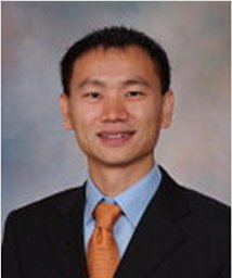 Shuai Leng合作论文数Mayo Clinic21
 David F. Kallmes合作论文数Department of Radiology, Mayo Clinic;Comprehensive Cancer Center, Mayo Clinic20Robert J Mcdonald合作论文数19Jennifer Mcdonald合作论文数19
 Mackram F Eleid合作论文数15Jacobo Kirsch合作论文数15Matthew W. Martinez合作论文数14James F. Glockner合作论文数14
 Rakesh Suri合作论文数Cleveland Clinic Abu Dhabi13
 Jeremy D Collins合作论文数Feinberg School of Medicine, Northwestern University;Department of Radiology, Mayo Clinic12
 Kevin Greason合作论文数11
 Jae K. Oh合作论文数Division of Structural Heart Disease, Department of Cardiovascular Medicine, Mayo Clinic;Division of Cardiovascular Ultrasound, Department of Cardiovascular Medicine, Mayo Clinic10Richard W. Katzberg合作论文数10
 Prabhakar Rajiah合作论文数9Carter Rickey E合作论文数9Mayra Guerrero合作论文数8
 Joel G. Fletcher合作论文数Department of Radiology, Mayo Clinic8Nkomo Vuyisile T合作论文数8
 Charanjit S. Rihal合作论文数Division of Cardiovascular Diseases, Department of Internal Medicine, Mayo Clinic;Division of Interventional Cardiology, Department of Cardiovascular Medicine, Mayo Clinic8Nikkole M Weber合作论文数7
 Terri Vrtiska合作论文数7
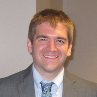 Peter Brady合作论文数Los Alamos National Laboratory7Nandan S. Anavekar合作论文数7
 Imran Syed合作论文数Cardiovascular Diseases, Mayo Clinic Mankato Campus7Jerome F. Breen合作论文数7Michael F. Morris合作论文数6L Eckel合作论文数6
 Jeremy J. Thaden合作论文数Department of Cardiovascular Medicine, Mayo Clinic6Jentoft Mark合作论文数6DaLi Feng合作论文数6Gurpreet S. Sandhu合作论文数6Crystal R. Bonnichsen合作论文数6David L Murray合作论文数5Karin E Dill合作论文数5
 Apurva B. Challa合作论文数5Pr Julsrud合作论文数5
 Panagiotis Korfiatis合作论文数Med. Phys. Dept., Univ. of Patras;c;Med. Phys. Dept., Univ. of Patras5Thielen Kent R合作论文数5
 Ryan K Funk合作论文数5
 Maurice Sarano合作论文数Minneapolis Heart Institute Foundation;Valve Science Center5
 John C. Burnett Jr.合作论文数Division of Circulatory Failure, Department of Cardiovascular Medicine, Mayo Clinic5Rajiv Gulati合作论文数5
 Nadia N. Laack合作论文数Department of Radiation Oncology, Mayo Clinic5Andrew N. Primak合作论文数5
 Sorin V. Pislaru合作论文数Department of Cardiovascular Medicine, Mayo Clinic5




