Supplemental Appendix 6. Summary of ddPCR mutations to yield highest sensitivity for ctDNA detection.
Supplemental Appendix 7: Paired pre- and post- treatment SNaPshot molecular profiling available for 11 patients.
AbstractPurpose: We performed a NCI-sponsored, prospective study of neoadjuvant FOLFIRINOX followed by chemoradiation with carboplatin/paclitaxel followed by surgery in patients with locally advanced gastric or gastroesophageal cancer. Patients and Methods: The primary objective was to determine completion rate of neoadjuvant FOLFIRINOX × 8 followed by chemoradiation. Secondary endpoints were toxicity and pathologic complete response (pCR) rate. Exploratory analysis was performed of circulating tumor DNA (ctDNA) to treatment response. Results: From October 2017 to June 2018, 25 patients were enrolled. All patients started FOLFIRINOX, 92% completed all eight planned cycles, and 88% completed chemoradiation. Twenty (80%) patients underwent surgical resection, and 7 had a pCR (35% in resected cohort, 28% intention to treat). Tumor-specific mutations were identified in 21 (84%) patients, of whom 4 and 17 patients had undetectable and detectable ctDNA at baseline, respectively. Presence of detectable post-chemoradiation ctDNA (P = 0.004) and/or postoperative ctDNA (P = 0.045) were associated with disease recurrence. Conclusions: Here we show neoadjuvant FOLFIRINOX followed by chemoradiation for locally advanced gastroesophageal cancer is feasible and yields a high rate of pCR. ctDNA appears to be a promising predictor of postoperative recurrence. See related commentary by Catenacci, p. 6281
547 Background: Metastatic biliary tract cancer (mBTC) is a lethal malignancy with median 5 year OS of less than 10%. Immunotherapy, particularly single agent anti-PD-1/PD-L1, has limited efficacy in mBTC with ORR~9-15%. Recently presented data shows responses in metastatic MSS pancreatic or colon cancer with combination anti-PD-1/CTLA-4 and radiation (XRT) to produce systemic response (abscopal effect) (Parikh A, GI ASCO 2019, ASCO 2019.). We evaluate safety and efficacy of dual PD-1/CTLA-4 inhibition with XRT in MSS mBTC. Methods: 15 of a planned 15 mBTC patients were enrolled. Eligible pts had histologically-confirmed mBTC, ECOG-PS 0/1, and must have progressed on at least one line of previous therapy or refused standard therapy. Safety cohort of 6 pts of durva 1500 mg/treme 75 mg q4w was enrolled. If > 2 DLTs, patients were enrolled subsequently to dose level -1 (durva 1125 mg/ treme 75 mg q4w). 3 fractions of 8 Gy of radiation at C2D1 every other day to a single metastatic site. Durva/treme continued for 4 cycles, followed by 4 cycles of maintenance durva until progressive disease, discontinuation or withdrawal. Endpoints include disease control rate (DCR (SD+PR+CR)), PFS and OS and safety. Radiological evaluations were done q2 mo. Results: 15 mBTC pts enrolled and evaluable from May 2018 to March 2019. Median age 63 years (range 48-75), 47% male. DLTs occurred in 3 patients during the safety run-in. One patient experienced DLT at dose level -1 and subsequent expansion. 3 patients did NOT reach radiation therapy. DCR was 27% with a 13% PR and 7% CR. Of those who reached radiation, DCR was 33% with a 17% PR and 8% CR. At time of analysis, median PFS was 54 days for ITT mBTC. Duration of response for 4 patients with DCR was 26, 52, 122, 254+ days. Treatment-related adverse events were reported in 12/15 patients (80%). Grade ≥3 toxicities were seen in 9/15 pts (60%) with lymphopenia (5 grade 3) and elevated LFTs (2 grade 4 and 4 grade 2) being the main adverse events. All patients with disease control were not MSI. Conclusions: Combination of durva/treme XRT is feasible and shows preliminary activity in metastatic BTC. An expansion cohort is being planned to confirm activity. Clinical trial information: NCT03482102.
4057 Background: We performed a single-arm pilot study of total neoadjuvant approach including FOLFIRINOX and chemoradiation (CRT) with concurrent carboplatin/taxol (C/T) followed by surgery in patients with locally advanced gastric or gastroesophageal junction (GEJ) cancer. Methods: Patients were enrolled on a NCI sponsored, prospective, single arm study (NCT03279237). Key eligibility criteria included: histologically confirmed T3/4 or lymph node (LN) positive gastric or GEJ cancer, ECOG PS ≤1, age 18+, life expectancy > 3 months. Exclusion criteria included: visceral metastases, prior chemotherapy or RT, or prior targeted therapy. Extensive LN disease beyond the surgical field (supraclavicular or para-aortic) was permitted if deemed feasible to be encompassed within a RT field. Laparoscopy was not required. Pts were treated with neoadjuvant FOLFIRINOX x 8, restaging, CRT (45 Gy for gastric, 50.4 Gy for GEJ) with concurrent C/T, restaging, followed by surgical resection. Dose reductions were at discretion of the treating physician. The primary objective was to determine the rate of completion of FOLFIRINOX x 8 followed by CRT delivered in the preoperative setting. Secondary endpoints included: 1) acute toxicity and 2) pathologic complete response (pCR). Results: From Oct 2017 to June 2018, 25 pts were enrolled. Median age was 60 (range:30-76), 17 pts were male (68%). All pts started FOLFIRINOX; 23 (92%) pts completed all 8 planned cycles. Two pts did not complete the planned 8 cycles due to metastatic progression. Rates of grade 3+ overall, gastrointestinal, and hematologic toxicities were 28%, 12%, and 28% respectively. Of the entire cohort, 23 (92%) pts started chemoRT and 22 (88%) pts completed chemoRT (1 pt died during CRT due to pulseless electrical activity arrest). All 22 pts (88%) who completed CRT went for surgical exploration, of whom 2 pts were found with intraoperative metastases. Therefore, 20 (80%) pts underwent surgical resection. At time of abstract, 1 pt’s pathology is in process; 7 pts had a pCR (37% in resected cohort, 28% in ITT cohort), all with R0 resection. Conclusions: Total neoadjuvant FOLFIRINOX followed by CRT is feasible with acceptable rates of treatment completion and grade 3+ toxicity. In our small series, the rate of pCR is promising and a follow-up study is currently planned. Clinical trial information: NCT03279237.
Abstract Purpose: The aims of this study were to investigate the correlation between MAPK activation and somatic mutation in GNAQ and GNA11 genes in uveal melanoma (UM) and to investigate the potential utility of MEK inhibition as a target for therapy of UM in tumors with GNAQ and GNA11 mutations. Methods: Sequencing and restriction fragment length polymorphism (RFLP) were utilized for detection of activating mutations in codon 209 of the GNAQ and GNA11 genes in 44 primary UMs. The expression of phospho p44/42 MAPK (pERK1/2) was assessed by immunohistochemistry in 44 UMs. In addition, the expression of pERK1/2 and pMEK 1/2 were studied using Western blot analysis in 17 primary UMs (14 with GNAQ or GNA11 mutations) and five UM cell lines (C918, 92.1, OCM3, MEL202, MEL270). Three of the UM cell lines (92.1, MEL202 and MEL270) have GNAQ codon 209 mutations. The effect of three different selective MEK inhibitors (U126, PD98059 and PD184352) on UM cell proliferation and apoptosis were studied. Results: The majority (30/44, 68.2%) of the primary UM tumors showed somatic mutation in codon 209 of either the GNAQ or the GNA11 genes. With the exception of one tumor, the mutations in either gene were mutually exclusive. Of the 30 UM primary tumors with GNAQ and/or GNA11 mutation, pERK1/2 expression was absent in 26.7%, weak in 33.3%, moderate in 23.3%, and high in only 16.7%. Western blot showed no pMEK1/2 expression in 6/14 (42.9%) of UM with either GNAQ or GNA11 mutations. Two of the UM cell lines with GNAQ mutation showed weak pERK1/2 expression. In addition one showed no pMEK1/2 expression while the other one showed very weak pMEK1/2 expression. Selective MEK inhibitors slowed UM cell proliferation but didn't induce significant apoptosis in UM cell lines regardless of GNAQ status. Conclusions: Our results indicate that a significant number of UM with somatic mutations in GNAQ or GNA11 showed no MAPK pathway activation. In UM, MEK inhibition is mostly cytostatic suggesting that selective MEK inhibitors alone may not be sufficient to control UM. Citation Format: {Authors}. {Abstract title} [abstract]. In: Proceedings of the 102nd Annual Meeting of the American Association for Cancer Research; 2011 Apr 2-6; Orlando, FL. Philadelphia (PA): AACR; Cancer Res 2011;71(8 Suppl):Abstract nr 1127. doi:10.1158/1538-7445.AM2011-1127
Follicular dendritic cells are the major supporting cell of the germinal center microenvironment. The major function of follicular dendritic cells is to present antigen to B cells in secondary lymphoid tissues. Through cell-cell interactions, FDCs are hypothesized to be central to the regulation of normal B cell growth and differentiation. The major receptor-ligand pair which mediates B cell-FDC adhesion is the beta 1 integrin VLA-4, present on B cells and VCAM-1 expressed on FDCs. Follicular non-Hodgkin's lymphomas similarly employ this mechanism to bind to neoplastic germinal centers. The VCAM-1 molecule can exist as a 6 or 7 immunoglobulin domain form. The major form of VCAM-1 on activated endothelium is the 7 domain form. In this report we have determined by polymerase chain reaction of purified FDCs that they express predominantly mRNA for 7 domain VCAM-1. It is likely that the two forms of VCAM-1 are associated with distinct functions, therefore the expression of 7 domain VCAM-1 may be important in normal and neoplastic B cell-FDC interactions.
ELAM1 is a leukocyte adhesion molecule induced on human umbilical vein endothelial cells (HUVECs) by inflammatory cytokines. Balb/C mice were immunized with COS cells transiently expressing cell-surface ELAM1 after transfection with ELAM1 cDNA. After fusion, ELAM1-specific monoclonal antibodies (Mabs) were identified by selective adhesion to ELAM1-expressing, but not control, CHO cells, and to cytokine-treated but not untreated HUVECs. One Mab, designated BB11, binds to and immunoprecipitates ELAM1 expressed on HUVECs, COS and CHO cells. BB11 blocks the interaction of ELAM1 with human PMN, the human myelomonocytic cell line HL60, and the human colon carcinoma line HT29.
 Christine Ulysse合作论文数Massachusetts Gen Hosp, Dept Med, Div Biostat, Boston, MA 02114 USA10Drapek Lorraine C合作论文数10Parikh Aparna R合作论文数Department of Medicine, University of California San Francisco10
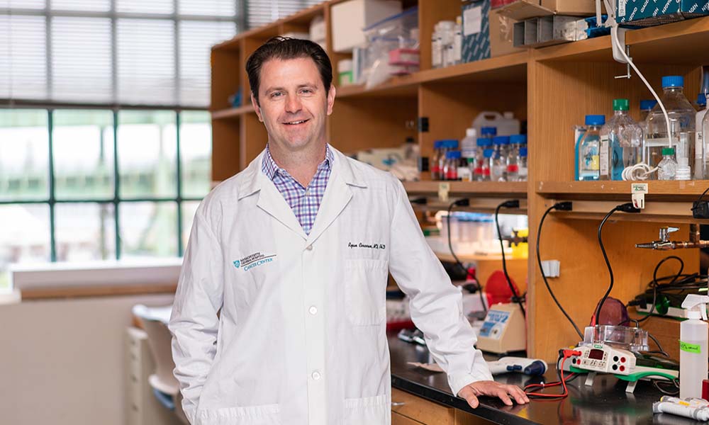 Ryan B. Corcoran合作论文数Department of Medicine, Harvard Medical School10
 Jill Allen合作论文数Stereotyping and Objectification Action Research Lab, Drake University;Department of Psychology and Neuroscience, College of Arts & Sciences, Drake University10
 Beow Yeap合作论文数Cancer Center, Massachusetts General Hospital;Massachusetts General Hospital Biostatistics;Harvard Medical School10
 Theodore Hong合作论文数Tucker Gosnell Center for Gastrointestinal Cancers, Massachusetts General Hospital;Harvard Medical School10
 Jennifer Wo合作论文数Mass General Research Institute, Massachusetts General Hospital;Harvard Medical School10Melin J Khandekar合作论文数9
 Ryan David P合作论文数9Keane Florence K合作论文数School of Medicine, Yale University9
 Eric Roeland合作论文数Knight Cancer Institute, Oregon Health and Science University;Division of Hematology, School of Medicine, Oregon Health and Science University9
 John Mullen合作论文数Department of Surgery, Massachusetts General Hospital;Harvard University9
 Michael Lanuti合作论文数Division of Thoracic Surgery, Massachusetts General Hospital;Harvard Medical School9Christopher R. Morse合作论文数8Christian Baglini合作论文数8Heather A. Shahzade合作论文数8Emily E Van Seventer合作论文数8Isobel J Fetter合作论文数8
 Mari Mino-Kenudson合作论文数Harvard Medical School;Massachusetts General Hospital8
 Jeffrey W Clark合作论文数Harvard Cancer Center, Harvard Medical School;Dana-Farber/Partners Cancer Care, Massachusetts General Hospital;Cancer Center Protocol Office, Massachusetts General Hospital8
 Sam Klempner合作论文数Harvard Medical School, Harvard University;Mass General Cancer Center, Massachusetts General Hospital, Mass General Brigham8Christine E. Eyler合作论文数8
 Rebecca Heist合作论文数Massachusetts General Hospital, Harvard Medical School;Cancer Center, Mass General Research Institute7Heist Rebecca S合作论文数2
 Mohamed Abdelrahman合作论文数2
 Jeffrey Clark合作论文数Mass General Cancer Center, Massachusetts General Hospital;Harvard Medical School2Laurelee Osborn合作论文数2
 Frederick Davidorf合作论文数2
 Colleen M Cebulla合作论文数Miller School of Medicine, University of Miami2K Rhynhart合作论文数1catherine hession合作论文数1Barbara J. Newman合作论文数1Susan Goelz合作论文数1James Massengill合作论文数1Roy R. Lobb合作论文数1J. Svahn合作论文数1
 Arnold S. Freedman合作论文数Harvard Medical School;Department of Medical Oncology, Dana-Farber Cancer Institute;Brigham and Women's Hospital1
 Andrew X. Zhu (朱秀轩)合作论文数Jiahui International Cancer Center, Jiahui International Hospital (Shanghai)1
 Lipika Goyal合作论文数Division of Oncology, Department of Medicine, School of Medicine, Stanford University;Stanford Cancer Institute1
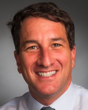 Jeffrey A. Meyerhardt合作论文数Colon and Rectal Cancer Center, Dana-Farber Cancer Institute;Harvard Medical School1Ryan David合作论文数1Verma Vishal合作论文数1Margaret D. Rosa合作论文数1Ming-Jer Huang合作论文数1Irene Dougas合作论文数1Kathy Mccarthy合作论文数1
 Getachew Boru合作论文数1Lawrence S. Blaszkowsky合作论文数1




