STAT3 phosphorylation and transcriptional activity promote breast cancer growth and metastasis. We have previously reported that the transcriptional adaptor LMO2 is required for metastasis in breast cancer and promotes STAT3-JAK2 interaction, leading to STAT3 phosphorylation in metastasis-initiating cells. Here, we find that constitutively activated STAT3 is insufficient to drive metastasis in the absence of LMO2. Mechanistically, we find that LMO2 is required not only for STAT3 phosphorylation in the cytoplasm but also for STAT3 translocation to the nucleus. These data suggest that LMO2 promotes STAT3 activation and localization to the nucleus in breast cancer cells.
Cellular plasticity in mammary epithelial cells enables dynamic cell state changes essential for normal development but can be hijacked by breast cancer cells to drive tumor progression and metastasis. However, the molecular factors that maintain cellular plasticity through the regulation of a hybrid cell state (epithelial/mesenchymal) are not fully defined. As LMO2 has been previously shown to regulate metastasis in breast cancer, here we determine the role of LMO2 in normal mammary epithelial cells. Using lineage tracing and knockout mouse models, we find that Lmo2 lineage-traced cells are present in the luminal and basal layer of the mammary gland but have limited proliferative potential. Lmo2 loss does not impact mammary gland development, but acute deletion decreases in vivo reconstitution. Moreover, LMO2 knockdown in mouse and human mammary epithelial cells (MECs) reduces organoid formation. We find that LMO2 regulates the epithelial cell state in MECs and LMO2 knockdown promotes mesenchymal differentiation. Transcriptional profiling of LMO2 knockdown cells reveals significant enrichment in the epithelial-mesenchymal transition (EMT) pathway and upregulation of MCAM, a mesenchymal marker and negative regulator of regenerative capacity in the mammary gland. Altogether, we show that LMO2 plays a role in maintaining cellular plasticity in MECs, adding insight into the normal differentiation programs hijacked by cancer cells to drive tumor progression.
Abstract Breast cancer is the most common cancer in women and the second-leading cause of cancer-related deaths in women. Breast cancer tumors are highly heterogeneous, with small populations of stem-like tumor-initiating cells that exhibit high cellular plasticity and often display both epithelial and mesenchymal characteristics (hybrid). These plastic stem-like cells contribute to tumorigenesis, so identifying and targeting these populations can lead to improved patient survival. ROBO1-4 proteins are transmembrane receptors that, with their SLIT1-3 ligands, regulate axon guidance, cell adhesion, mammary gland development, and angiogenesis. This signaling pathway is tightly regulated and has a wide variety of roles in normal development, but cancer is defined as abnormal development and frequently has alterations in signaling pathways. SLIT-ROBO signaling has been implicated in cell adhesion regulation in breast, lung, and colorectal cancer, but there is conflicting evidence on whether it is tumor-promoting or tumor-suppressive. Previous studies have shown that ROBO4 expression in endothelial cells suppresses breast cancer growth; however, ROBO4 has not been investigated in tumor epithelial cells. We have found that ROBO4 is expressed in epithelial breast cancer cells, and a high ROBO4+ epithelial fraction in basal-like tumor cells predicts worse survival in patients. ROBO4 knockdown in breast cancer cells reduces colony formation and proliferation, and increases apoptosis. Using patient-derived xenografts (PDXs) in vivo, constitutive ROBO4 knockdown reduced tumorigenesis, and inducible knockdown halted further tumor growth in pre-established tumors. Transcriptomic and proteomic analyses reveal enrichment of cancer stem cell and plasticity markers in control cells compared to ROBO4 knockdown, including ALDH3A1, MSI2, CD44, and S100A4. Flow cytometry analysis of tumor cells shows that knocking down ROBO4 reduces hybrid cells as measured by co-expression of CD44 (mesenchymal cells) and CD104 (epithelial cells). Preliminary results from ROBO4 overexpression tumors show an increase in hybrid CD44+/CD104+ cells, suggesting that ROBO4 maintains stem and hybrid populations in breast cancer. Gene Set Enrichment Analysis (GSEA) suggests Rho GTPase, Wnt/β-catenin, and Notch signaling are downregulated in ROBO4 knockdown cells. These findings reveal a novel role for ROBO4 in maintaining stem-like and hybrid cell populations in breast cancer, with ROBO4's location on the cell surface providing an attractive target for potential therapeutic applications. Citation Format: Isobel J. Fetter, Veronica Haro Acosta, Paloma Medina, Shaheen S. Sikandar. Dissecting ROBO4 function in regulating breast cancer cell plasticity [abstract]. In: Proceedings of the American Association for Cancer Research Annual Meeting 2026; Part 1 (Regular Abstracts); 2026 Apr 17-22; San Diego, CA. Philadelphia (PA): AACR; Cancer Res 2026;86(7 Suppl):Abstract nr 2199.
Abstract Introduction: Previously we discovered that a novel metastasis-initiating cell (MIC) population is marked by LMO2, a transcriptional adaptor protein in hematopoietic stem cells, and a T-cell oncogene. Knocking down LMO2 did not affect the growth of primary tumors, but did significantly reduce the number of circulating tumor cells and lung metastases. Transcriptional analysis of LMO2 knockdown shows the HALLMARK_DNA_REPAIR pathway is upregulated, suggesting that LMO2 is involved in regulating DNA repair in breast cancer cells. Interestingly, a previous study reported that in diffuse large B-cell lymphoma high LMO2 expression results in defective repair of double-stranded DNA breaks (Parvin et al., Cancer Cell, 36, 3 [2019]). We hypothesize that in breast cancer LMO2 expression is regulating the choice between homology-directed repair (HDR), an error-free method of DNA repair, and non-homologous end-joining (NHEJ), a more error-prone method. Cells that use NHEJ to repair DNA are dependent on poly (ADP-ribose) polymerase (PARP) and can be targeted with PARP inhibitors. LMO2 could thus be used as a new biomarker for benefit from PARP inhibition. Methods: LMO2 was identified using single-cell RNAseq data from human breast cancer patients. In vitro and in vivo assays using breast cancer cell lines and patient-derived xenografts were used to characterize the effect of LMO2 on metastasis. To study the role of LMO2 in the DNA damage response, we used MDA-MB-468 and HCC1806 cells with control and 2 independent shRNAs targeting LMO2. Immunofluorescent staining for γH2AX, 53BP1, and RAD51 was performed to measure changes in DNA damage and repair markers. Control and LMO2 knockdown cells were treated with olaparib, with cellular proliferation measured with a WST-1 assay and colony formation measured with crystal violet staining. Results and Conclusion: Knocking down LMO2 expression results in decreased NHEJ and increased HDR in MDA-MB-468 and HCC1806 cells. There is no significant difference in DNA damage overall as measured by γH2AX foci/nucleus. LMO2 knockdown in MDA-MB-468 cells reduces sensitivity to PARP inhibition with olaparib, as measured by an increase in the IC50 values. Our preliminary data suggest that LMO2 expression likely regulates the choice between NHEJ and HDR. LMO2 expression introduces a bias towards NHEJ, which sensitizes cells to PARP inhibition. This suggests that LMO2+ cells could be targeted with PARP inhibition, which could thus be used to target metastasis-initiating cells. Citation Format: Isobel Fetter, Veronica Haro-Acosta, Shaheen Sikandar. The role of LMO2 in DNA damage repair pathway choice in metastatic breast cancer [abstract]. In: Proceedings of the AACR Special Conference in Cancer Research: Advances in Breast Cancer Research; 2023 Oct 19-22; San Diego, California. Philadelphia (PA): AACR; Cancer Res 2024;84(3 Suppl_1):Abstract nr B052.
Abstract Purpose: MEK inhibitors (MEKi) lack monotherapy efficacy in most RAS-mutant cancers. BCL-xL is an anti-apoptotic protein identified by a synthetic lethal shRNA screen as a key suppressor of apoptotic response to MEKi. Patients and Methods: We conducted a dose escalation study (NCT02079740) of the BCL-xL inhibitor navitoclax and MEKi trametinib in patients with RAS-mutant tumors with expansion cohorts for: pancreatic, gynecologic (GYN), non–small cell lung cancer (NSCLC), and other cancers harboring KRAS/NRAS mutations. Paired pretreatment and day 15 tumor biopsies and serial cell-free (cf)DNA were analyzed. Results: A total of 91 patients initiated treatment, with 38 in dose escalation. Fifty-eight percent had ≥3 prior therapies. A total of 15 patients (17%) had colorectal cancer, 19 (11%) pancreatic, 15 (17%) NSCLC, and 32 (35%) GYN cancers. The recommended phase II dose (RP2D) was established as trametinib 2 mg daily days 1 to 14 and navitoclax 250 mg daily days 1 to 28 of each cycle. Most common adverse events included diarrhea, thrombocytopenia, increased AST/ALT, and acneiform rash. At RP2D, 8 of 49 (16%) evaluable patients achieved partial response (PR). Disease-specific differences in efficacy were noted. In patients with GYN at the RP2D, 7 of 21 (33%) achieved a PR and median duration of response 8.2 months. No PRs occurred in patients with colorectal cancer, NSCLC, or pancreatic cancer. MAPK pathway inhibition was observed in on-treatment tumor biopsies. Reductions in KRAS/NRAS mutation levels in cfDNA correlated with clinical benefit. Conclusions: Navitoclax in combination with trametinib was tolerable. Durable clinical responses were observed in patients with RAS-mutant GYN cancers, warranting further evaluation in this population.
Although U2AF1 S34F is a recurrent splicing factor mutation in lung adenocarcinoma (ADC), U2AF1 S34F alone is insufficient for producing tumors in previous models. Because lung ADCs with U2AF1 S34F frequently have co-occurring KRAS mutations and smoking histories, we hypothesized that tumor-forming potential arises from U2AF1 S34F interacting with oncogenic KRAS and environmental stress. To elucidate the effect of U2AF1 S34F co-occurring with a second mutation, we generated human bronchial epithelial cells (HBEC3kt) with co-occurring U2AF1 S34F and KRAS G12V . Transcriptome analysis revealed that co-occurring U2AF1 S34F and KRAS G12V differentially impacts inflammatory, cell cycle, and KRAS pathways. Subsequent phenotyping found associated suppressed cytokine production, increased proliferation, anchorage-independent growth, and tumors in mouse xenografts. Interestingly, HBEC3kts harboring only U2AF1 S34F display increased splicing in stress granule protein genes and viability in cigarette smoke concentrate. Our results suggest that U2AF1 S34F may potentiate transformation by granting precancerous cells survival advantage in environmental stress, permitting accumulation of additional mutations like KRAS G12V , which synergize with U2AF1 S34F to transform the cell.
Supplemental Appendix 6. Summary of ddPCR mutations to yield highest sensitivity for ctDNA detection.
Supplemental Appendix 7: Paired pre- and post- treatment SNaPshot molecular profiling available for 11 patients.
While BRAF inhibitor combinations with EGFR and/or MEK inhibitors have improved clinical efficacy in BRAF V600E colorectal cancer (CRC), response rates remain low and lack durability. Preclinical data suggest that BRAF/MAPK pathway inhibition may augment the tumor immune response. We performed a proof-of-concept single-arm phase 2 clinical trial of combined PD-1, BRAF and MEK inhibition with sparatlizumab (PDR001), dabrafenib and trametinib in 37 patients with BRAF V600E CRC. The primary end point was overall response rate, and the secondary end points were progression-free survival, disease control rate, duration of response and overall survival. The study met its primary end point with a confirmed response rate (24.3% in all patients; 25% in microsatellite stable patients) and durability that were favorable relative to historical controls of BRAF-targeted combinations alone. Single-cell RNA sequencing of 23 paired pretreatment and day 15 on-treatment tumor biopsies revealed greater induction of tumor cell-intrinsic immune programs and more complete MAPK inhibition in patients with better clinical outcome. Immune program induction in matched patient-derived organoids correlated with the degree of MAPK inhibition. These data suggest a potential tumor cell-intrinsic mechanism of cooperativity between MAPK inhibition and immune response, warranting further clinical evaluation of optimized targeted and immune combinations in CRC. ClinicalTrials.gov registration: NCT03668431.
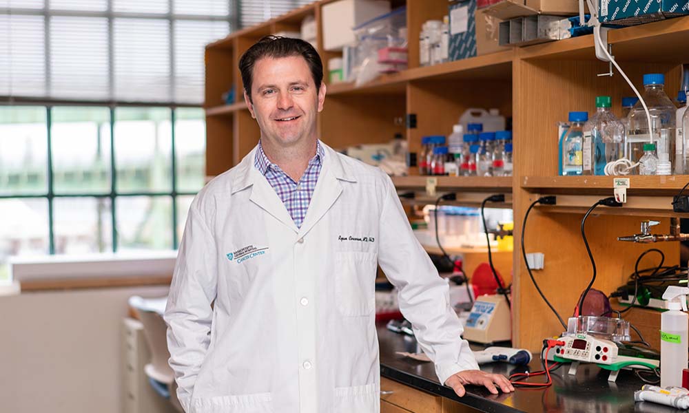 Ryan B. Corcoran合作论文数Department of Medicine, Harvard Medical School29
 Emily E Van Seventer合作论文数24Parikh Aparna R合作论文数Department of Medicine, University of California San Francisco23
 Jill Allen合作论文数Stereotyping and Objectification Action Research Lab, Drake University;Department of Psychology and Neuroscience, College of Arts & Sciences, Drake University22
 Jeffrey W Clark合作论文数Harvard Cancer Center, Harvard Medical School;Dana-Farber/Partners Cancer Care, Massachusetts General Hospital;Cancer Center Protocol Office, Massachusetts General Hospital19
 Theodore Hong合作论文数Tucker Gosnell Center for Gastrointestinal Cancers, Massachusetts General Hospital;Harvard Medical School19Heather A. Shahzade合作论文数18
 Giulia Siravegna合作论文数Haystack Oncology17
 Eric Roeland合作论文数Knight Cancer Institute, Oregon Health and Science University;Division of Hematology, School of Medicine, Oregon Health and Science University17
 Ryan David P合作论文数15
 Lipika Goyal合作论文数Division of Oncology, Department of Medicine, School of Medicine, Stanford University;Stanford Cancer Institute15
 Jennifer Wo合作论文数Mass General Research Institute, Massachusetts General Hospital;Harvard Medical School14Brandon Nadres合作论文数Massachusetts Gen Hosp, Canc Ctr11
 Katie Kanter合作论文数Harvard Med Sch, Boston, MA 02115 USA10
 Colin Weekes合作论文数Mass General Research Institute, Massachusetts General Hospital;Harvard Medical School;University of Colorado Health Sciences Center10
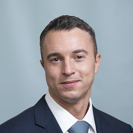 Sam Klempner合作论文数Harvard Medical School, Harvard University;Mass General Cancer Center, Massachusetts General Hospital, Mass General Brigham10
 Rebecca Heist合作论文数Massachusetts General Hospital, Harvard Medical School;Cancer Center, Mass General Research Institute10
 Lawrence Blaszkowsky合作论文数Mass General Cancer Center, Newton-Wellesley Hospital;Massachusetts General Hospital9
 Mari Mino-Kenudson合作论文数Harvard Medical School;Massachusetts General Hospital9
 Joe Lennerz合作论文数BostonGene9
 Andrew X. Zhu (朱秀轩)合作论文数Jiahui International Cancer Center, Jiahui International Hospital (Shanghai)9Christine E. Eyler合作论文数9
 Bruce Giantonio合作论文数Massachusetts General Hospital9Christopher R. Morse合作论文数8Christian Baglini合作论文数8Melin J Khandekar合作论文数8Christine Ulysse合作论文数Massachusetts Gen Hosp, Dept Med, Div Biostat, Boston, MA 02114 USA8Benjamin Christopher合作论文数8
 Anthony Iafrate合作论文数Department of Pathology, Massachusetts General Hospital Cancer Center;Harvard Medical School8Keane Florence K合作论文数School of Medicine, Yale University8Drapek Lorraine C合作论文数8
 Beow Yeap合作论文数Cancer Center, Massachusetts General Hospital;Massachusetts General Hospital Biostatistics;Harvard Medical School8
 John Mullen合作论文数Department of Surgery, Massachusetts General Hospital;Harvard University8
 Michael Lanuti合作论文数Division of Thoracic Surgery, Massachusetts General Hospital;Harvard Medical School8Kathryn Fosbenner合作论文数6
 Shaheen Sikandar合作论文数Stanford Institute for Stem Cell Biology and Regenerative Medicine, Stanford University6
 Ryan Nipp合作论文数The Hematology Oncology Section, Department of Internal Medicine, College of Medicine, The University of Oklahoma6
 Gad A. Getz合作论文数Harvard Medical School;Department of Pathology, Massachusetts General Hospital;Broad Institute of Harvard and MIT6Fish Madeleine合作论文数6Amirkasra Mojtahed合作论文数Massachusetts Gen Hosp, Dept Radiol, 55 Fruit St,White 270, Boston, MA 02114 USA5
 Islam Baiev合作论文数Harvard Med Sch, Canc Ctr, Massachusetts Gen Hosp, Boston, MA 02115 USA5Liudmila Elagina合作论文数5Murphy Janet E合作论文数5
 Ferran Fece de la Cruz合作论文数5
 Alberto Bardelli合作论文数Department of Oncology, Medical School, University of Turin;The AIRC Institute of Molecular Oncology, IFOM ETS;Molecular Biotechnology Center, University of Turin5
 David T Ting合作论文数Krantz Family Center for Cancer Research, Massachusetts General Hospital;Harvard Medical School5
 Dejan Juric合作论文数MiNDNET;Frankfurt School of Finance & Management;JK GmbH5Jaime L Schneider合作论文数4Srivatsan Raghavan合作论文数Dana Farber Canc Inst, Dept Med Oncol, Boston, MA 02115 USA4




