To meet the brain's moment-to-moment energy demand, neural activation rapidly increases local blood flow. This process, known as neurovascular coupling, involves rapid, coordinated vasodilation of the brain's arterial network. Here, we demonstrate that endothelial gap junction coupling enables long-range propagation of vasodilation signals through the vasculature during neurovascular coupling. The molecular composition of these gap junctions is zonated along the arterio-venous axis, with arteries being the most strongly coupled segment. Using optogenetics and visual stimuli in awake mice, we found that acute, arterial endothelial cell type-specific deletion of Cx37 and Cx40 abolishes arterial gap junction coupling and results in impaired vasodilation. Specifically, we demonstrated that arterial endothelial gap junction coupling determines both the speed and the spatial extent of vasodilation propagation elicited by neural activity. These findings indicate that endothelial gap junctions serve as a signaling highway for neurovascular coupling, enabling flexible and efficient distribution of limited energetic resources.
Prion disease is a fatal neurodegenerative disease caused by the misfolding of prion protein (PrP) encoded by the PRNP gene. While there is currently no cure for the disease, depleting PrP in the brain is an established strategy to prevent or stall templated misfolding of PrP. Here we developed in vivo cytosine and adenine base strategies delivered by adeno-associated viruses to permanently modify the PRNP locus to achieve PrP knockdown in the mouse brain. Systemic injection of dual-adeno-associated virus PHP.eB encoding BE3.9max and single guide RNA installing PRNP R37X resulted in 37% average installation of the desired edit, 50% reduction of PrP in the mouse brain and 52% extension of lifespan in transgenic human PRNP mice inoculated with pathogenic human prion isolates representing the most common sporadic and genetic subtypes of prion disease. We further engineered base editing systems to achieve improved in vivo potency and reduced base editor expression in nontargeting tissues, resulting in 63% average PrP reduction in the mouse brain from a 6.7-fold lower viral dose, with no detected off-target editing of anticipated clinical significance observed in either human cells or mouse tissues. These findings support the potential of in vivo base editing as one-time treatment for prion disease.
Prion disease is caused by misfolding of the prion protein (PrP) into pathogenic self-propagating conformations, leading to rapid-onset dementia and death. However, elimination of endogenous PrP halts prion disease progression. In this study, we describe Coupled Histone tail for Autoinhibition Release of Methyltransferase (CHARM), a compact, enzyme-free epigenetic editor capable of silencing transcription through programmable DNA methylation. Using a histone H3 tail-Dnmt3l fusion, CHARM recruits and activates endogenous DNA methyltransferases, thereby reducing transgene size and cytotoxicity. When delivered to the mouse brain by systemic injection of adeno-associated virus (AAV), Prnp -targeted CHARM ablates PrP expression across the brain. Furthermore, we have temporally limited editor expression by implementing a kinetically tuned self-silencing approach. CHARM potentially represents a broadly applicable strategy to suppress pathogenic proteins, including those implicated in other neurodegenerative diseases.
Broadening gene therapy applications requires manufacturable vectors that efficiently transduce target cells in humans and preclinical models. Conventional selections of adeno-associated virus (AAV) capsid libraries are inefficient at searching the vast sequence space for the small fraction of vectors possessing multiple traits essential for clinical translation. Here, we present Fit4Function, a generalizable machine learning (ML) approach for systematically engineering multi-trait AAV capsids. By leveraging a capsid library that uniformly samples the manufacturable sequence space, reproducible screening data are generated to train accurate sequence-to-function models. Combining six models, we designed a multi-trait (liver-targeted, manufacturable) capsid library and validated 88% of library variants on all six predetermined criteria. Furthermore, the models, trained only on mouse in vivo and human in vitro Fit4Function data, accurately predicted AAV capsid variant biodistribution in macaque. Top candidates exhibited production yields comparable to AAV9, efficient murine liver transduction, up to 1000-fold greater human hepatocyte transduction, and increased enrichment relative to AAV9 in a screen for liver transduction in macaques. The Fit4Function strategy ultimately makes it possible to predict cross-species traits of peptide-modified AAV capsids and is a critical step toward assembling an ML atlas that predicts AAV capsid performance across dozens of traits.
Spatially charting molecular cell types at single-cell resolution across the three-dimensional (3D) volume of the brain is critical for illustrating the molecular basis of the brain anatomy and functions. Single-cell RNA sequencing (scRNA-seq) has profiled molecular cell types in the mouse brain, but cannot capture their spatial organization. Here, we employed an in situ sequencing technique, STARmap PLUS, to map 1.09 million high-quality cells across the whole adult mouse brain and the spinal cord, profiling 1,022 genes at subcellular resolution with a voxel size of 194 X 194 X 345 nm3 in 3D. We developed computational pipelines to segment, cluster, and annotate 230 molecular cell types by single-cell gene expression and 106 molecular tissue regions by spatial niche gene expression. Joint analyses of molecular cell types and molecular tissue regions enabled a systematic molecular spatial cell type nomenclature and allowed the identification of tissue architectures previously undefined in established brain anatomy. To create a transcriptome-wide spatial atlas, we further integrated the STARmap PLUS measurements with a published scRNA-seq atlas, imputing 11,844 genes at the single-cell level. Finally, we delineated the tropisms of a brain-wide transgene delivery tool, AAV-PHP.eB5,6, across the molecular cell types and tissue regions of the whole mouse brain. Together, this annotated dataset provides a comprehensive, high-resolution, single-cell resource that integrates a spatial molecular atlas, cell taxonomy, brain anatomy, and genetic manipulation accessibility of the mammalian central nervous system (CNS).
Crossing the blood–brain barrier in primates is a major obstacle for gene delivery to the brain. Adeno-associated viruses (AAVs) promise robust, non-invasive gene delivery from the bloodstream to the brain. However, unlike in rodents, few neurotropic AAVs efficiently cross the blood–brain barrier in non-human primates. Here we report on AAV.CAP-Mac, an engineered variant identified by screening in adult marmosets and newborn macaques, which has improved delivery efficiency in the brains of multiple non-human primate species: marmoset, rhesus macaque and green monkey. CAP-Mac is neuron biased in infant Old World primates, exhibits broad tropism in adult rhesus macaques and is vasculature biased in adult marmosets. We demonstrate applications of a single, intravenous dose of CAP-Mac to deliver functional GCaMP for ex vivo calcium imaging across multiple brain areas, or a cocktail of fluorescent reporters for Brainbow-like labelling throughout the macaque brain, circumventing the need for germline manipulations in Old World primates. As such, CAP-Mac is shown to have potential for non-invasive systemic gene transfer in the brains of non-human primates.
Developing vehicles that efficiently deliver genes throughout the human central nervous system (CNS) will broaden the range of treatable genetic diseases. We engineered an adeno-associated virus (AAV) capsid, BI-hTFR1, that binds human transferrin receptor (TfR1), a protein expressed on the blood-brain barrier. BI-hTFR1 was actively transported across human brain endothelial cells and, relative to AAV9, provided 40 to 50 times greater reporter expression in the CNS of human TFRC knockin mice. The enhanced tropism was CNS-specific and absent in wild-type mice. When used to deliver GBA1 , mutations of which cause Gaucher disease and are linked to Parkinson’s disease, BI-hTFR1 substantially increased brain and cerebrospinal fluid glucocerebrosidase activity compared with AAV9. These findings establish BI-hTFR1 as a potential vector for human CNS gene therapy.
Adeno-associated viruses (AAVs) promise robust gene delivery to the brain through non-invasive, intravenous delivery. However, unlike in rodents, few neurotropic AAVs efficiently cross the blood-brain barrier in non-human primates (NHPs). Here we describe AAV.CAP-Mac, an engineered variant identified by screening in adult marmosets and newborn macaques with improved efficiency in the brain of multiple NHP species: marmoset, rhesus macaque, and green monkey. CAP-Mac is neuron-biased in infant Old World primates, exhibits broad tropism in adult rhesus macaques, and is vasculature-biased in adult marmosets. We demonstrate applications of a single, intravenous dose of CAP-Mac to deliver (1) functional GCaMP for ex vivo calcium imaging across multiple brain areas, and (2) a cocktail of fluorescent reporters for Brainbow-like labeling throughout the macaque brain, circumventing the need for germline manipulations in Old World primates. Given its capabilities for systemic gene transfer in NHPs, CAP-Mac promises to help unlock non-invasive access to the brain.
Viruses have evolved the ability to bind and enter cells through interactions with a wide variety of cell macromolecules. We engineered peptide-modified adeno-associated virus (AAV) capsids that transduce the brain through the introduction of de novo interactions with 2 proteins expressed on the mouse blood-brain barrier (BBB), LY6A or LY6C1. The in vivo tropisms of these capsids are predictable as they are dependent on the cell- and strain-specific expression of their target protein. This approach generated hundreds of capsids with dramatically enhanced central nervous system (CNS) tropisms within a single round of screening in vitro and secondary validation in vivo thereby reducing the use of animals in comparison to conventional multi-round in vivo selections. The reproducible and quantitative data derived via this method enabled both saturation mutagenesis and machine learning (ML)-guided exploration of the capsid sequence space. Notably, during our validation process, we determined that nearly all published AAV capsids that were selected for their ability to cross the BBB in mice leverage either the LY6A or LY6C1 protein, which are not present in primates. This work demonstrates that AAV capsids can be directly targeted to specific proteins to generate potent gene delivery vectors with known mechanisms of action and predictable tropisms.
 Ken Chan合作论文数37
 Viviana Gradinaru合作论文数Tianqiao and Chrissy Chen Institute for Neuroscience, California Institute of Technology;Center for Molecular and Cellular Neuroscience, California Institute of Technology;Capsida Biotherapeutics, Inc.28Qin Huang合作论文数20Pamela Brauer合作论文数15
 Chan Y Alina合作论文数14Qingxia Zheng合作论文数14Simon Pacouret合作论文数13
 Fatma-Elzahraa Eid合作论文数Stanley Ctr Psychiat Res, Broad Inst MIT & Harvard13
 Jang Min Jee合作论文数Department of Bio and Brain Engineering, KAIST11Thomas Beddow合作论文数10Isabelle G. Tobey合作论文数10Albert T. Chen合作论文数9
 Sripriya Ravindra Kumar合作论文数California Institute of Technology9Jason Wu合作论文数9Casey Keyes合作论文数8
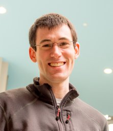 Andrew J. Barry合作论文数DNA Sequencing Operations Group, Broad Institute of MIT and Harvard8
 Xinhong Chen合作论文数CALTECH, Div Biol & Biol Engn, Pasadena, CA 91125 USA7Flytzanis Nicholas C合作论文数7Yujia Alina Chan合作论文数6Jencilin Johnston合作论文数6Binhui Zhao合作论文数6
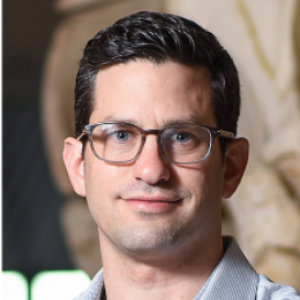 Alon Greenbaum合作论文数Joint Department of Biomedical Engineering, College of Arts and Sciences, The University of North Carolina at Chapel Hill6Goeden Nick合作论文数5Yang Bin合作论文数Division of Biology and Biological Engineering, California Institute of Technology5
 Luo Yicheng合作论文数Division of Biology, California Institute of Technology;Division of Biology, California Institute of Technology5
 Albert Chen合作论文数GE Healthcare Toronto5
 Andrew S. Fox合作论文数Waisman Laboratory for Brain Imaging and Behavior, University of Wisconsin-Madison5Timothy F. Miles合作论文数4Kristina M. Roxas合作论文数4Roy J. Blackburn合作论文数4Lillian J. Campos合作论文数4Sj Weintraub合作论文数4Gabrielle Clouse合作论文数4Janet Winchester合作论文数4Megan Powell合作论文数4James Pickel合作论文数4Julia Vendemiatti合作论文数4Cynthia Arokiaraj合作论文数4Keith Beadle合作论文数4
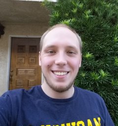 David Brown合作论文数Division of Biology and Biological Engineering, California Institute of Technology4
 Tatyana Dobreva合作论文数CALTECH, Div Biol & Biol Engn, Pasadena, CA 91125 USA4
 John D Tompkins合作论文数Neurocardiol Res Program Excellence, Univ Calif Los Angeles4
 Rajendran Pradeep S合作论文数David Geffen School of Medicine, University of California, Los Angeles;David Geffen School of Medicine, UCLA;and.;David Geffen School of Medicine, University of California, Los Angeles4
 Challis Collin合作论文数‡Neuroscience Graduate Group, University of Pennsylvania Perelman School of Medicine4
 J Christopher Octeau合作论文数4
 Guoping Feng (冯国平)合作论文数McGovern Institute for Brain Research, Massachusetts Institute of Technology;Stanley Center for Psychiatric Research, Broad Institute4Miguel R. Chuapoco合作论文数4Xiaozhe Ding合作论文数3Petur H. Einarsson合作论文数3




