Background Feline coronavirus infection causes feline infectious peritonitis in a subset of cats, but can also result in persistent infection. The tissue reservoirs of feline coronavirus and the role of viral persistence in pathogenesis are poorly understood. Aims This study aimed to identify sites of feline coronavirus persistence in a naturally infected cat, identify disease correlates and characterise within-host viral evolution. Methods The study followed a 5-year-old Bengal cat for 6 years and collected non-invasive samples, including faeces and conjunctival, oropharyngeal and saliva swabs. At 11-years-old, the patient was euthanised as a result of respiratory distress, and tissue samples were collected. The authors used hybridisation capture and next-generation sequencing methodologies focused on the feline coronavirus S gene, along with RNA in-situ hybridisation. Results During the study, the patient was diagnosed with inflammatory bowel disease, alimentary small cell lymphoma, chronic rhinitis and mitral valve regurgitation. Feline coronavirus was detected in the nasal cavity, intestine, faeces and conjunctiva in 2017, and in the intestine, faeces and heart in 2022. Sequence analysis showed that the virus adapted to tissue reservoirs over time. Conclusions This study identifies potential feline coronavirus reservoirs. The relationship of persistent feline coronavirus infection to chronic conditions warrants further investigation.
Feline coronavirus type 1 (FCoV-1) is widely known for causing feline infectious peritonitis (FIP), a systemic infection that is often fatal, with the virus known as the FIPV biotype. However, subclinical disease also occurs, in which cats may not show signs and intermittently shed the virus, including in feces, possibly for long periods of time. This virus is known as the FECV biotype. Progression of FECV to FIPV has been linked to several genomic changes, however a specific region of the viral spike protein at the interface of the spike S1 and S2 domains has been especially implicated. In this study, we followed a cat (#576) for six years from 2017, at which time FCoV-1 was detected in feces and conjunctival swabs, until 2022, when the animal was euthanized based on a diagnosis of alimentary small cell lymphoma. Over this time period, the cat was clinically diagnosed with inflammatory bowel disease and chronic rhinitis, and cardiac problems were also suspected. Using hybridization capture targeting the spike (S) gene of FCoV followed by next-generation sequencing, we screened 27 clinical samples. We detected FCoV-1 in 4 samples taken in 2017 (intestine and nasal tissue, feces, and conjunctiva), and 3 samples taken in 2022 (feces, and intestinal and heart tissue), but not in fecal samples taken in 2019 and 2020. Next, we focused on the S1/S2 region within S, which contains the furin cleavage site (FCS), a key regulator of viral transmission and pathogenesis. We show that the FCoV-1 variants obtained from feces in 2017 and 2022 were identical, while the ones from conjunctiva (2017), heart (2022), and intestine (2017 and 2022) were distinct. Sequence comparison of all the variants obtained showed that most of the non-synonymous changes in the S1/S2 region occur within the FCS. In the heart, we found two variants that differed by a single nucleotide, resulting in distinct FCS motifs that differ in one amino acid. It is predicted that one of these FCS motifs will down-regulate spike cleavability. The variant from the conjunctiva (2017) had a 6-nucleotide in-frame insertion that resulted in a longer and more exposed S1/S2 loop, which is predicted to be more accessible to the furin protease. Our studies indicate that FCoV-1 can independently persist in the gastrointestinal tract and heart of a cat over a long period of time without evidence of typical FIP signs, with intermittent viral shedding from the gastrointestinal and respiratory tracts.
Feline infectious peritonitis (FIP) is a systemic disease of cats caused by a highly pathogenic variant of feline coronavirus, or FCoV. Two distinct genotypes of FCoV exist (also referred to as serotypes): Type 1 viruses constitute the vast majority of FIP cases, while type 2 viruses are responsible for the remaining infections. Immunohistochemistry (IHC) currently serves as the gold standard for diagnosis of FIP; however, IHC is limited by variations in sensitivity. RNA in situ hybridization (RNA ISH) has an established foothold in infectious disease diagnostics and presents a potentially improved method for detection of FIP. This proof-of-concept study evaluated the efficacy of RNA ISH probes targeted to FCoV, as compared to IHC using monoclonal antibody FIP 3-70. Formalin-fixed paraffin-embedded tissues from FIP-positive cats were used for ISH, with the presence of RNA determined chromogenically. ISH tissue slides were then compared to their IHC counterparts, with efficacy determined based on metrics including staining intensity and abundance. Positive ISH staining on tissue was found to be both more intense and abundant than for IHC, suggesting that ISH serves as a highly sensitive method for the detection of FCoV/FIP in comparison to IHC - a finding that awaits further validation.
The emergence of severe acute respiratory syndrome 2 (SARS-CoV-2) has led the medical and scientific community to address questions surrounding the pathogenesis and clinical presentation of COVID-19; however, relevant clinical models outside of humans are still lacking. In felines, a ubiquitous coronavirus, described as feline coronavirus (FCoV), can present as feline infectious peritonitis (FIP)—a leading cause of mortality in young cats that is characterized as a severe, systemic inflammation. The diverse extrapulmonary signs of FIP and rapidly progressive disease course, coupled with a closely related etiologic agent, present a degree of overlap with COVID-19. This paper will explore the molecular and clinical relationships between FIP and COVID-19. While key differences between the two syndromes exist, these similarities support further examination of feline coronaviruses as a naturally occurring clinical model for coronavirus disease in humans.
In the context of the ongoing COVID-19 pandemic, comparative medicine can be a powerful approach to help refine our understanding of coronavirus pathophysiology, vaccine efficacy and pharmaceutical interventions. With feline infectious peritonitis (FIP), in particular, coronaviruses have been recognized to be important causes of disease in animal species for many decades, in some cases being associated with the enigmatic outcomes now recognized for COVID-19 in humans. Cats and other animals have also risen to our attention as hosts for severe acute respiratory syndrome coronavirus 2 (SARS-CoV-2). Endotheliitis and vasculitis are becoming established as a component of systemic disease caused by SARSCoV-2, including in children who develop multisystem inflammatory syndrome (MIS-C).1 A case series has also described an equivalent multisystem inflammatory syndrome in adults (MIS-A), which has included a variety of extrapulmonary disease manifestations.2 By analogy, feline coronavirus (FCoV) is well known for its ability to cause FIP, which is also a multisystem inflammatory syndrome of cats – although classically defined as either effusive (fluid accumulation in any body cavity) or noneffusive (development of granulomatous to pyogranulomatous lesions across body systems).3 The multisystem signs are only more recently becoming recognized as connected to FCoV infection.4 FIP most frequently occurs in younger cats, though all age groups are susceptible. A defining feature of FIP is the invasion of the macrophage, where mutations in the spike protein contribute to the cellular tropism of the virus. Antibody-dependent enhancement (ADE) is considered a possible mechanism underlying the development of FIP. ADE has not been considered a main mechanism driving COVID-19 pathology and SARS-CoV-2 replication in macrophages appears limited;5,6 however, these do remain open and unresolved questions. Cats are also susceptible to SARS-CoV-2, both experimentally and in the community.7,8 Feline infections may be quite common, but clinical signs appear to consist of only mild respiratory signs. Cats seroconvert and can transmit to other animals, but the role of cats in spreading infection in the community appears to remain minor. Another set of susceptible animals are mustelids, including ferrets and mink. In particular, the spread of the virus between mink and humans has been observed – termed ‘spillback’ – causing a public health crisis in Denmark.9 Exposure of mink to SARS-CoV-2 from handlers in farms led to widespread infection. In many cases, the mink developed severe interstitial pneumonia and diffuse alveolar damage with systemic signs, including ‘hepatic lipidosis, chronic nephritis, sepsis, dystocia and urolithiasis’.10 Experimental challenge of the closely related domestic ferret species has failed to yield severe signs, with ferrets typically responding like cats. As with cats, both mink and ferrets harbor their own coronaviruses, with ferrets showing a notable systemic outcome consisting of an FIP-like disease, but with less vasculitis.11 The impact of coinfection with human and animal coronaviruses in this set of susceptible species, including clinical presentation or viral recombination, is presently unclear. The term ‘One Medicine’ was coined by Rudolf Virchow in the late 19th century12 and this remains a cornerstone of 21st century pathology. Virchow is perhaps better associated with the eponymous ‘triad’ comprising the components of venous thrombosis, including venous stasis, activation of blood coagulation and endothelial damage. This triad is a contributor to the multisystem signs and symptoms in both COVID-19 and FIP. One Medicine also forms the foundation of the widely publicized ‘One Health’ triad of people, animals and the environment. The environment is key to SARS-CoV-2 spillover events, whether this is local (eg, in mink farms) or global (eg, in bats and their intermediate host species). As with Virchow in the 19th century, we should today examine coronaviruses of both humans and animals using a comparative approach spanning human, veterinary and wildlife medicine – incorporating the two distinct triads that Virchow inspired (Figure 1). This may allow us to more fully draw the broad comparisons necessary to solve the public health crisis of COVID-19 and prepare for future pandemics. One Medicine: a comparative approach to investigating human and animal coronavirus infections 998904 JFM This editorial was handled and processed by the European Editorial Office (ISFM) for publication in JFMSJournal of Feline Medicine and SurgeryStout et al
ABSTRACT Emerging canine coronavirus (CCoV) variants that are associated with systemic infections have been reported in the European Union; however, CCoV-associated disease in the United States is incompletely characterized. The purpose of this study was to correlate the clinicopathological findings and viral antigen distribution with the genotypic characteristics of CCoV in 11 puppies from nine premises in five states that were submitted for diagnostic investigation at Cornell University between 2008 and 2013. CCoV antigen was found in epithelial cells of small intestinal villi in all puppies and the colon in 2 of the 10 puppies where colon specimens were available. No evidence of systemic CCoV infection was found. Comparative sequence analyses of viral RNA extracted from intestinal tissues revealed CCoV-II genotype in 9 out of 11 puppies. Of the nine CCoV-IIs, five were subtyped as group IIa and one as IIb, while three CCoVs could not be subtyped. One of the CCoV-IIa variants was isolated in cell culture. Infection with CCoV alone was found in five puppies, of which two also had small intestinal intussusception. Concurrent infections with either parvovirus (n = 1), attaching-effacing Escherichia coli (n = 4), or protozoan parasites (n = 3) were found in the other six puppies. CCoV is an important differential diagnosis in outbreaks of severe enterocolitis among puppies between 4 days and 21 weeks of age that are housed at high population density. These findings will assist with the rapid laboratory diagnosis of enteritis in puppies and highlight the need for continued surveillance for CCoV variants and intestinal viral diseases of global significance.
Canine enteric coronavirus (CCoV) is an alphacoronavirus infecting dogs that is closely related to enteric coronaviruses of cats and pigs. While CCoV has traditionally caused mild gastro-intestinal clinical signs, there are increasing reports of lethal CCoV infections in dogs, with evidence of both gastrointestinal and systemic viral dissemination. Consequently, CCoV is now considered to be an emerging infectious disease of dogs. In addition to the two known serotypes of CCoV, novel recombinant variants of CCoV have been found containing spike protein N-terminal domains (NTDs) that are closely related to those of feline and porcine strains. The increase in disease severity in dogs and the emergence of novel CCoVs can be attributed to the high level of recombination within the spike gene that can occur during infection by more than one CCoV type in the same host.
Feline infectious peritonitis (FIP) is associated with mutations in the feline coronavirus (FCoV) genome that are thought to convert the subclinical feline enteric coronavirus (FECV) into the lethal feline infectious peritonitis virus (FIPV). A key feature of FIPV, not shared with FECV, is the productive infection of macrophages. Therefore mutations in proteins that govern cell tropism, such as the spike glycoprotein, may play an important role in FIP progression. In a prior study, involving a limited number of samples, we have shown an association of FIP with mutations in the protease cleavage-activation site located between the receptor-binding and fusion domains of the FCoV spike (S1/S2). Here, we extend these studies to investigate a larger sample set and to obtain a more refined analysis of the mutations at this S1/S2 site. Our larger data set more clearly shows that the mutations acquired by FIPV at S1/S2 are also accompanied by additional mutations at a second protease cleavage-activation site located in the fusion domain (S2'), adjacent to the viral fusion peptide. Overall, our data indicate a pattern of mutations across the two protease recognition sites that results in substitutions, and/or altered recognition, of critical basic/polar amino acid residues needed for virus activation in the enteric tract. Typically, FIPVs have substitutions of non-polar, aliphatic or aromatic residues in the protease recognition sites. These changes likely modulate the proteolytic activation of the virus and its ability to productively infect macrophages in vivo.
Feline coronaviruses (FCoV) exist as 2 biotypes: feline enteric coronavirus (FECV) and feline infectious peritonitis virus (FIPV). FECV causes subclinical infections; FIPV causes feline infectious peritonitis (FIP), a systemic and fatal disease. It is thought that mutations in FECV enable infection of macrophages, causing FIP. However, the molecular basis for this biotype switch is unknown. We examined a furin cleavage site in the region between receptor-binding (S1) and fusion (S2) domains of the spike of serotype 1 FCoV. FECV sequences were compared with FIPV sequences. All FECVs had a conserved furin cleavage motif. For FIPV, there was a correlation with the disease and >1 substitution in the S1/S2 motif. Fluorogenic peptide assays confirmed that the substitutions modulate furin cleavage. We document a functionally relevant S1/S2 mutation that arises when FIP develops in a cat. These insights into FIP pathogenesis may be useful in development of diagnostic, prevention, and treatment measures against coronaviruses.
Canine alphacoronaviruses (CCoV) exist in two serotypes, type I and II, both of which can cause severe gastroenteritis. Here, we characterize a canine alphacoronavirus, designated CCoV-A76, first isolated in 1976. Serological studies show that CCoV-A76 is distinct from other CCoVs, such as the prototype CCoV-1-71. Efficient replication of CCoV-A76 is restricted to canine cell lines, in contrast to the prototypical type II strain CCoV-1-71 that more efficiently replicates in feline cells. CCoV-A76 can use canine aminopeptidase N (cAPN) receptor for infection of cells, but was unable to use feline APN (fAPN). In contrast, CCoV-1-71 can utilize both. Genomic analysis shows that CCoV-A76 possesses a distinct spike, which is the result of a recombination between type I and type II CCoV, that occurred between the N- and C-terminal domains (NTD and C-domain) of the S1 subunit. These data suggest that CCoV-A76 represents a recombinant coronavirus form, with distinct host cell tropism.
Coronaviruses are enveloped positive-stranded RNA viruses that replicate in the cytoplasm. To deliver their nucleocapsid into the host cell, they rely on the fusion of their envelope with the host cell membrane. The spike glycoprotein (S) mediates virus entry and is a primary determinant of cell tropism and pathogenesis. It is classified as a class I fusion protein, and is responsible for binding to the receptor on the host cell as well as mediating the fusion of host and viral membranes-A process driven by major conformational changes of the S protein. This review discusses coronavirus entry mechanisms focusing on the different triggers used by coronaviruses to initiate the conformational change of the S protein: receptor binding, low pH exposure and proteolytic activation. We also highlight commonalities between coronavirus S proteins and other class I viral fusion proteins, as well as distinctive features that confer distinct tropism, pathogenicity and host interspecies transmission characteristics to coronaviruses.
 Gary Whittaker合作论文数Department of Microbiology & Immunology, College of Veterinary Medicine, Cornell University10
 Nicole M André合作论文数Dept Microbiol & Immunol, Cornell Univ5Alison E Stout合作论文数3
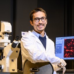 Millet Jean Kaoru合作论文数Veterinary Medical Center, Cornell University3
 Gerald Duhamel合作论文数Biomedical Sciences, College of Veterinary Medicine, Cornell University3Arjun Sweet合作论文数2Regan Andrew D合作论文数2
 Vera D. Rinaldi合作论文数Department of Biomedical Sciences, Cornell University2
 Olarte-Castillo Ximena A合作论文数Leibniz Institute for Zoo and Wildlife Research2
 Edward Dubovi合作论文数Department of Population Medicine and Diagnostic Sciences, College of Veterinary Medicine, Cornell University2Brian S Hamilton合作论文数1
 Maria A Sierra合作论文数Coll Dent, NYU1
 Javier A. Jaimes合作论文数College of Veterinary Medicine, Cornell University1
 Christopher Mason合作论文数Institute for Computational Biomedicine, Cornell University;Weill Cornell Medicine Brain & Mind Institute, Cornell University;Weill Cornell Medicine Meyer Cancer Center, Cornell University1
 Victor Tse合作论文数1Belouzard Sandrine合作论文数1
 Chris Town合作论文数J. Craig Venter Institute1Lee Donald W合作论文数School of Chemical and Biomolecular Engineering, Cornell University1Chillag Zach合作论文数1
 Laura B Goodman合作论文数Department of Microbiology and Immunology, Cornell University1




