Very rare cells often have outsized physiologic functions. However, their low abundance presents a technical challenge for studying their function. Here, we generate a murine model, which enables the profiling of secreted proteomes (secretomes) from physiologically relevant low-abundance cells in complex tissues, without the use of viral vectors. We then deploy this model to profile the secretomes of two rare innate immune cell types: Kupffer macrophages from whole liver and the rare airway microfold (M) cells in cultures of differentiated airway epithelia. The method we have developed is characterized by remarkable sensitivity, capturing cell-specific proteins that were not captured in single-cell RNA sequencing (scRNA-seq) atlases. We have made this genetic secretome mouse model available to the community to empower future studies of cell-to-cell communication, secreted protein function, and biomarker discovery for any given cell type in vitro or in vivo.
Transthyretin (TTR) amyloid cardiomyopathy (ATTR-CM) is a restrictive cardiac disease caused by the deposition of TTR in the heart. TTR is synthesized by hepatocytes and circulates as a homotetramer carrying complex for thyroxine and retinol. Mutations in TTR destabilize the tetramer promoting its dissociation into monomers that can enter the heart and aggregate into amyloid fibrils driving cytotoxicity, fibrosis, impaired electrical conductivity, and progressive cardiac dysfunction. Mechanistic studies and therapeutic development have been limited by the lack of physiologically relevant human preclinical models. Here we describe the creation of two in vitro models of ATTR-CM using human iPSC-derived cells. First, a custom microphysiological chip containing co-cultured hepatic and cardiac organoids in isolated chambers connected by microchannels enabling free passage, including hepatocyte synthesized TTR 122VI , which diffuses to and is taken up by cardiac organoids recapitulating pathologic hepatic-cardiac crosstalk. Cardiac organoids exposed to TTR V122I exhibited increased oxidative stress, elevated cytotoxicity, and upregulation of fibronectin. Second, engineered heart tissues cultured with monomeric TTR V122I demonstrated TTR uptake, reduced contractility, and prolonged relaxation time that mirrors cardiac dysfunction observed clinically. These novel human-based models recapitulate key biochemical, cellular, and functional features of ATTR-CM, providing translational platforms to elucidate disease mechanisms and evaluate novel therapeutic strategies.
IntroductionMouse models share significant genetic similarities with humans and have expanded our understanding of how embryonic tissue-specific genes influence disease states. By improved analyses of temporal, transcriptional data from these models, we can capture unique tissue codon usage patterns and determine how deviations from these patterns can influence developmental disorders.MethodsWe analyzed transcriptomic-weighted data from four mouse strains across three different germ layer tissues (liver, heart, and eye) and through embryonic stages. Applying a multifaceted approach, we calculated relative synonymous codon usage, reduced the dimensionality, and employed machine learning clustering techniques.Results and discussionThese techniques identified relative synonymous codon usage differences/similarities among strains and deviations in codon usage patterns between healthy and disease-linked genes. Original transcriptomic mouse data and RefSeq gene sequences can be found at the associated Mouse Embryo CoCoPUTs (codon and codon pair usage tables) website. Future studies can leverage this resource to uncover further insights into the dynamics of embryonic development and the corresponding codon usage biases that are paramount to understanding disease processes of embryologic origin.
We recently described a new stratified airway epithelial structure called the airway hillock. Hillocks are squamous barrier cells that shield a unique population of underlying hillock basal stem cells and resist a multitude of injuries including influenza, acid, freezing, and the toxin naphthalene. Hillocks are present in both mouse and human airways and demonstrate many conserved properties, including injury resistance. Following injury, hillock basal stem cells undergo massive clonal expansion and resurface denuded airway. We find that this regenerated epithelium demonstrates profound plasticity and converts into a classic airway epithelium with all six different airway epithelial cell types, including ciliated cells. Modulating retinoic acid concentrations, a known regulator of ciliogenesis, has dramatic effects on the formation of ciliated cells from hillock basal stem cells after injury. Inhibiting retinoic acid metabolism reduces the formation of ciliated cells, while a pan-retinoic acid receptor agonist increases ciliated cell differentiation from the hillock regenerated epithelium after injury. Finally, using a novel bead transport assay, we find that mucociliary transport is appropriately restored after injury in regions containing ciliated cells derived from hillocks, while non-hillock derived regions form disorganized areas of mucociliary transport in “hurricanes”. We extend these findings to human airways, identifying “hurricanes” in patient samples with bronchiectasis, while non-diseased samples show mucociliary transport in the expected distal to proximal orientation. Overall, this work suggests that the restoration of the ciliated epithelium from hillocks after injury is sensitive to retinoic acid. Furthermore, we find that hillocks functionally regenerate appropriate mucociliary transport in murine airways after injury, which suggests that non-hillock derived epithelium may be a source for the mucociliary transport seen as “hurricanes” in diseases like bronchiectasis.
Background: The coagulopathy of liver cirrhosis remains incompletely understood. Intrahepatic microthrombi formation is integral to the progression of liver fibrosis while macrovascular complications—such as hepatic artery and portal vein thrombosis—frequently complicate orthotopic liver transplantation and contribute to early graft failure. Pro-thrombotic ultralarge multimers of von Willebrand Factor (VWF) have been identified in patients with liver cirrhosis. Deficiency of ADAMTS13, the VWF-cleaving protease, is also observed in cirrhotic patients and is associated with thrombotic events and shortened overall survival. Despite its prognostic significance, the mechanistic basis of ADAMTS13 deficiency in this patient population remains poorly defined. Furthermore, there are no established strategies to enhance physiological VWF cleavage, reduce thrombotic risk, or improve microvascular perfusion in patients with liver cirrhosis. Methods: VWF and ADAMTS13 function were characterized in a cohort of patients with liver cirrhosis (n = 12) in a clinically stable state. Primary human hepatic stellate cells and whole liver lysates were obtained from healthy controls and patients with liver cirrhosis to investigate the biogenesis of ADAMTS13. Hepatic stellate cells were activated in vitro to model a fibrotic phenotype and analyzed alongside primary human endothelial cells. The relationship between intracellular proteolysis of ADAMTS13 and its secretion was further examined using ADAMTS13-expressing cell lines. Results: In patients with liver cirrhosis, VWF activity was elevated (mean: 599% of control, P < 0.001), while ADAMTS13 activity (mean: 61%) and antigen levels (mean: 43%) were both significantly reduced (P < 0.001). A strong correlation was observed between the level of ADAMTS13 antigen and activity (r = 0.91), whereas ADAMTS13 autoantibody levels in plasma showed no correlation with ADAMTS13 activity (r = - 0.14). Activated hepatic stellate cells exhibited markedly impaired secretion of ADAMTS13 when compared to endothelial cells, despite similar intracellular expression levels. Proteolysis of the ADAMTS13 metalloprotease domain was detected in activated hepatic stellate cells and in whole liver lysates of patients with chronic hepatitis and cirrhosis. In model cell lines, intracellular proteolysis of ADAMTS13 was similarly associated with impaired cellular secretion. Conclusions: Patients with liver cirrhosis consistently exhibit elevated VWF levels and reduced plasma ADAMTS13 activity, resulting in hemostatic imbalance. Activation of hepatic stellate cells during liver fibrogenesis impairs ADAMTS13 secretion, likely due to intracellular proteolysis of its metalloprotease domain. These findings provide mechanistic insight into ADAMTS13 deficiency observed in cirrhosis, which contributes to VWF:ADAMTS13 imbalance, promotes microvascular thrombosis, and may drive the progression of liver fibrosis.
Despite promising initial results in targeting the RAF-MEK-ERK cascade, resistance to BRAF/MEK inhibitors remains a critical challenge in nearly 50% of melanoma patients. Our study demonstrates that robust YAP1 activation in metastatic melanoma correlates with poor survival and drives transcriptional programs linked to therapeutic resistance. Mechanistically, YAP1 predominantly remodels the chromatin landscape in resistant tumors by partnering with BRD4 and TEAD, creating a permissive transcriptional state that sustains oncogenic signaling. Clinical validation in biopsies from resistant melanoma confirms elevated expression of YAP1 target genes. Furthermore, pharmacological inhibition of BRD4 or TEAD reduces YAP1-driven transcription and reactivates antitumor immunity programs. TEAD specific inhibitors (and not verteporfin which is a highly non-specific inhibitor) synergize with immune checkpoint blockade in in vivo model system by promoting increased CD8⁺ T cell infiltration and prolonged survival in the melanoma mouse model. Collectively, these findings reveal a chromatin-centric vulnerability in BRAF/MEK inhibitor-resistant melanoma and propose TEAD specific inhibitors as a promising dual strategy to overcome resistance and reinvigorate the immune response, offering a novel therapeutic avenue for patients.
The olfactory epithelium (OE) contains stem cells capable of generating olfactory sensory neurons throughout life, making it a valuable model for studying epithelial neurogenesis. We present a protocol to develop a highly robust 3D mouse organoid model from OE cells. We describe a workflow for dissecting murine OE and subsequent organoid culturing. We also provide guidance on how to perform immunostaining of the organoids. This protocol has been validated for mice and does not require fluorescence-activated cell purification. For complete details on the use and execution of this protocol, please refer to Gameiro et al.1.
Human airways contain specialized rare epithelial cells including CFTR-rich ionocytes that regulate airway surface physiology and chemosensory tuft cells that produce asthma-associated inflammatory mediators. Here, using a lung cell atlas of 311,748 single cell RNA-Seq profiles, we identify 687 ionocytes (0.45%). In contrast to prior reports claiming a lack of ionocytes in the small airways, we demonstrate that ionocytes are present in small and large airways in similar proportions. Surprisingly, we find only 3 mature tuft cells (0.002%), and demonstrate that previously annotated tuft-like cells are instead highly replicative progenitor cells. These tuft-ionocyte progenitor (TIP) cells produce ionocytes as a default lineage. However, Type 2 and Type 17 cytokines divert TIP cell lineage in vitro, resulting in the production of mature tuft cells at the expense of ionocyte differentiation. Our dataset thus provides an updated understanding of airway rare cell composition, and further suggests that clinically relevant cytokines may skew the composition of disease-relevant rare cells.
Current strategies for optimizing gene therapeutics and recombinant protein production typically rely on universal host codon usage indices. However, there is a growing shift toward incorporating gene-specific traits to enhance therapeutic characteristics. In this study, we investigate position-specific variations in codon and adjacent codon-pair usage biases (CPUBs), offering potential for more tailored gene engineering approaches. We focus our analysis on the coding sequences of four coagulation factors: ADAMTS13, von Willebrand factor, factor VIII, and factor IX, which have been used in therapeutic applications. By aligning transcript homologs with human sequences for each gene using Discontiguous Megablast and MACSE, we assess "sequence-position-specific" codon and CPUBs; 157 homologous sequences for ADAMTS13, 148 for F8, 96 for F9, and 202 for VWF. Species with homologs ranged from Primates and Artiodactyla (Even-toed Ungulates) to Testudines. Statistically significant, position-specific positive CPUBs were observed that contrasted with conventional, alignment-specific negative CPUBs. Moreover, we observed that codon and codon-pair usages are highly associated at sequence positions despite little or no association in conventional-position-agnostic analyses. The distinct biases observed at different positions/functionally critical domains in coding sequences highlight the importance of considering position-specific effects in codon optimization strategies.
The mammalian olfactory neuronal lineage is regenerative, and accordingly, maintains a population of pluripotent cells that replenish olfactory sensory neurons and other olfactory cell types during the life of the animal. Moreover, in response to acute injury, the early transit amplifying cells along the olfactory sensory neuronal lineage are able to de-differentiate to shift resources in support of tissue restoration. In order to further explore plasticity of various cellular stages along the olfactory sensory neuronal lineage, we challenged the epigenetic stability of olfactory placode-derived cell lines that model immature olfactory sensory neuronal stages. We found that perturbation of the Ehmt2 chromatin modifier transformed the growth properties, morphology, and gene expression profiles toward states with several stem cell characteristics. This transformation was dependent on continued expression of the large T-antigen, and was enhanced by Sox2 over-expression. These findings may provide momentum for exploring inherent cellular plasticity within early cell types of the olfactory lineage, as well as potentially add to our knowledge of cellular reprogramming.
Stem cells are known to provide signals that contribute to the maintenance and function of neighboring cells. We demonstrate that Notch signaling arising from airway basal stem cells is necessary for the function of a unique population of intraepithelial airway macrophages (IAMs) in the murine trachea. Without this stem cell signaling, IAMs lose MHC II expression, which in turn prevents antigen-induced allergic inflammation. Distal murine airways do not harbor basal stem cells, and, in this region of the lung, allergic inflammation proceeds unperturbed. We speculate that the functional coupling of specific anatomically restricted stem cell populations and adjacent immune cells is one mechanism for ensuring that inflammatory responses are compartmentalized to regions of injury. Basal stem cells are found throughout the human airway tree and we demonstrate the existence of human IAM-like cells, suggesting that their interaction may influence airways disease.
Cardiovascular disease is the leading cause of mortality in the United States. Studies suggest a role for environmental exposures in the etiology of cardiovascular disease, including exposure to arsenic through drinking water. Arsenic exposure during pregnancy has been shown to have effects on offspring, but few studies have examined impacts on maternal cardiovascular health. Although our prior work documented the detrimental effect of arsenic on the maternal heart during pregnancy, our current study examines the effect of gestational arsenic exposure on the maternal heart postpartum. Timed-pregnant wild-type (C57BL/6J) mice were exposed to 0, 100, or 1,000 µg/L sodium arsenite (NaAsO2) via drinking water from embryonic day 2.5 until parturition. Postpartum heart structure and function was assessed via transthoracic echocardiography and gravimetric measurement. Hypertrophic markers were probed via qRT-PCR and Western blot. Isolated cardiomyocyte Ca2+-handling and contraction were also assessed, along with the expression of with Ca2+-handling and contractile proteins. Interestingly, we found that exposure to either 100 or 1,000 µg/L sodium arsenite increased postpartum heart size at postpartum day 12 vs. nonexposed postpartum controls. At the cellular level, we found altered cardiomyocyte Ca2+-handling and contraction, along with expression changes of key contractile proteins, including α-actin and cardiac myosin binding protein C (cMyBP-c). Together, these findings suggest that gestational arsenic exposure impacts the postpartum maternal heart, possibly inducing long-term cardiovascular changes. Furthermore, these findings highlight the importance of reducing arsenic exposure during pregnancy, and the need for more research on the impact of arsenic on maternal heart health and adverse pregnancy events.NEW & NOTEWORTHY Gestational exposure to sodium arsenite at environmentally relevant doses (100 and 1,000 µg/L) increases postpartum heart size, and induces dysregulated Ca2+ homeostasis and impaired shortening in isolated cardiomyocytes. This is the first study to demonstrate that gestational arsenic exposure impacts postpartum heart structure and function beyond the exposure period.
The olfactory epithelium contains two basal stem cell populations that facilitate the usually life-long ability for neuronal regeneration that is required for maintaining our sense of smell. Horizontal basal cells (HBCs) are generally quiescent and only become active after direct injury to the epithelium that eliminates more than just the olfactory sensory neurons (OSNs). Globose basal cells (GBCs) lie apical to HBCs and are solely responsible for the generation of olfactory neurons in the undamaged epithelium. Understanding how these two neurogenic stem cell populations are regulated as OSNs are replenished is hampered by a lack of robust culture models. Here, we report the development of a 3D mouse organoid model that recapitulates the neurogenic cascade, forming immature OSNs while maintaining both HBCs and GBCs in culture. We use this model to demonstrate that, despite their relative quiescence, HBCs form a critical niche for the emergence and composition of the organoid.
A "universal strategy" replacing the full-length CFTR cDNA may treat >99% of people with cystic fibrosis (pwCF), regardless of their specific mutations. Cas9-based gene editing was used to insert the CFTR cDNA and a truncated CD19 (tCD19) enrichment tag at the CFTR locus in airway basal stem cells. This strategy restores CFTR function to non-CF levels. Here, we investigate the safety of this approach by assessing genomic and regulatory changes after CFTR cDNA insertion. Safety was first assessed by quantifying genetic rearrangements using CAST-seq. After validating restored CFTR function in edited and enriched airway cells, the CFTR locus open chromatin profile was characterized using ATAC-seq. The regenerative potential and differential gene expression in edited cells was assessed using scRNA-seq. CAST-seq revealed a translocation in ∼0.01% of alleles primarily occurring at a nononcogenic off-target site and large indels in 1% of alleles. The open chromatin profile of differentiated airway epithelial cells showed no appreciable changes, except in the region corresponding to the CFTR cDNA and tCD19 cassette, indicating no detectable changes in gene regulation. Edited stem cells produced the same types of airway cells as controls with minimal alternations in gene expression. Overall, the universal strategy showed minor undesirable genomic changes.
Airway hillocks are stratified epithelial structures of unknown function1. Hillocks persist for months and have a unique population of basal stem cells that express genes associated with barrier function and cell adhesion. Hillock basal stem cells continually replenish overlying squamous barrier cells. They exhibit dramatically higher turnover than the abundant, largely quiescent classic pseudostratified airway epithelium. Hillocks resist a remarkably broad spectrum of injuries, including toxins, infection, acid and physical injury because hillock squamous cells shield underlying hillock basal stem cells from injury. Hillock basal stem cells are capable of massive clonal expansion that is sufficient to resurface denuded airway, and eventually regenerate normal airway epithelium with each of its six component cell types. Hillock basal stem cells preferentially stratify and keratinize in the setting of retinoic acid signalling inhibition, a known cause of squamous metaplasia2,3. Here we show that mouse hillock expansion is the cause of vitamin A deficiency-induced squamous metaplasia. Finally, we identify human hillocks whose basal stem cells generate functional squamous barrier structures in culture. The existence of hillocks reframes our understanding of airway epithelial regeneration. Furthermore, we show that hillocks are one origin of ‘squamous metaplasia’, which is long thought to be a precursor of lung cancer. In the lungs, recently identified epithelial structures known as hillocks can act as injury-resistant reservoirs of stem cells.
Clinical trials delivering high doses of adeno-associated viruses (AAVs) expressing truncated dystrophin molecules (microdystrophins) are underway for Duchenne muscular dystrophy (DMD). We examined the efficiency and efficacy of this strategy with 4 microdystrophin constructs (3 in clinical trials and a variant of the largest clinical construct), in a severe mouse model of DMD, using AAV doses comparable with those in clinical trials. We achieved high levels of microdystrophin expression in striated muscles with cardiac expression approximately 10 -fold higher than that observed in skeletal muscle. Significant, albeit incomplete, correction of skeletal muscle disease was observed. Surprisingly, a lethal acceleration of cardiac disease occurred with 2 of the microdystrophins. The detrimental cardiac effect appears to be caused by variable competition (dependent on microdystrophin design and expression level) between microdystrophin and utrophin at the cardiomyocyte membrane. There may also be a contribution from an overloading of protein degradation. The significance of these observations for patients currently being treated with AAV-microdystrophin therapies is unclear since the levels of expression being achieved in the DMD hearts are unknown. However, these findings suggest that microdystrophin treatments need to avoid excessively high levels of expression in the heart and that cardiac function should be carefully monitored in these patients.
Mouse (Mus musculus) models have been heavily utilized in developmental biology research to understand mammalian embryonic development, as mice share many genetic, physiological, and developmental characteristics with humans. New explorations into the integration of temporal (stage-specific) and transcriptional (tissue-specific) data have expanded our knowledge of mouse embryo tissue-specific gene functions. To better understand the substantial impact of synonymous mutational variations in the cell-state-specific transcriptome on a tissue’s codon and codon pair usage landscape, we have established a novel resource—Mouse Embryo Codon and Codon Pair Usage Tables (Mouse Embryo CoCoPUTs). This webpage not only offers codon and codon pair usage, but also GC, dinucleotide, and junction dinucleotide usage, encompassing four strains, 15 murine embryonic tissue groups, 18 Theiler stages, and 26 embryonic days. Here, we leverage Mouse Embryo CoCoPUTs and employ the use of heatmaps to depict usage changes over time and a comparison to human usage for each strain and embryonic time point, highlighting unique differences and similarities. The usage similarities found between mouse and human central nervous system data highlight the translation for projects leveraging mouse models. Data for this analysis can be directly retrieved from Mouse Embryo CoCoPUTs. This cutting-edge resource plays a crucial role in deciphering the complex interplay between usage patterns and embryonic development, offering valuable insights into variation across diverse tissues, strains, and stages. Its applications extend across multiple domains, with notable advantages for biotherapeutic development, where optimizing codon usage can enhance protein expression; one can compare strains, tissues, and mouse embryonic stages in one query. Additionally, Mouse Embryo CoCoPUTs holds great potential in the field of tissue-specific genetic engineering, providing insights for tailoring gene expression to specific tissues for targeted interventions. Furthermore, this resource may enhance our understanding of the nuanced connections between usage biases and tissue-specific gene function, contributing to the development of more accurate predictive models for genetic disorders.
Exposure to chemical phenols, which can act as tyrosine analogues and result in anti‐melanocyte autoimmunity, has been associated with vitiligo. Acetaminophen (N‐acetyl‐p‐aminophenol) is an over‐the‐counter analgesic of phenolic origin. The risk of vitiligo with systemic exposure to acetaminophen has not yet been evaluated.
 James Schwob合作论文数Department of Developmental, Molecular & Chemical Biology, School of Medicine, Tufts University16
 David Kass合作论文数Department of Biomedical Engineering, Whiting School of Engineering, Johns Hopkins University;Institute of CardioScience, School of Medicine, Johns Hopkins University14
 Jayaraj Rajagopal合作论文数Department of Molecular and Cellular Biology, Harvard University14
 Waghray Avinash合作论文数The Black Family Stem Cell Institute, Icahn School of Medicine at Mount Sinai;The Graduate School of Biomedical Sciences, Icahn School of Medicine at Mount Sinai;Department of Developmental and Regenerative Biology, Icahn School of Medicine at Mount Sinai;Icahn School of Medicine at Mount Sinai, Icahn School of Medicine at Mount Sinai12
 Kimchi-Sarfaty Chava合作论文数11
 Sakthivel Sadayappan合作论文数Division of Molecular Cardiovascular Biology, Cincinnati Children’s Hospital Medical Center9
 Gary C. Curhan合作论文数Division of Renal Medicine, Brigham and Women's Hospital;Harvard Medical School8Jesse Peterson合作论文数McLean Hospital, Harvard Medical School8Julie H Coleman合作论文数7
 Katneni Upendra K合作论文数7
 Anton A Komar合作论文数Center for Gene Regulation in Health and Disease, Cleveland State University7
 Elliott D. Kozin合作论文数Department of Surgery, University of Pennsylvania7
 Konstantina Stankovic合作论文数6
 Alan W. Partin合作论文数Department of Urology, Johns Hopkins Medicine6
 Han Misop合作论文数Department of Epidemiology, Johns Hopkins Bloomberg School of Public Health;Department of Urology and the James Buchanan Brady Urological Institute, Johns Hopkins University School of Medicine6Bar Haim Y合作论文数Cornell University6
 Herrick Daniel B合作论文数School of Medicine, Johns Hopkins University6Hamasaki-Katagiri N合作论文数6
 Eva Andres-Mateos合作论文数McKusick-Nathans Institute of Genetic Medicine, Johns Hopkins University School of Medicine6Jang Woochan合作论文数6
 Susan Redline合作论文数Division of Sleep Medicine, Harvard Medical School;Division of Sleep and Circadian Disorders, Departments of Medicine and Neurology, Brigham and Women's Hospital;Division of Pulmonary Medicine, Department of Medicine, Beth Israel Deaconess Medical Center6
 Huang Tianyi合作论文数Norwich Medical School, University of East Anglia;the Department of Epidemiology, Harvard School of Public Health;and the Channing Division of Network Medicine, Department of Medicine, Brigham and Women's Hospital and Harvard Medical School, Boston, MA (TH, MSR, EBR, and SST).;Harvard Medical School, University of East Anglia6
 Ryan Hunt合作论文数6
 Shelley Tworoger合作论文数Division of Oncological Sciences, School of Medicine, Oregon Health & Science University;Knight Cancer Institute, School of Medicine, Oregon Health & Science University6
 Meir Stampfer合作论文数Department of Epidemiology, Harvard T.H. Chan School of Public Health;Department of Nutrition, Harvard T.H. Chan School of Public Health;Brigham and Women's Hospital, Harvard Medical School5Michal Slyper合作论文数Broad Inst MIT & Harvard, Klarman Cell Observ, Cambridge, MA 02142 USA5
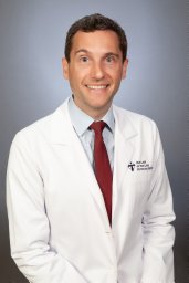 Samuel R Barber合作论文数Department of Otolaryngology – Head and Neck Surgery, University of Arizona5
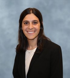 Julia Waldman合作论文数Broad Inst MIT & Harvard, Klarman Cell Observ, Cambridge, MA 02142 USA5
 Suraj Kannan合作论文数Johns Hopkins Univ, Dept Med, Div Cardiol, Baltimore, MD USA5
 Phillip Pierorazio合作论文数Perelman School of Medicine, University of Pennsylvania;Hospital of the University of Pennsylvania;Division of Urology, Penn Presbyterian Medical Center, University of Pennsylvania5
 Chulan Kwon合作论文数Department of Otolaryngology-Head and Neck Surgery, School of Medicine, Johns Hopkins University5
 Srinivas Vinod Saladi合作论文数Stealth Mode;Department of Cell and Cancer Biology, College of Medicine and Life Sciences, The University of Toledo5JiaWei Sun合作论文数Tsinghua Univ, Tsinghua Univ.5Muus Christoph合作论文数5
 Frank B. Hu (胡丙长)合作论文数Department of Epidemiology, Harvard T.H. Chan School of Public Health;Department of Nutrition, Harvard T.H. Chan School of Public Health5
 Patrick C. Walsh合作论文数School of Medicine, Johns Hopkins University5
 Matthew Tirrell合作论文数Pritzker School of Molecular Engineering, The University of Chicago;Institute for Molecular Engineering, University of Chicago;Eckhardt Research Center, University of Chicago5
 Eric H. Holbrook合作论文数Division of Rhinology, Massachusetts Eye and Ear, Harvard Medical School, Harvard University;Department of Otolaryngology Head and Neck Surgery, Harvard Medical School, Harvard University5
 Christian Pavlovich合作论文数Department of Urology, James Buchanan Brady Urological Institute, Johns Hopkins University School of Medicine;Johns Hopkins Bayview Medical Center, Johns Hopkins University School of Medicine5Sharon G. Curhan合作论文数5
 Nicole Higgins合作论文数4
 Jessica Tantivit合作论文数4
 Kasidet Manakongtreecheep合作论文数Broad Inst Massachusetts Inst Technol, MIT & Harva4Adam Haber合作论文数4
 Sumita Mishra合作论文数johns hopkins university4
 Bruce Trock合作论文数Departments of Urology, Epidemiology, and Oncology, Johns Hopkins School of Medicine;Division of Epidemiology, Brady Urological Institute, The Johns Hopkins University4
 Ranek Mark J合作论文数Division of Basic Biomedical Sciences, University of South Dakota4Heimberg Graham合作论文数4Nadan Wang合作论文数4




