CONTEXT.—Breast cancer biomarker assessment is critical in determining treatment and prognosis. In Tanzania, immunohistochemistry (IHC) is limited to surgical specimens and core biopsies. However, performing IHC on fine-needle aspiration biopsy cell blocks would offer numerous advantages.OBJECTIVE.—To compare the performance between estrogen receptor (ER) IHC performed at Muhimbili National Hospital (MNH) in Tanzania and ER IHC performed at University of California, San Francisco (UCSF), to demonstrate feasibility of performing IHC using cell blocks in Tanzania.DESIGN.—Patients with breast masses were recruited prospectively from the fine-needle aspiration biopsy clinic at MNH. Estrogen receptor IHC results on cell blocks, performed at both MNH and UCSF, and corresponding tissue blocks, performed at MNH, were compared to determine concordance.RESULTS.—Eighty-six cell blocks were evaluated by ER IHC at MNH, with 41 of 86 (47.7%) positive and 45 of 86 (52.3%) negative. Among 65 UCSF and MNH cell block pairs, overall ER IHC concordance was 93.8% (61 of 65) and positive concordance was 93.5% (29 of 31) (κ = 0.88, P > .99). Among 43 paired UCSF cell blocks and MNH tissue blocks, overall ER IHC concordance was 88.3% (38 of 43) and positive concordance was 90.5% (19 of 21) (κ = 0.77, P > .99). We compared 62 MNH cell block and tissue block pairs. Overall ER IHC concordance was 90.3% and positive concordance was 87.9% (κ = 0.81, P = .69).CONCLUSIONS.—Pairwise comparisons between ER IHC at MNH, on cell blocks and tissue blocks, with ER IHC at UCSF on cell blocks showed excellent concordance. We demonstrate that ER IHC on fine-needle aspiration biopsy specimens can be implemented in resource-constrained settings.
Background: Prior studies evaluating thyroid fine needle aspiration biopsies (FNABs) have limited the calculation of risk of malignancy (ROM) to cytologic specimens with corresponding histologic specimens, and clinical follow-up for those patients who do not undergo immediate surgery has been largely disregarded. Moreover, there is marked variability in how researchers have approached thyroid FNAB statistical analyses. This study addresses the urgent need for information from a large cohort of patients with long-term clinical follow-up to more accurately determine the performance of thyroid FNAB and ROM for each diagnostic category. Methods: A retrospective review of the University of California, San Francisco (UCSF), pathology database for thyroid FNABs from January 1, 1997, to December 31, 2004, was performed. Diagnoses were coded using the 2017 The Bethesda System for Reporting Thyroid Cytopathology (TBSRTC), and patients were matched to both the UCSF cancer registry and California Cancer Registry. Data were analyzed using the Kaplan-Meier method, and stratified by TBSRTC diagnostic category. Kaplan-Meier curves were used to estimate incidence rates of malignancy, stratified by FNAB category. Cox proportional hazards models were used to determine the instantaneous ROM. Results: Initial FNABs from 2207 patients were included. Median follow-up period after the first thyroid FNAB was 13.9 years (range: 10.5-18.4 years). During follow-up, there were 279 confirmed diagnoses of thyroid malignancy. Estimates derived from Kaplan-Meier curves demonstrated that the risk of having a thyroid malignancy was low for nondiagnostic and benign categories, intermediate for atypia of undetermined significance (AUS), follicular lesion of undetermined significance (FLUS), AUS/FLUS combined, and follicular neoplasm, and high for suspicious and malignant categories. A total of 52/1575 false-negative cases (3.2%) were identified. Excluding papillary microcarcinomas, the false-negative rate was 1.5% (23/1575). No patients with a false-negative diagnosis died of thyroid cancer during the follow-up period. Conclusions: Asymptomatic patients with low-risk clinical and radiologic features and initially benign or unsatisfactory biopsy are unlikely to develop thyroid malignancy and highly unlikely to die of thyroid cancer. FNAB is highly accurate in detecting malignancy. Additional studies evaluating similar large data sets after the adoption of TBSRTC and the integration of molecular testing are needed.
BACKGROUND This retrospective study evaluated and compared the diagnostic accuracy and suitability of tissue specimens for advanced molecular diagnostic testing obtained via 2 different techniques for percutaneous biopsy of primary and metastatic liver tumors. PATIENTS AND METHODS Samples from 137 patients with liver masses who underwent concurrent fine-needle aspiration biopsy with cell block (FNAB-CB) and core needle biopsy (CNB) at 2 hospitals were assessed for diagnostic accuracy, tumor fraction, and tumor cellularity. A subset of FNAB-CBs, that were deemed to have less or equal tumor cellularity compared with CNBs, had level sections performed and were reassessed for tumor cellularity. RESULTS Diagnostic accuracy was 96% for FNAB and 93% for CNB (P=.267). In FNAB-CBs, tumor fraction was significantly higher than in CNB samples (67% vs 36%; P<.0001), whereas nontumor components were significantly lower (stromal component, 7% vs 29%; P<.0001; background benign hepatocytes, 25% vs 36%; P=.003). Additionally, in 44% of cases, FNAB-CB tumor cellularity was equal to or greater than that of the concurrent CNB. CONCLUSIONS In the current age of personalized medicine, a minimally invasive, safe approach to obtaining adequate tissue for myriad molecular testing is paramount. We have shown that FNAB sampling is diagnostically accurate and produces higher tumor fractions than CNB. Thus, FNAB should be strongly considered as an initial sampling modality, especially for patients in whom molecular tests will determine management.
Cancer CytopathologyVolume 127, Issue 10 p. 615-617 Bridging the GapFree Access Teaching and learning FNA biopsy: An update for the modern audience Soo-Ryum Yang MD, Soo-Ryum Yang MD orcid.org/0000-0002-1051-502X Department of Pathology, University of California at San Francisco, San Francisco, CaliforniaSearch for more papers by this authorBritt-Marie Ljung MD, Britt-Marie Ljung MD orcid.org/0000-0002-9751-1848 Department of Pathology, University of California at San Francisco, San Francisco, CaliforniaSearch for more papers by this author Soo-Ryum Yang MD, Soo-Ryum Yang MD orcid.org/0000-0002-1051-502X Department of Pathology, University of California at San Francisco, San Francisco, CaliforniaSearch for more papers by this authorBritt-Marie Ljung MD, Britt-Marie Ljung MD orcid.org/0000-0002-9751-1848 Department of Pathology, University of California at San Francisco, San Francisco, CaliforniaSearch for more papers by this author First published: 21 August 2019 https://doi.org/10.1002/cncy.22169Citations: 5 Bridging the Gap represents the opinions and views of the author and does not reflect any policy or opinion of the American Cancer Society, Cancer Cytopathology, or Wiley unless this is clearly specified. AboutSectionsPDF ToolsRequest permissionExport citationAdd to favoritesTrack citation ShareShare Give accessShare full text accessShare full-text accessPlease review our Terms and Conditions of Use and check box below to share full-text version of article.I have read and accept the Wiley Online Library Terms and Conditions of UseShareable LinkUse the link below to share a full-text version of this article with your friends and colleagues. Learn more.Copy URL Share a linkShare onFacebookTwitterLinkedInRedditWechat The idea for the original instructional video regarding fine-needle aspiration biopsy (FNAB) came about in the late 1980s during a conversation between Dr. Ljung and Dr. Leopold Koss.1 As a contributor to Koss' Diagnostic Cytology and Its Histopathologic Bases, Dr. Ljung submitted a chapter dedicated to FNAB methods. Soon after, Dr. Koss called Dr. Ljung on the telephone, explaining that he did not understand her section concerning smearing techniques. To clarify, Dr. Ljung sent Dr. Koss a video demonstrating the smearing process. After receiving the video, Dr. Koss stated that he now understood what Dr. Ljung was trying to describe. Since then, the video series has been expanded to include other aspects of FNAB, and continues to serve as an important educational resource. Since its inception, the FNAB video has been integrated formally into the curriculum for residents and fellows at the University of California at San Francisco (UCSF). At the beginning of the cytopathology rotation and fellowship, trainees are expected to review the video and practice their technical skills on the bench. During the practical sessions, the trainees receive feedback from instructors and refine their skills for approximately 2 days before performing FNAB on patients under faculty supervision. For the past 30 years, the video has been used by learners outside of UCSF and around the world. During this time, new advances in imaging techniques and minimally invasive biopsy methods have emerged, thus encouraging our clinical colleagues in interventional radiology, pulmonology, and gastroenterology to embrace FNAB techniques. Hence, the clinical practice of FNAB has changed with the increasing application of image-guided FNAB and a growing emphasis on biomarker testing on FNAB material. To update the contents and incorporate newer concepts, a second edition of the video has been produced for the modern audience, including pathologists and other interventional subspecialists who perform FNAB.2 Herein, we address both the advantages and limitations of various strategies for learning FNAB and offer suggestions for how this new video may be incorporated into an effective training program. Importance of Hands-On Learning and Feedback There is no doubt that trainees gain important insights from viewing the video alone. However, the skill gained from observational learning alone is limited.3 One major issue is that learning motor skills is fundamentally different from learning facts. Typically, fact-based learning involves memorization and the understanding of concepts. In contrast, the development of motor skills requires a strong foundation of knowledge and a rigorous, hands-on application of that knowledge. For beginners, becoming proficient in FNAB procurement is similar to learning a new piece of music. Mastery of the concepts and theory is not sufficient. Rather, extensive practice with repeated feedback and correction is needed to achieve proficiency. Often, observational learning can provide a false sense of competency. A recent study examined dancers learning a new choreography through watching a video.4 Specifically, dancers who watched the video 20 times had a higher estimation of their skill levels compared with those who watched it only once. However, the group who watched the video 20 times did not outperform those who watched the video once, thereby highlighting the limitations of pure observational learning. Similarly, hands-on application without proper feedback can instill a false sense of confidence and perpetuate poor habits. In a study of medical residents, those who had extensive practical experience in cardiopulmonary resuscitations, albeit without feedback, had significantly higher confidence in their skills compared with those with less exposure.5 However, the actual skill levels between the experienced and novice groups were not different. In addition, our group conducted a comprehensive study evaluating the accuracy of breast FNAB performed by operators with varying levels of experience.6 The study examined 1043 consecutive breast FNABs performed at 3 different institutions with a minimum of 3 years of follow-up. We found that the operators' level of training in sample procurement was the only factor that determined the sensitivity of FNAB. The effect was striking. Operators with formal training in FNAB achieved a sensitivity of 98%, whereas those without training reached a sensitivity of 75%. It is interesting to note that 3 operators who were lacking formal training but performing a large number of FNABs in their practice expressed high confidence in their skills but demonstrated a sensitivity of 74%. These findings underscore the value of dedicated training for FNAB, in which practice, evaluation, and feedback are integrated into the educational experience. Practical Steps for Teaching and Learning FNAB Techniques There are many effective strategies for teaching and learning FNAB techniques. Herein, we present a model that has been implemented at UCSF for training fellows in cytopathology. These instructions offer practical steps that can be customized to different learning environments. Step 1: watch the video, a live demonstration, or both Observational learning builds the conceptual foundation for performing FNAB. Viewing another person perform a set of tasks activates “mirror neurons” in the learner, triggering the learning process.7, 8 For beginners, it will be nearly impossible to absorb all the details in the first viewing. Revisiting the video and/or demonstration at different stages of the learning process will result in new insights and reinforce key concepts. Step 2: practice in a simulated setting Combining observational learning with physical practice is essential for cultivating motor-based skills.9 Beef liver provides an excellent model for practicing palpation-guided FNAB. It is best to use beef liver wrapped tightly in a double layer of disposable gloves, as demonstrated in the video. Previously frozen liver can be used, but fresh liver is preferred. It is helpful to outline a target on the surface of the glove and to designate the depth of the target to prompt the learner to practice entering the skin at the optimal angle. For practical purposes, the liver may be refrigerated between sessions and will last for approximately 3 days. For ultrasound-guided FNAB, there are many commercially available models for practice. In the initial stages, learners may benefit from practicing “internal focus” (ie, focusing on the positioning and movement of the hand). As the technical maneuvers become more routine, learners should adopt “external focus” (ie, focusing on the location of the needle and its movement within the target) for optimizing FNAB procurement.9 Step 3: evaluation and feedback Trainees should receive frequent evaluation and feedback from instructors who are skilled in performing and teaching FNAB techniques. Providing and receiving feedback can be challenging for instructors and learners. When delivering feedback to beginners, it may be helpful to begin with the accomplishments before discussing areas that require improvement. After assessment, trainees should assimilate and apply the suggested changes in their bench sessions. This iterative process of evaluation, feedback, and adjustment should be repeated until trainees achieve profiency in the simulated learning environment. Step 4: FNAB performed in patients under supervision Performing FNAB on patients marks a critical step for trainees. To ensure an optimal learning environment, it is important to establish a collaborative partnership with patients. There are several ways to facilitate cooperation. For example, trainees should be encouraged to explain the procedure to patients and obtain informed consent before the biopsy without the instructor being present. This initial interaction creates a unique bond between patients and trainees that promotes trust and confidence. During the procedure, instructors should ensure that patient safety and the success of the biopsy remain the top priorities. This reassurance can mitigate the fear that patients may have when undergoing procedures performed by less experienced operators. In addition, application of a local anesthetic can be helpful, particularly when trainees are performing FNAB. Nearly all patients will agree to participate in an educational experience for a trainee if given sufficient support and reassurance. For trainees without prior experience, it is best to begin with targets that are simple and straightforward. In these cases, trainees should perform the first biopsy attempt or “pass.” If unsuccessful, instructors should perform the next pass to secure sufficient material for diagnosis. In cases that are more challenging, instructors may choose to perform the first pass. Once adequate material has been obtained, trainees may perform subsequent passes for education and the acquisition of additional material. As a trainee becomes more skilled, the instructor should grant more autonomy and encourage him or her to perform FNAB with indirect supervision in select cases. In our cytopathology fellowship program, we expect a fellow to become independent and perform FNAB with faculty oversight for the majority of cases toward the end of their training. New learners should not only become proficient in obtaining adequate biopsy material, but also in performing rapid on-site assessment (ROSE). ROSE is a powerful technique that allows for rapid diagnosis, the evaluation of sample adequacy, and the triage of biopsy material for ancillary testing.10, 11 In addition, ROSE offers real-time feedback regarding the accuracy and efficacy of the biopsy technique and serves an important ongoing educational opportunity for beginners as well as more experienced operators. Conclusions Similar to other interventional procedures, the clinical usefulness of FNAB is highly dependent on the technical skills of the operator. When performed optimally, FNAB can provide important diagnostic, prognostic, and therapeutic insights using minimally invasive methods that are safe, rapid, and inexpensive. Alternatively, ineffective FNAB techniques can jeopardize patient care and undermine the value of FNAB as a clinical tool. The endorsement of core needle biopsy over FNAB in published clinical guidelines is based on reports of a low diagnostic yield for FNAB.12 Hence, it is critical that FNAB operators have achieved proficiency and use best practices for optimal performance. To ensure competency, trainees learning FNAB should be immersed in a learning environment that incorporates both observational learning and hands-on practice, with frequent evaluation and feedback from skilled instructors. Sophocles once stated that “One learns by doing a thing; for though you think you know it, you have no certainty until you try.” We hope that the video and our guided suggestions provide a useful framework for training and inspiring the next generation of interventional pathologists and subspecialists. Biographies Soo-Ryum Yang is a fellow in molecular genetic pathology at Memorial Sloan Kettering Cancer Center in New York City. He received his medical degree from Vanderbilt University in Nashville, Tennessee, and completed his residency in anatomic and clinical pathology at Stanford University in Stanford, California, as well as a fellowship in cytopathology at the University of California at San Francisco. Britt-Marie Ljung is Professor Emerita at the University of California at San Francisco. She is past director of the division of cytology and the cytology fellowship program. She has maintained a special focus on the importance of quality fine-needle aspiration biopsy specimens and the teaching of procurement techniques. As a part of this effort, she has produced a widely used video as a teaching aid, with an updated version having recently been released and published free of charge. References 1 Papanicolaou Society for Cytopathology; Ljung BM. Fine needle aspiration (FNA) biopsy techniques. A series of 13 fine needle aspiration (FNA) educational videos from the DVD by Britt-Marie Ljung, MD. Published 1990. Accessed June 25, 2019. https://www.youtube.com/playlist?list=PLn1z_EFdhDPE2SCEFKeuvuLUNLZMp69pg 2 USCAP Your Academy; Ljung BM. Fine needle aspiration biopsy (FNA) techniques-Dr. Britt Marie Ljung. Published 2018. Accessed June 25, 2019. https://www.youtube.com/watch?v=mXh9en_nCBU 3Scully D, Newell K. Observational learning and the acquisition of motor skills-toward a visual-perception perspective. J Hum Move Studies. 1985; 11: 169- 186. 4Kardas M, O’Brien E. Easier seen than done: merely watching others perform can foster an illusion of skill acquisition. Psychol Sci. 2018; 29: 521- 536. 5Marteau TM, Wynne G, Kaye W, Evans TR. Resuscitation: experience without feedback increases confidence but not skill. BMJ. 1990; 300: 849- 850. 6Ljung BM, Drejet A, Chiampi N, et al. Diagnostic accuracy of fine-needle aspiration biopsy is determined by physician training in sampling technique. Cancer. 2001; 93: 263- 268. 7McCullagh P, Weiss MR. Modeling: considerations for motor skill performance and psychological responses. In: RN Singer, HA Hausenblas, CM Janelle, eds. Handbook of Sport Psychology. John Wiley & Sons; 2001: 205- 238. 8Cook R, Bird G, Catmur C, Press C, Heyes C. Mirror neurons: from origin to function. Behav Brain Sci. 2014; 37: 177- 192. 9Wulf G, Shea C, Lewthwaite R. Motor skill learning and performance: a review of influential factors. Med Educ. 2010; 44: 75- 84. 10Jeffus SK, Joiner AK, Siegel ER, et al. Rapid on-site evaluation of EBUS-TBNA specimens of lymph nodes: comparative analysis and recommendations for standardization. Cancer Cytopathol. 2015; 123: 362- 372. 11Witt BL, Schmidt RL. Rapid onsite evaluation improves the adequacy of fine-needle aspiration for thyroid lesions: a systematic review and meta-analysis. Thyroid. 2013; 23: 428- 435. 12Fatheree LA, Fitzgibbons PL, Rao P, Thomas NE, Zhou M, Tambouret R. Localized renal masses: comment on recent American Urological Association Guideline. Arch Pathol Lab Med. 2019; 143: 659. Citing Literature Volume127, Issue10October 2019Pages 615-617 This article also appears in:Bridging the Gap: Educational Content ReferencesRelatedInformation
Biceps tendon rupture is generally a clinical and radiographic diagnosis, and only rarely presents to the cytopathologist for fine needle aspiration biopsy. We present a case of ruptured biceps tendon associated with a cystic mass of the upper arm that was diagnosed using fine needle aspiration biopsy, and confirmed with subsequent MRI scan. We describe the clinical presentation, cytomorphology, and immunohistochemical profile of the marked chronic inflammatory infiltrate within the synovial fluid. We also provide a discussion of the differential diagnosis for a cystic mass associated with the biceps tendon on cytology.
INTRODUCTION:The Accreditation Council for Graduate Medical Education requires residents to examine 1500 cytology specimens by the end of residency. Cytology cases of the week (COWs) were instituted in 2010-2011 in an effort to increase trainee exposure to cytology. MATERIALS AND METHODS:Images of 2 to 5 cases with basic clinical information are sent to residents weekly. Residents have 1 week to respond by e-mail; after which, correct answers are e-mailed. Cytology resident in-service examination (RISE) scores were used to assess the effectiveness of COWs. Additionally, a feedback survey was distributed to trainees to determine the perception of COWs as a teaching tool. RESULTS:An unpaired two-sided t test showed residents who participated in COWs scored 15.4% higher on the RISE than residents who participated minimally or not at all over the 5-year period (P < 0.05). In 2014-2015 and 2015-2016, when COWs were minimally and not at all offered, we saw a significant decrease in average cytology RISE scores compared with prior years when COWs were offered (P < 0.05). There was no correlation between percentage of correctly submitted answers for COWs and RISE scores. The vast majority (83%) of trainees reported participating in COWs for self-study, and the majority (86%) felt participation in COWs increased their cytology knowledge. Major reasons for not participating included technical challenges and time limitations. CONCLUSIONS:COWs are an effective educational tool that increase resident fund of knowledge in cytology. Residents who participate in COWs perform higher on the RISE, regardless of percentage of correctly submitted answers.
For many decades, basic principles of surgical oncology have advised against manipulation of tumors to prevent local and systemic spread of cancer cells. McGuirt and McCabe demonstrated that open biopsy of metastatic cancer in the neck before definitive treatment increased the risk of both local recurrence and distant metastasis.1 Furthermore, Roussel and colleagues conducted a meta-analysis focused on intra-abdominal and intrathoracic lesions and calculated that increasing the diameter of the needle by a factor of 2 increased the risk of tumor implantation by a factor of 60.2 Experimental studies have demonstrated such an event in animal models.3 Fine-needle aspiration biopsy (FNAB) was used fairly extensively for diagnosis of breast lesions during the 1970s, 1980s, and 1990s. Since then, core-needle biopsy (CNB) has been widely adopted. The reasons are multiple. Two main motivators were the often high nondiagnostic rates as well as concerns about false-negative and false-positive diagnoses. This was mainly because of the lack of adequate training in specimen procurement. FNAB was perceived as a simple and easy to perform procedure requiring only readily available, simple tools and minimal training. Studies have indicated that there is wide variability in the accuracy of breast FNAB4 and that the main impetus for accuracy is specimen quality.5 In addition, interpretation of FNAB specimens is different from interpretation of histologic specimens and requires specific training.6 Pathologists without adequate training in FNAB interpretation may be inclined to report excessive numbers of cases as “atypical,” significantly reducing the usefulness of the FNAB procedure. An additional concern is the need to assess hormone receptor and human epidermal growth factor receptor status in patients with cancer. A recent study demonstrates that FNAB-generated cell blocks serve as a reliable substrate for such testing.7 A shift in practice driven by mammography screening and increased use of other breast imaging modalities has transitioned the sampling of breast lesions to radiologists who routinely use imaging guidance, even when targeting palpable lesions. CNB and Mammotome procedures (Devicor Medical Products, Inc., Leica Biosystems, Buffalo Grove, IL) have been widely marketed to radiologists, and reimbursement is significantly higher than for FNAB. However, there are institutions that have maintained FNAB as part of the armamentarium of breast cancer diagnosis.8 In this issue of Cancer Cytopathology, Sennerstam and colleagues9 report how FNAB is associated with a lower rate of distant metastases than CNB in a comparison between 2 rigorously matched cohorts. The difference in the physics of tumor disruption between the 2 procedures, combined with the difference in needle diameter, provides a rational explanation for this result. Therefore, if we accept that the samples obtained from each of these modalities are on par, given adequate training in both procurement and interpretation, then Sennerstam et al make a very strong case for shifting the pendulum back to using FNAB as the first modality in the diagnosis of breast lesions. Keep in mind, however, that their study is retrospective and compares noncontemporary cohorts. The authors did exclude patients who received adjuvant chemotherapy, including estrogen receptor blockers, in the 1990s group, who were diagnosed using CNB, to adjust for the difference in treatment modalities over time. Moreover, if the reported finding of an increased rate of distant metastases associated with CNB is not sufficient to convince the reader of the benefit of FNAB over CNB, then the marked difference in cost alone of 1 modality (CNB) over the other (FNAB) may be an impetus for a shift in practice. CNBs charges are > $10,000 per site, including facility fees, physician's fees, etc, whereas FNAB charges usually are approximately a few hundred dollars, rarely exceeding $1500. The concern for mounting medical costs, combined with the ubiquity of breast lesions requiring biopsy, provides a strong rationale for FNAB over CNB. To make a stronger case for using FNAB, the study by Sennerstam et al should have included the number of passes performed for each procedure, not just the gauge of the biopsy needles, a number frequently difficult to ascertain upon review of medical records. Some studies have addressed the identification of tumor cells in axillary lymph nodes after CNB,10 revealing no difference in the presence of displaced tumor cells in lymph nodes after either core biopsies or FNAB. However, to our knowledge, the study by Sennerstam and coworkers is the first report of long-term follow-up of patients comparing the different biopsy techniques. Some clearly defined clinical situations may require CNB, including sclerotic lesions, subsets of papillary tumors and lobular carcinomas, nonpalpable radiologic lesions without a mass, and cytologic/radiologic discrepancies, as the authors mention. However, these situations represent a small subset of total biopsies. Many practicing pathologists have witnessed, and some have published11 reports on, the rather prominent and extensive reactive (traumatic) tissue changes observed after the use of a spring-loaded CNB, in which the needle moves forward with the speed and associated energy similar to those of a projectile. There may be ways to alleviate the tissue trauma caused by CNB with the use of radiofrequency ablation at the time of the core biopsy, alternative spring systems, or modifying the needle to a smaller size. Still, many years of follow-up would be needed to properly analyze the results of these potential modifications. Breast cancer is quite frequently a slow-growing tumor, and therapeutic interventions delay the appearance of metastases, so the usual short-term follow-up has only limited value. The study by Sennerstam and colleagues provides a strong case for revisiting and potentially changing current practice to include FNAB as the initial biopsy procedure in patients with breast masses. Additional studies, including contemporary FNAB and CNB with defined different treatment scenarios, need to be performed. Comparing the presence and numbers of circulating tumor cells and tumor DNA before, immediately after, and at defined points of time after these 2 different procedures is a logical next step. What is clear is that, given this information, we must reflect on what is the obligation of any physician: “First, do no harm.” No specific funding was disclosed. The authors made no disclosures.
The change in nomenclature from noninvasive encapsulated follicular variant of papillary thyroid carcinoma (EFVPTC) to noninvasive follicular thyroid neoplasm with papillary-like nuclear features (NIFTP) has been proposed to more accurately reflect the indolent behavior of this neoplasm and has implications for diagnosis and treatment. However, the ability to recognize NIFTP and reliably distinguish it from infiltrative follicular variant of papillary thyroid carcinoma (IFVPTC) presents a significant challenge for cytopathologists. To identify cytologic features useful in this distinction, all cases of NIFTP and IFVPTC with a preceding diagnostic FNA were reviewed. Twenty-two cases of NIFTP and twenty cases of IFVPTC were identified. The cytomorphologic features of NIFTP and IFVPTC were compared. The majority of NIFTP cases were diagnosed as either follicular neoplasm/lesion (FN/L) (11/22) or atypia of undetermined significance/follicular lesion of undetermined significance (AUS/FLUS) (10/22). None were diagnosed as suspicious for malignancy (SUS) and only 1 of 22 was diagnosed as PTC on cytology. In contrast, the majority of the IFVPTC cases were diagnosed as PTC on FNA (12/22) ( P = 0.0004). Among the IFVPTCs, 5/20 were classified as FN/L, 2/20 as SUS, and only 1/20 was diagnosed as AUS/FLUS. No particular cytomorphologic features could reliably distinguish NIFTP from IFVPTC; however, the presence of microfollicular architecture ( P = 0.03) and absence of pseudoinclusions ( P = 0.008) were significantly associated with NIFTP. Nuclear crowding and overlapping ( P = 0.03) as well as grooves and irregular nuclear contours ( P = 0.0004) were significantly associated with IFVPTC. Eight IFVPTC cases were positive for BRAFV600E mutation, while all tested NIFTP cases were negative. Additional studies are required to further explore cytomorphologic features and molecular signatures that may assist in the preoperative diagnosis of NIFTP.
We read with great interest the timely and thoughtful commentary regarding rapid on-site evaluation (ROSE) by Dr. Zakowski.1 The author noted that if an operator has a high accuracy rate without ROSE, there may be little opportunity for him or her to improve, which suggests that highly proficient operators do not benefit as much (or at all) compared with those with lower proficiency rates. Intuitively, the closer one is to 100% adequate samples without ROSE, the less room there is for improvement. However, the role of ROSE in training and improving proficiency was not mentioned in the commentary. Our experience has been that immediate, well-communicated feedback provided to the operator in the form of ROSE at the time of sampling serves as an effective educational tool that enhances specimen adequacy and quality over time. Dr. Zakowski noted that “the use of ROSE was found to increase the yield of material for the genotyping of lung cancer” compared with samples obtained without ROSE.1 This in and of itself is an important reason to apply ROSE given the increasing emphasis on genotyping in the quest for effective therapeutic targets, not only for lung cancer but more and more for many other types of cancer as well. Data from the interim analysis of the National Cancer Institute-Molecular Analysis for Therapy Choice (NCI-MATCH) phase 2 precision medicine trial demonstrated that cytology specimens were used for analysis in 19 cases in which core needle biopsy specimens were unusable. Furthermore, the analysis predicted that if cytology material was collected for every case, an additional 84 patients would have undergone genotyping, which represents an increase in complete tumor testing from 87% of patients with core needle biopsy alone to 98.6% with the addition of cytology. Based on these data, the trial now requires cytology samples for all cases.2 Dr. Zakowski argues that evaluation for adequacy is separate and different from rendering a diagnosis. We agree that there should be a distinction. However, reducing adequacy evaluation to a simple assessment of the amount of material is not optimal in many situations. To judge adequacy most effectively, preliminary consideration of the differential diagnosis is needed. For example, a differential diagnosis that includes lymphoma should prompt the triaging of material for flow cytometry and, likewise, the presence of malignant cells in a new primary tumor or potentially metastatic site that requires an immunohistochemistry workup should prompt robust collection for cell block processing. Even in situations in which ancillary studies are not required, the differential diagnosis must be considered. For example, if the initial material from a thyroid sample demonstrates features that raise concern for a possible papillary carcinoma or a follicular neoplasm, then additional material often will enable a definitive evaluation rather than reporting “atypia of unknown significance,” “suspicious for papillary carcinoma,” etc. Although certainly distinct, adequacy assessment and rendering a diagnosis are closely linked in many cases. We argue that ROSE plays an important role in securing material for complete diagnosis, including increasingly important testing such as genotyping, and that ROSE also serves as an important tool in the training of operators performing fine-needle aspiration biopsies and in maintaining the quality of sampling over time. No specific funding was disclosed. The authors made no disclosures. Britt-Marie Ljung, MD Joshua R. Menke, MD Department of Pathology University of California at San Francisco San Francisco, California
Background Antibodies against programmed death 1 (PD-1) receptor and cytotoxic T-lymphocyte-associated antigen 4 (CTLA-4) have transformed the systemic treatment of melanoma and many other cancers. Understanding the spectrum of benign findings and atypical response patterns seen in immune checkpoint blockade is important for accurately assessing treatment response as these immunotherapies become more widely used.Case presentation We report a 63-year-old man with metastatic melanoma successfully treated with combination CTLA-4 and PD-1 blockade (ipilimumab and nivolumab), after non-response to pembrolizumab monotherapy. The initial impression of disease progression, based on cutaneous and PET/CT findings of increased fluoro-2-deoxy-D-glucose (FDG) uptake in benign lymphoid tissue, proved to be erroneous after assiduous review of radiographic imaging and correlative pathology.Conclusions These findings indicate that increased FDG uptake in benign lymphoid tissue seen on PET/CT may be a surrogate marker of immune activation and treatment response. Prospective studies will be invaluable in validating immune-related radiographic findings as a prognostic biomarker of response in cancer patients being treated with immune checkpoint blockade.
OBJECTIVES:Nodular fasciitis (NF) is a self-limited, mass-forming, fibrous proliferation that can occur in the head and neck and may mimic malignancy. Fine-needle aspiration biopsy (FNAB) is a minimally invasive, rapid, accurate method of obtaining diagnostic material from head and neck masses. In this study, we verify the usefulness of FNAB in obtaining a definitive diagnosis of NF.METHODS:Cases were identified from our laboratory information system. Cytology slides were reviewed to note morphologic features and confirm diagnoses. Clinical history was obtained to document the case presentations and outcomes.RESULTS:All 9 cases were found to have clinical presentations and common distinguishing morphologic features consistent with NF. Two cases were excised surgically, and the remainder regressed spontaneously. There were no recurrences.CONCLUSIONS:FNAB can produce a definitive diagnosis of NF, providing an opportunity to avoid surgical excision in patients with a typical clinical presentation.
Cell block (CB) techniques for fine‐needle aspiration biopsies (FNABs) vary. A direct comparison of CB techniques with statistical validation was performed to identify the best method.
BACKGROUNDMolecular analysis represents an increasingly important component of the pathologic examination of tumor specimens. Notably, the characterization of estrogen receptor (ER), progesterone receptor (PR), and human epidermal growth factor receptor 2 (HER2) expression in breast cancer specimens provides critical prognostic and predictive information. The objective of the current study was to compare the concordance of these markers as determined on fine‐needle aspiration (FNA) cell blocks compared with tissue blocks prepared from surgical specimens.METHODSA total of 134 cases of breast carcinoma were identified from 2002 through 2014 with both FNA cell blocks (fixed in 10% formalin) and corresponding available tissue blocks and ER, PR, and HER2 were characterized in both specimens. Negative and positive concordances were determined for ER and PR in cell blocks compared with tissue blocks, and for HER2 immunohistochemistry on cell blocks and tissue blocks versus the corresponding reference method, fluorescence in situ hybridization (FISH).RESULTSConcordance for ER expression evaluated on a cell block compared with the corresponding tissue block was 96.2%. Concordance for PR expression was 77.5%. Overall agreement of HER2 FISH testing between cell blocks and tissue blocks was 96.7%. For both cell blocks and tissue blocks, HER2 expression by immunohistochemistry demonstrated ≥98% positive and negative concordance with the FISH reference method.CONCLUSIONSER, PR, and HER2 determination on FNA‐acquired cell block (fixed exclusively in 10% formalin) showed excellent agreement for ER and HER2 and moderate agreement for PR with the corresponding tissue block. These findings support the equivalency of ER and HER2 evaluation performed on FNA cell blocks compared with surgical tissue blocks. Cancer Cytopathol 2016;124:828‐35. © 2016 American Cancer Society.
 Karen Chew合作论文数28Helene S. Smith合作论文数25Frederic M. Waldman合作论文数17
 Benjamin O. Anderson合作论文数School of Medicine, University of Washington14Shanaz H. Dairkee合作论文数14Sandy Devries合作论文数14
 Harold Burstein合作论文数Dana-Farber Cancer Institute;Harvard Medical School;Brigham and Women's Hospital13
 Clifford A. Hudis合作论文数American Society of Clinical Oncology;Conquer Cancer, The ASCO Foundation13
 Antonio C. Wolff合作论文数School of Medicine, Johns Hopkins University13John H. Ward合作论文数12Elizabeth C. Reed合作论文数12
 George Somlo合作论文数12
 Robert W. Carlson合作论文数Division of Oncology, Department of Medicine, School of Medicine, Stanford University12William L Farrar合作论文数Cancer Stem Cell Section, Laboratory of Cancer Prevention, Center for Cancer Research, National Cancer Institute at Frederick12
 Daniel F. Hayes合作论文数Rogel Cancer Center, University of Michigan;Medical School, University of Michigan12
 Lori Pierce合作论文数Department of Radiation Oncology, University of Michigan12Lori J. Goldstein合作论文数12Mary Lou Smith合作论文数11
 Paul Kelly Marcom合作论文数Department of Medicine, Duke University School of Medicine11Beryl Mccormick合作论文数11
 Ingrid A. Mayer合作论文数Breast and Gynecological Cancers, AstraZeneca11Elham Khanafshar合作论文数UCSF Comprehensive Cancer Center at Mount Zion, University of California San Francisco11
 Goodson William合作论文数11
 Stephen Edge合作论文数Department of Surgical Oncology, Roswell Park Comprehensive Cancer Center;Department of Cancer Prevention and Control, Roswell Park Comprehensive Cancer Center10D. Craig Allred合作论文数10Theodore R. Miller合作论文数9
 Joe W. Gray合作论文数Department of Biomedical Engineering, School of Medicine, Oregon Health & Science University;Center for Spatial Systems Biomedicine, Oregon Health & Science University;University of California, San Francisco9
 William J Gradishar合作论文数Division of Hematology and Oncology, Department of Medicine, Feinberg School of Medicine, Northwestern University;Robert H. Lurie Comprehensive Cancer Center, Northwestern University9WL Kuo合作论文数8
 Dan Moore合作论文数Department of Geography, Faculty of Arts, The University of British Columbia8Christopher C Benz合作论文数8I Cha合作论文数8
 Sharon H. Giordano合作论文数Department of Health Services Research, Division of OVP, Cancer Prevention and Population Sciences, The University of Texas MD Anderson Cancer Center;Department of Breast Medical Oncology, Division of Cancer Medicine, The University of Texas MD Anderson Cancer Center;Graduate School of Biomedical Sciences, The University of Texas MD Anderson Cancer Center8W. B. Carter合作论文数7Ng Dianna合作论文数7John K. Erban合作论文数Department of Medicine, New England Medical Center7KL CHEW合作论文数6
 Annemieke van Zante合作论文数6
 Richard Zellars合作论文数6
 Richard L. Theriault合作论文数University of Texas M.D. Anderson Cancer Center6Jerry Waisman合作论文数6
 Laura J. Esserman合作论文数Helen Diller Family Comprehensive Cancer Center, University of California San Francisco;Breast Care Center, University of California San Francisco;UCSF Medical Center;The Wisdom Study;Athena Breast Health Network6
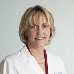 Martha Pitman合作论文数Medical School, Harvard University;Cytopathology Laboratory, Massachusetts General Hospital6Ling-Chun Chen合作论文数6Brian H. Mayall合作论文数6
 Karla Kerlikowske合作论文数Helen Diller Family Comprehensive Cancer Center, University of California, San Francisco;Department of Medicine, School of Medicine, University of California, San Francisco6Andres Forero合作论文数6
 Eric Winer合作论文数School of Medicine, Yale University;Smilow Cancer Hospital6B Mayall合作论文数5




