Lithium, introduced 75 years ago by John Cade1, remains the most effective mood stabilizer for bipolar disorder2. Lithium is proposed to modulate an array of cellular pathways, many ubiquitous to all cells, with pleiotropic roles unlinked to bipolar disorder or lithium responsiveness in genome wide association studies3,4. These mechanisms cannot explain lithium's specific effects on mood and behaviour. We demonstrate that lithium's primary action is in the periphery, not in the brain itself. Lithium acts in the gut to trigger behavioural and physiological changes, akin to those associated with a torpor-like state, that protect individuals from ingested toxins. Lithium activates gastrointestinal enterochromaffin (EC) cells via their Trpm2 cation channels to modulate afferent vagal and area postrema inputs to the brain. Eliminating these inputs by focal brain lesions eliminates lithium's effects, as does ablation of EC cells or their Trpm2 expression. Lithium's Trpm2-dependent activation of EC cells also occurs in human gut tissue, providing translational relevance for our discovery. These findings challenge the prevailing perception that lithium acts directly on the brain. Via a previously unsuspected gut-brain pathway, lithium engages brain circuitry that reduces arousal and interaction with the external world, therapeutic goals in the manic phase of bipolar disorder. ### Competing Interest Statement The authors have declared no competing interest. National Health and Medical Research Council, https://ror.org/011kf5r70 Baszucki Brain Research Fund
In the gastrointestinal tract, spinal afferent and sympathetic efferent neurons are key regulators of motility, secretion, and sensation. Strong colocalization of the afferent markers calcitonin gene-related peptide (CGRP) and TRPV1 is established in mouse gut. However, the expression of these markers in sympathetic axons-identifiable by immunoreactivity for tyrosine hydroxylase (TH), the rate-limiting enzyme for noradrenaline synthesis- has not yet been characterized in the colon. Using multiple-label immunohistochemistry, we quantified the coexistence of CGRP, TRPV1, and TH in varicose axons in the myenteric plexus of the mouse proximal and mid colon. Approximately half of CGRP-immunoreactive (IR) varicosities were also TRPV1-IR in the proximal (48.5 ± 3.5) and mid colon (50.3 ± 3.5) and the great majority of TRPV1-IR varicosities were CGRP-IR in the proximal (87 ± 1.1) and mid colon (90 ± 3.3). In the proximal and mid colon, TH-IR varicosities were neither CGRP- or TRPV1-IR, and the converse was also true; CGRP- and TRPV1-IR varicosities were not TH-IR. Our observations reveal that while CGRP and TRPV1 expression largely occurs together in axons in the proximal and mid colon, there is a population of CGRP axons that do not express TRPV1. In varicose axons, TH expression is mutually exclusive from expression of CGRP and TRPV1. Together, the results support the notion that CGRP together with TRPV1 and TH serve as respective "neurochemical markers" to distinguish spinal afferents from sympathetic efferents in the murine GI tract.
BACKGROUND:The enteric nervous system plays a key role in the coordination of gastrointestinal motility together with sympathetic, parasympathetic, and extrinsic sensory pathways. In some cases, abnormalities in neural activity in these pathways contribute to disorders of gut motility. Where this is associated with damage or death of enteric neurons, usually detected by microscopy, this is considered a gut neuropathy. PURPOSE:This review summarizes recent advances in the identification of neuropathies in a range of gastrointestinal motility disorders.
Indirect immunofluorescence is usually restricted to 3-5 markers per preparation, limiting analysis of coexistence. A solution containing 2-mercaptoethanol and sodium dodecyl sulfate (2-ME/SDS) can elute indirect immunofluorescence labelling (i.e. primary antisera followed by fluorophore-conjugated secondary antisera) and has been used for sequential staining of sections. The aim of this study was to test whether 2-ME/SDS is effective for eluting indirect immunofluorescent staining (with primary antisera visualised by fluorophore-coupled secondary antisera) in wholemount preparations. We also analysed how 2-ME/SDS may work and used this understanding to devise additional uses for immunofluorescence in the nervous system. 2-ME/SDS appears to denature unfixed proteins (including antisera used as reagents) but has much less effect on antigenicity of formaldehyde-fixed epitopes. Moieties linked by strong biotin-streptavidin bonds are highly resistant to elution by 2-ME/SDS. Two primary antisera raised in the same species can be applied without spurious cross-reactivity, if a specific order of labelling is followed. The first primary antiserum is followed by a biotinylated secondary, then a tertiary of fluorophore-conjugated streptavidin. The preparation is then exposed to 2-ME/SDS, which has minimal impact on labelling by the first primary/secondary/tertiary combination. However, when this is followed by a second primary antiserum (raised in the same species), followed by a fluorophore-conjugated secondary antiserum, the intervening 2-ME/SDS exposure prevents cross-reactivity between primary and secondary antisera of the two layers. A third property of 2-ME/SDS is that it reduces lipofuscin autofluorescence, although it also raises background fluorescence and strongly enhances autofluorescence of erythrocytes. In summary, 2-ME/SDS is easy to use, cost-effective and does not require modified primary antisera. It can be used as the basis of a multi-layer immunohistochemistry protocol and allows 2 primary antisera raised in the same species to be used together.
Cannabinoid agonists can potentially ameliorate lower urinary tract symptoms (LUTS), including pain associated with interstitial cystitis/bladder pain syndrome (IC/BPS). This study aims to determine the contributions of the cannabinoid 1 receptors (CB1Rs) and CB2Rs in regulating the activity of different functional classes of afferents, comparing normal healthy bladder with bladders from guinea pigs with protamine/zymosan-induced cystitis. The mechanosensitivity of different functional afferent classes was determined by ex vivo single-unit extracellular recordings. Peripherally restricted CB1R preferential agonists, ACEA and PrNMI and peripherally restricted CB2R selective agonists, 4Q3C and olorinab all reduced the mechanosensitivity of mucosal bladder afferents. The potency and efficacy of these synthetic cannabinoid agonists were significantly increased in cystitis compared to controls. Combined application of CB1R agonists, ACEA or PrNMI with the CB2R agonist, 4Q3C produced additive inhibitory effects. ACEA and PrNMI also inhibited the stretch-induced firing of high-threshold muscular bladder afferents in animals with cystitis. In contrast, low- and high-threshold muscular-mucosal bladder afferents were unaffected by the CB1R and CB2R agonists in control and cystitis. Our data indicated that peripherally restricted CB1R and CB2R agonists effectively reduce the sensitisation of probable nociceptive afferents in the bladder in cystitis. The findings also suggest a potential benefit of simultaneously targeting both the CB1Rs and CB2Rs to ameliorate LUTS in cystitis.
The enteric nervous system (ENS) consists of an extensive network of neurons and glial cells embedded within the wall of the gastrointestinal (GI) tract. Alterations in neuronal distribution and function are strongly associated with GI dysfunction. Current methods for assessing neuronal distribution suffer from undersampling, partly due to challenges associated with imaging and analyzing large tissue areas, and operator bias due to manual analysis. We present the Gut Analysis Toolbox (GAT), an image analysis tool designed for characterization of enteric neurons and their neurochemical coding using two-dimensional images of GI wholemount preparations. GAT is developed in Fiji, has a user-friendly interface, and offers rapid and accurate segmentation via custom deep learning (DL)-based cell segmentation models developed using StarDist, as well as a ganglia segmentation model in deepImageJ. We apply proximal neighbor- based spatial analysis to reveal differences in cellular distribution across gut regions using a public dataset. In summary, GAT provides an easy-to-use toolbox to streamline routine image analysis tasks in ENS research. GAT enhances throughput, allowing rapid unbiased analysis of larger tissue areas, multiple neuronal markers and numerous samples.
Background and AimsViscerofugal neurons (VFNs) have cell bodies in the myenteric plexus and axons that project to sympathetic prevertebral ganglia. In animals they activate sympathetic motility reflexes and may modulate glucose metabolism and feeding. We used rapid retrograde tracing from colonic nerves to identify VFNs in human colon for the first time, using ex vivo preparations with multi-layer immunohistochemistry.MethodsColonic nerves were identified in isolated preparations of human colon and set up for axonal tracing with biotinamide. After fixation, labeled VFN cell bodies were subjected to multiplexed immunohistochemistry for 12 established nerve cell body markers.ResultsBiotinamide tracing filled 903 viscerofugal nerve cell bodies (n = 23), most of which (85%) had axons projecting orally before entering colonic nerves. Morphologically, 97% of VFNs were uni-axonal. Of 215 VFNs studied in detail, 89% expressed ChAT, 13% NOS, 13% calbindin, 9% enkephalin, 7% substance P and 0 of 123 VFNs expressed CART. Few VFNs contained calretinin, VIP, 5HT, CGRP, or NPY. VFNs were often surrounded by dense baskets of axonal varicosities, probably reflecting patterns of connectivity; VAChT+ (cholinergic), SP+ and ENK+ varicosities were most abundant around them. Human VFNs were diverse; showing 27 combinations of immunohistochemical markers, 4 morphological types and a wide range of cell body sizes. However, 69% showed chemical coding, axonal projections, soma-dendritic morphology and connectivity similar to enteric excitatory motor neurons.ConclusionViscerofugal neurons are present in human colon and show very diverse combinations of features. High proportions express ChAT, consistent with cholinergic synaptic outputs onto postganglionic sympathetic neurons in prevertebral ganglia.
It is with sadness that we announce that Marcello Costa, our dear friend and colleague, passed away peacefully at his home in Seacliff (Adelaide, South Australia) on Sunday 14 April 2024. Professor Marcello Costa (AO, FAA) was a pioneer and leader in the field of autonomic neuroscience, specifically the intrinsic neural circuits of the enteric nervous system. From the mid-1970s to 2023 he led a series of studies characterising enteric neuronal pathways, particularly those related to gastrointestinal motility. His work traversed many disciplines including anatomy, histology, physiology and pharmacology through to electrophysiology. His papers often combined multiple approaches well before 'multi-disciplinary research' became fashionable. In later years he led the development of methods to objectively analyse gastrointestinal motility patterns, combining multi-dimensional maps of diameter, length and pressure with recordings of smooth muscle electrical activity. His quantitative accounts of complex motility patterns far surpassed the descriptive summaries that had previously characterised this field. His work was driven by a goal to demystify how cells of the gut work together to give rise to simple, adaptive behaviour patterns. Marcello was born on 9 January 1940 in Turin, Italy. In his childhood, he spent a lot of time at the family's apartment in the High Susa Valley to the west of Turin. Here, he developed a life-long love of mountains. He and his family (mother: Verbena, stepfather: Augusto and older brother: Giorgio) moved to Buenos Aires, Argentina, in 1949. He attended the High School San Martin and the public Italian High School of Buenos Aires, finishing school in early 1960. Later that year, Marcello moved back to Turin to study medicine. However, between these moves he fitted in joining an expedition to explore the continental icepacks of Patagonia, venturing into barely mapped territory. A few months later, Marcello undertook another trip with his brother Giorgio, climbing mountains in Peru and descending the Ucayali and Amazon rivers. His time at the Universita di Torino was intense. Not only did he become heavily involved in student politics (elected as Leader of the Student Centre Left Party) but he also trained in mountain climbing with the Gervasutti School of the Torino Alpine Club, played guitar in a jazz band and joined the student choir. He also undertook a research internship in the Departments of Anatomy and Histology under the supervision of Giorgio Gabella. Using a newly discovered technique (the 'Falckh–Hillarp method') to visualise monoamine transmitters (Falckh et al., 1962), Marcello undertook microscopy studies that led to 11 publications between 1965 and 1970 and award of his MD (Gabella & Costa, 1968). After graduating in Medicine and Surgery in 1967, Marcello did compulsory military service, finishing in 1969. Shortly thereafter, at a scientific meeting in Venice, he got talking to Professor Geoff Burnstock. An offer soon arrived by mail, inviting Marcello for an 18 month post-doctoral fellowship in Geoff's lab at Melbourne University. Marcello married his girlfriend Daniela Tuffanelli in May 1970 and shortly after set out for Australia; Daniela followed a few months later. On arriving at Melbourne University, Marcello met John Furness and they discovered many shared interests and ideas. Together, they extended studies on peripheral aminergic innervation. This was the start of a long and productive collaboration which continued until 1988. There was a brief interlude when Marcello's funding ran out and he had to return to Europe in 1973. Fortunately, he was soon invited back to Melbourne, within the year, but in 1974 Geoff Burnstock announced that he would be moving to University College, London. The group at Melbourne University dispersed; John Furness was offered a lectureship in Anatomy and Histology in the new Flinders University School of Medicine in Adelaide. In 1975 Marcello accepted a lectureship in Human Physiology also at Flinders University, where he remained for the next 47 years. He retired as Mathew Flinders Distinguished Professor in 2021, at the age of 81, retaining his links as Emeritus Professor. At a young age, Marcello was given a small microscope; this opened up an intriguing new world of tiny objects. As a teenager he sold his beloved bicycle in order to buy a better microscope, capable of resolving bacteria. He spent many hours entranced by different types of protozoa moving in pond water. This early love of microscopy may have prepared him for his research as an intern under the critical eye of Professor Giorgio Gabella. He carried out an extensive series of histochemical studies localising amine transmitters in the autonomic nervous system, working with Giorgio Gabella (Gabella & Costa, 1969), then John Furness (Costa & Furness, 1971) and, during a sabbatical at the University of Helsinki, with Olavi Erankö (Costa et al., 1974). These studies were notable for being more than simple descriptions – crushes and lesions were applied to identify the projections of aminergic nerves. For the first 10 years of his career, Marcello's publications were dominated by studies of aminergic pathways. After his move to Flinders University, Marcello's head of department was Laurie Geffen. Robert Rush joined the department shortly after Marcello. Together with Bruce Livett at Monash, Geffen and Rush had published one of the first studies using antibodies to localise macromolecules in nerve cells (Livett et al., 1971). With Marcello's interest in localisation of neurotransmitters, the stage was set. In 1977 Marcello and colleagues obtained an antiserum that bound the neuropeptide somatostatin. They used it to reveal varicose axons and cell bodies immunoreactive for this neuropeptide in the gut wall (Costa et al., 1977). They concluded, with remarkable insight, that somatostatin may be expressed by a population of interneurons in the enteric nervous system – a proposal that was confirmed many years later. Work on amines continued, but a new series of studies commenced, using immunohistochemical methods to reveal neuropeptides and other molecules including Substance P (Franco et al., 1979), VIP (vasoactive intestinal peptide) (Furness & Costa, 1979), tyrosine hydroxylase and dopamine beta hydroxylase (Furness et al., 1979), 5-hydroxytryptamine (Costa et al., 1982), enkephalin (Furness et al., 1983), GRP (gastrin-releasing peptide) (Costa et al., 1984), galanin (Furness et al., 1987) and calbindin (Furness et al., 1988). Costa, Furness and colleagues combined multi-labelling immunohistochemistry with methods to study projections of axons, including specialised operations. Enteric neural pathways were lesioned with a cut made around the circumference of the intestine ('myotomy') or by removal of a segment of myenteric plexus from the full circumference ('myectomy'). From the degeneration of axons and accumulation of axonal cytoplasm proximal to damage, the length and polarity of projections could be estimated. The publication of a method to optimise histochemical and immunohistochemical labelling in peeled whole-mount preparations further improved the analysis of projections and soma-dendritic morphology (Costa et al., 1980). For many antigens, immunohistochemical localisation was supplemented by biochemical measurement of the antigens in tissue, adding a quantitative validation of methods. The discovery that many immunohistochemically detected molecules were expressed by subsets of enteric neurons led to the idea that they could be considered as 'markers'. Marcello and colleagues provided early reports showing that single neurons could express multiple neuropeptide transmitters (Costa et al., 1988). Their wide-ranging immunohistochemical analysis firmly established the reputation of Marcello and his colleagues as leaders in the field of enteric neurobiology. A review published in 1986 introduced the concept that combinations of markers could be used to distinguish different functional classes of autonomic neurons (Costa et al., 1986): 'the results point to the principle that the enteric neurons, and other autonomic neurons, are subdivided into groups with well-defined combinations of chemical messengers (chemical coding), well-defined projections (ie: origins, terminations and connections) and well-defined functions.' The influential concept of chemical coding of neurons was established. A few years later, Joel Bornstein joined the laboratory, bringing expertise in electrophysiological recording which he combined with intracellular dye filling and immunohistochemical labelling to create a multi-faceted analysis of enteric neurobiology (Bornstein, Costa, et al., 1984). It quickly became apparent that not only were immunohistochemical markers selectively expressed, but so were electrophysiological features and soma-dendritic morphology. This methodology was then combined with selective lesions (Bornstein, North, et al., 1984) to investigate the pathways responsible for the mysterious slow excitatory post-synaptic potentials discovered by Jackie D. Wood in 1978 (Wood & Mayer, 1978). A series of innovative studies followed, concentrating on the intrinsic and extrinsic innervation of the gut, mostly in guinea pigs. Findings included the identification of substance P and CGRP (calcitonin gene-related peptide) coexisting in a subset of spinal nociceptors (Gibbins et al., 1985). Sympathetic neurons projecting to the gut were shown to express different combinations of neuropeptides depending on their targets and function (Macrae et al., 1986). These approaches were expanded to human tissue obtained from elective surgery in the Flinders Medical Centre by David Wattchow and Janet Keast (Keast et al., 1984; Wattchow et al., 1988). Later, methods for maintaining the intestine for several days ex vivo in organ culture were developed by Marcello and his colleague Simon Brookes, allowing the application of retrograde tracing of neurons in both animal and human tissue (Brookes & Costa, 1990). This allowed more precise analysis of projections and pathways of enteric neurons in human and animal intestine in a series of papers. These studies accumulated a huge amount of information about the enteric nervous system and its organisation, but the data were very fragmented. Marcello was determined to combine these data into a single, cohesive account by exhaustively studying many combinations of markers and quantifying their overlap. In a landmark paper in 1996, Marcello distinguished 14 classes of myenteric neurons, on the basis of co-localisation of nine different markers which could be combined with four classes of submucosal neurons identified previously. This made the enteric neurons of the guinea pig small intestine one of the most comprehensively characterised innervated preparations of any mammal and provided a foundation for interpretation of many later studies (Costa et al., 1996). With this major goal achieved, Marcello used this account as a foundation to characterise the basis of motility patterns of the small intestine. With Marcello Tonini from the University of Pavia he measured smooth muscle activity in ex vivo preparations (Tonini & Costa, 1990) with novel watertight partitions so that drugs could be added selectively to specific parts of motility reflex pathways. This method has been widely used by a number of groups internationally. In the late 1990s, Marcello led several studies of peristalsis in the guinea pig small intestine (Waterman et al., 1992; Waterman, Costa & Tonini, 1994; Waterman, Tonini & Costa, 1994). A PhD student, Grant Hennig, was charged with measuring diameter changes from video recordings. This was time-consuming and demanding, but Grant had programming skills and wrote a routine to count the total number of pixels across the gut wall using NIH Image software. This method created unmanageably large datasets of measurements. Grant tried converting the numbers to greyscale pixels, which elegantly visualised the data. When Marcello saw this image he instantly recognised its potential. The greyscale images showed pale, angled streaks of propagating contractions and darker areas where content was accumulating. A casual glance could distinguish peristalsis from other motor patterns. It was easy to quantify the wavelength, amplitude, rate of propagation and frequency of events. Marcello used these 'spatio-temporal maps' as a powerful new way to quantify motility patterns for quantitative study; this was a huge advance over previous descriptive analyses (Cannon, 1902). Interestingly similar maps were developed at nearly the same time in two other laboratories (Bercik et al., 2000; Bouchoucha et al., 1999). In 2008, Nick Spencer joined Flinders University and soon after Marcello joined Nick's laboratory, for the next 13 years until his retirement in 2021. This was an incredibly productive and dynamic era – a time of great energy, vision and innovation. It was also Marcello's most productive publication period. It was a true honour having Marcello perform hands-on experiments in the laboratory of N.J.S., right up until he retired at 81 years of age. This period also involved extensive collaboration with Phil Dinning and Lukasz Wiklendt, where Marcello combined intraluminal pressure maps with spatio-temporal maps of wall movements and focal recordings of electrical activity (Fig. 1) (Costa et al., 2021; Dinning et al., 2011). During this time in Nick's laboratory, Marcello produced a series of at least 30 key papers, detailing interactions between neurogenic and myogenic mechanisms in the small and large bowel of several species with remarkable quantitative precision. Marcello had a clear vision on how to translate his research into the clinical world. Live specimens of human gut could be obtained (with appropriate permits) from surgeons operating in the same building. This pathway was established by David Wattchow, a colorectal surgeon who did his PhD with Marcello and John Furness. David's efforts made it possible for many studies in animal tissue to be tested in human tissue over ensuing decades. Specimens were taken from the healthy segment of the small or large intestine resected in the treatment of malignancy. In the laboratory they were studied anatomically under the microscope (Wattchow et al., 1995) or physiologically in organ baths (Carbone et al., 2013) in the labs of Simon Brookes and Nick Spencer. Several surgical trainees carried out PhD studies on these specimens, supervised by Marcello. Marcello was impressed by the work of Dr Anthony Bauer from Pittsburgh, USA, who had shown in animals that anti-inflammatory drugs were candidates to treat post-operative ileus (Bauer & Boeckxstaens, 2004). With David Wattchow and clinical colleagues, a large clinical trial of the use of anti-inflammatories in abdominal surgery was undertaken (Wattchow et al., 2009). Thus, Marcello was involved in study of the human enteric nervous system from the level of the cells, muscle strips and organ bath recordings through to the whole patient and therapeutic applications. He fostered the collaborations that made this happen. Marcello was extraordinarily well read in non-fiction. He had a collection of several thousand books by contemporary philosophers, mathematicians, modellers and scientists. His study of knowledge itself formed a strong underpinning for his research, and a firm foundation for his humanist beliefs. He was enthusiastic about sharing knowledge and this was evident in his lectures. Throughout his 47 years at Flinders University he held a mixed lecturer/researcher appointment. His lectures to students were eloquent, authoritative and carefully structured. Generations of medical students remember his teaching of neuroscience and physiology, delivered with his distinctive accent. Many young researchers encountered him at scientific meetings and benefited from his perceptive and focused questioning. He supported many junior scientists, going out of his way to provide encouragement and advice. His questions often had a long preamble, but were always followed by an incisive query or comment. Marcello would be the first to acknowledge that form-filling was not his forte. However, his enthusiasm for good causes more than made up for this. He played a key role in setting up the Centre for Neuroscience at Flinders in 1977 and in 1980−1981 was instrumental in turning the Australian Neuroscience Society into the vigorous formal entity that thrives to this day (renamed the Australasian Neuroscience Society, ANS). In 1994, Marcello served as President of the ANS. He was on the organising committee of 23 research conferences. He also founded the South Australian Neuroscience Institute (SANI) in 2003. Its goal was to unite and coordinate neuroscientific research, education, services and commercialisation. As co-chair, he took on many roles and organised over 20 public forums including several during International Brain Awareness Week. Other talks were held under the title of 'Science outside the Square'. SANI developed links with the Physical and Neurological Council of South Australia, and developed a Graduate Certificate in Neuroscience (Learning) for teachers. Marcello had a strong belief that leaders should be answerable for their decisions. He had a well-developed sense of fair play and was fearless in university meetings. He was equally outspoken about the misuse of science, especially the promotion of pseudoscience by unscrupulous operators. He was a founding member of Friends of Science in Medicine and openly challenged bogus claims, while championing the value of evidence-based medicine in numerous interviews on radio and television. In his childhood, Marcello showed a real talent for painting and drawing. This ability was later used in his scientific drawings, some of which have become discipline classics. After moving to Adelaide, he became a keen windsurfer, taking up the sport in its early days. He was noted for some of the long-distance journeys he made; windsurfing to Kangaroo Island, Yorke Peninsula and along much of the 100 km length of the Coorong lagoon. He appreciated art and created a series of YouTube videos on the history of art and perspective. Last, but not least, Marcello was also a family man. He and Daniela have a son, Andre, who is now a successful academic at the University of Adelaide. Marcello was also the father-in-law of Kat, grandfather of Harry, Orlando and Heidi, and brother of Giorgio. Marcello received many awards and accolades during his long career. These included appointment as Fellow of the Australian Academy of Sciences in 1989. In 1992, he was honoured with the title of 'Cavaliere della Repubblica Italiana'. In 2001, he was awarded the Australian Centenary Medal. In 2018, Marcello was awarded the inaugural Lifetime Achievement Award by the Federation of Neurogastroenterology and Motility Societies and, in 2020, he was appointed as an Officer of the Order of Australia. The latter honour is given by the Governor General of Australia for contributions to Australian society. However, perhaps his greatest accolade was the respect and affection of the many people who he personally interacted with, in his many areas of interest. In the world of university and science he influenced undergraduates, medical students and many leading scientists in neuroscience and neurogastroenterology. Many of his colleagues referred to him as a 'renaissance man' on the basis of his mastery of multiple fields of knowledge, combined with a deep culture and education. His belief in the value of scientific understanding was unbounded. His own work added enormously to that store of knowledge but, in addition, he encouraged many researchers, junior and senior, in their quest for discovering new knowledge. Marcello was not only a great scientist, ambassador, teacher, public advocate and family man, he was also our friend and inspiration. We miss him deeply. Please note: The publisher is not responsible for the content or functionality of any supporting information supplied by the authors. Any queries (other than missing content) should be directed to the corresponding author for the article. No competing interests declared. N.S.: Conception or design of the work; Drafting the work or revising it critically for important intellectual content; Final approval of the version to be published; Agreement to be accountable for all aspects of the work. S.B.: Conception or design of the work; Drafting the work or revising it critically for important intellectual content; Final approval of the version to be published; Agreement to be accountable for all aspects of the work. D.W.: Conception or design of the work; Drafting the work or revising it critically for important intellectual content; Final approval of the version to be published; Agreement to be accountable for all aspects of the work. Australian Research Council Centre of Excellence in Advanced Molecular Imaging (Imaging CoE): Nick J. Spencer, DP190103628; DHAC | National Health and Medical Research Council (NHMRC): Nick J. Spencer, 1156427.
This protocol explains how viscerofugal neurons can be studied in ex vivo specimens of human colon. Viscerofugal neurons are characterized by having cell bodies in myenteric ganglia and axons that projectout of the gut via colonic or mesenteric nerves. The protocol covers obtaining and handling live human colonic tissue specimens, biotinamide tracing of viscerofugal neuron cell bodies from axons in colonic nerves, tissue culture, fixation and immunohistochemical processing. This protocol was used in: Chen BN, Humenick A, Hibberd TJ, Yew WP, Wattchow DA, Dinning PG, Costa M, Spencer NJ, Brookes SJH (2024) Characterisation of viscerofugal neurons in human colon by retrograde tracing and multi-layer immunohistochemistry.Frontiers in Neuroscience 17:1313057. doi: 10.3389/fnins.2023.1313057
The autonomic nervous system that regulates the gut is divided into sympathetic (SNS), parasympathetic (PNS), and enteric nervous systems (ENS). They inhibit, permit, and coordinate gastrointestinal motility, respectively. A fourth pathway, "extrinsic sensory neurons," connect gut to the central nervous system, mediating sensation. The ENS resides within the gut wall and its activities are critical for life; ENS failure to populate the gut in development is lethal without intervention."Viscerofugal neurons" are a distinctive class of enteric neurons, being the only type that escapes the gut wall. They form a unique circuit: their axons project out of the gut wall and activate sympathetic neurons, which then project back to the gut, and inhibit gut movements.For 80 years viscerofugal/sympathetic circuits were thought to have a restricted role, mediating simple sensory-motor reflexes. New data shows viscerofugal and sympathetic neurons behaving unexpectedly, compelling a re-evaluation of these circuits: both viscerofugal and sympathetic neurons transmit higher order, synchronized firing patterns that originate within the ENS. This identifies them as driving long-range motility control between different gut regions.There is need for gut motor control over distances beyond the range of ENS circuits, yet no mechanism has been identified to date. The entero-sympathetic circuits are ideally suited to meet this need. Here we provide an overview of the structure and functions of these peripheral sympathetic circuits, including new data showing the firing patterns generated by enteric networks can transmit through sympathetic neurons.
Over 150 years ago, methods for quantitative analysis of gastrointestinal motor patterns first appeared. Graphic representations of physiological variables were recorded with the kymograph after the mid-1800s. Changes in force or length of intestinal muscles could be quantified, however most recordings were limited to a single point along the digestive tract. In parallel, photography and cinematography with X-Rays visualised changes in intestinal shape, but were hard to quantify. More recently, the ability to record physiological events at many sites along the gut in combination with computer processing allowed construction of spatiotemporal maps. These included diameter maps (DMaps), constructed from video recordings of intestinal movements and pressure maps (PMaps), constructed using data from high-resolution manometry catheters. Combining different kinds of spatiotemporal maps revealed additional details about gut wall status, including compliance, which relates forces to changes in length. Plotting compliance values along the intestine enabled combined DPMaps to be constructed, which can distinguish active contractions and relaxations from passive changes. From combinations of spatiotemporal maps, it is possible to deduce the role of enteric circuits and pacemaker cells in the generation of complex motor patterns. Development and application of spatiotemporal methods to normal and abnormal motor patterns in animals and humans is ongoing, with further technical improvements arising from their combination with impedance manometry, magnetic resonance imaging, electrophysiology, and ultrasonography.
Compared with healthy adults this study has shown a significant reduction in the prevalence of the postprandial retrograde cyclic motor pattern in the distal colon of patients with diarrhea-predominant irritable bowel syndrome. We hypothesize that this altered motility may allow for premature rectal filling which contributes to the postprandial urgency and diarrhea experienced by these patients.
Distinguishing and characterising the different classes of neurons that make up a neural circuit has been a long-term goal for many neuroscientists. The enteric nervous system is a large but moderately simple part of the nervous system. Enteric neurons in laboratory animals have been extensively characterised morphologically, electrophysiologically, by projections and immunohistochemically. However, studies of human enteric nervous system are less advanced despite the potential availability of tissue from elective surgery (with appropriate ethics permits). Recent studies using single cell sequencing have confirmed and extended the classification of enteric neurons in mice and human, but it is not clear whether an encompassing classification has been achieved. We present preliminary data on a means to distinguish classes of myenteric neurons in specimens of human colon combining immunohistochemical, morphological, projection and size data on single cells. A method to apply multiple layers of antisera to specimens was developed, allowing up to 12 markers to be characterised in individual neurons. Applied to multi-axonal Dogiel type II neurons, this approach demonstrated that they constitute fewer than 5% of myenteric neurons, are nearly all immunoreactive for choline acetyltransferase and tachykinins. Many express the calcium-binding proteins calbindin and calretinin and they are larger than average myenteric cells. This methodology provides a complementary approach to single-cell mRNA profiling to provide a comprehensive account of the types of myenteric neurons in the human colon.
Motor function of the colon is essential for health. Our current understanding of the mechanisms that underlie colonic motility are based upon a range of experimental techniques, including molecular biology, single cell studies, recordings from muscle strips, analysis of part or whole organ ex vivo through to in vivo human recordings. For the surgeon involved in the clinical management of colonic conditions this amounts to a formidable volume of material. Here, we synthesize the key findings from these various experimental approaches so that surgeons can be better armed to deal with the complexities of the colon.
Multiplexed immunohistochemistry for 12 markers was used to classify myenteric neurons of human colon. Twenty classes were distinguished with statistical validation. Cell morphology, soma size, and associations with axon terminals were quantified, providing a wide-ranging account of human myenteric plexus. BACKGROUND AND AIMS: Gut functions including motility, secretion, and blood flow are largely controlled by the enteric nervous system. Characterizing the different classes of enteric neurons in the human gut is an important step to understand how its circuitry is organized and how it is affected by disease. METHODS: Using multiplexed immunohistochemistry, 12 discriminating antisera were applied to distinguish different classes of myenteric neurons in the human colon (2596 neurons, 12 patients) according to their chemical coding. All antisera were applied to every neuron, in multiple layers, separated by elutions. RESULTS: A total of 164 combinations of immunohistochemical markers were present among the 2596 neurons, which could be divided into 20 classes, with statistical validation. Putative functions were ascribed for 4 classes of putative excitatory motor neurons (EMN1-4), 4 inhibitory motor neurons (IMN1-4), 3 ascending interneurons (AIN1-3), 6 descending interneurons (DIN1-6), 2 classes of multiaxonal sensory neurons (SN1-2), and a small, miscellaneous group (1.8% of total). Soma-dendritic morphology was analyzed, revealing 5 common shapes distributed differentially between the 20 classes. Distinctive baskets of axonal varicosities surrounded 45% of myenteric nerve cell bodies and were associated with close appositions, suggesting possible connectivity. Baskets of cholinergic terminals and several other types of baskets selectively targeted ascending interneurons and excitatory motor neurons but were significantly sparser around inhibitory motor neurons. CONCLUSIONS: Using a simple immunohistochemical method, human myenteric neurons were shown to comprise multiple classes based on chemical coding and morphology and dense clusters of axonal varicosities were selectively associated with some classes.
Inflammatory and functional gastrointestinal disorders such as irritable bowel syndrome (IBS) and obstructive bowel disorder (OBD) underlie the most prevalent forms of visceral pain. Although visceral pain can be generally provoked by mechanical distension/stretch, the mechanisms that underlie visceral mechanosensitivity in colon-innervating visceral afferents remain elusive. Here, we show that virally mediated ablation of colon-innervating TRPV1-expressing nociceptors markedly reduces colorectal distention (CRD)-evoked visceromotor response (VMR) in mice. Selective ablation of the stretch-activated Piezo2 channels from TRPV1 lineage neurons substantially reduces mechanically evoked visceral afferent action potential firing and CRD-induced VMR under physiological conditions, as well as in mouse models of zymosan-induced IBS and partial colon obstruction (PCO). Collectively, our results demonstrate that mechanosensitive Piezo2 channels expressed by TRPV1-lineage nociceptors powerfully contribute to visceral mechanosensitivity and nociception under physiological conditions and visceral hypersensitivity under pathological conditions in mice, uncovering potential therapeutic targets for the treatment of visceral pain.
BACKGROUND AND PURPOSE:Interstitial cystitis (=painful bladder syndrome) is a chronic bladder syndrome characterised by pelvic and bladder pain, urinary frequency and urgency, and nocturia. Transient receptor potential (TRP) channels are an attractive target in reducing the pain associated with interstitial cystitis. The current study aims to determine the efficacy of combination of TRP vanilloid 1 (TRPV1) and TRP melastatin 8 (TRPM8) channel inhibition in reducing the pain associated with experimental cystitis in guinea pigs.EXPERIMENTAL APPROACH:A novel animal model of non-ulcerative interstitial cystitis has been developed using protamine sulfate/zymosan in female guinea pigs. Continuous voiding cystometry was performed in conscious guinea pigs. Ex vivo "close-to-target" single unit extracellular recordings were made from fine branches of pelvic nerves entering the guinea pig bladder. Visceromotor responses in vivo were used to determine the effects of TRP channel antagonists on cystitis-induced bladder hypersensitivity.KEY RESULTS:Protamine sulfate/zymosan treatment evoked mild inflammation in the bladder and increased micturition frequency in conscious animals. In cystitis, high threshold muscular afferents were sensitised via up-regulation of TRPV1 channels, high threshold muscular-mucosal afferents were sensitised via TRPM8 channels, and mucosal afferents by both. Visceromotor responses evoked by noxious bladder distension were significantly enhanced in cystitis and were returned to control levels upon administration of combination of low doses of TRPV1 and TRPM8 antagonists.CONCLUSIONS AND IMPLICATIONS:The data demonstrate the therapeutic promises of combination of TRPV1 and TRPM8 antagonists for the treatment of bladder hypersensitivity in cystitis.
BACKGROUND:Ex vivo intracellular recordings and dye fills, combined with immunohistochemistry, are a powerful way to analyze the enteric nervous system of laboratory animals.METHODS:Myenteric neurons were recorded in isolated specimens of human colon. A key determinant of successful recording was near-complete removal of circular muscle from the surface of ganglia.KEY RESULTS:Treatment with a collagenase/neutral protease mix before dissection significantly improved recording success and reduced damage to the plexus. Carboxyfluorescein in microelectrodes allowed recorded neurons to be routinely labeled, analyzed, and subjected to multi-layer immunohistochemistry. Carboxyfluorescein revealed morphological details that were not detected by immunohistochemical methods. Of 54 dye-filled myenteric neurons (n = 22), 45 were uni-axonal and eight were multi-axonal. There was a significant bias toward recordings from large neural somata. The close association between morphology and electrophysiology (long after-hyperpolarizations and fast EPSPs) seen in mice and guinea pigs did not hold for human myenteric neuron recordings. No slow EPSPs were recorded; however, disruption to the myenteric plexus during dissection may have led the proportion of cells receiving synaptic potentials to be underestimated. Neurons immunoreactive for nitric oxide synthase were more excitable than non-immunoreactive neurons. Distinctive grooves were observed on the serosal and/or mucosal faces of myenteric neurons in 3D reconstructions. These had varicose axons running through them and may represent a preferential site of synaptic inputs.CONCLUSIONS:Human enteric neurons share many features with laboratory animals, but the combinations of features in individual cells appear more variable.
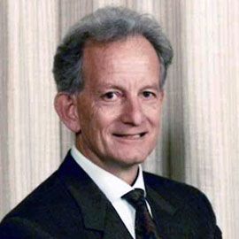 Marcello Costa合作论文数College of Medicine and Public Health, Flinders University134
 Spencer Nick J合作论文数100
 Phil G Dinning合作论文数Dept Surg, Flinders Univ S Australia72David Wattchow合作论文数52
 Vladimir P Zagorodnyuk合作论文数Flinders Health & Medical Research Institute, Flinders University42
 Lukasz Wiklendt合作论文数HeartAI42Tim Hibberd合作论文数Discipline of Surgery and Centre for Neuroscience, Flinders University Adelaide36Bao Nan Chen合作论文数23Adam Humenick合作论文数20
 John W. Arkwright合作论文数CSIRO Materials Science and Engineering17
 Melinda Kyloh合作论文数16Tiong Cheng Sia合作论文数12Bn Chen合作论文数10Wai Ping Yew合作论文数10
 Lauren Keightley合作论文数8
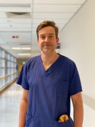 Paul T Heitmann合作论文数College of Medicine and Public Health, Flinders University8
 David Grundy合作论文数School of Biosciences, The University of Sheffield8
 Damien J Keating合作论文数Corning IntelliSense Corp.8
 David Smolilo合作论文数7
 Lee Travis合作论文数7
 Nan Chen合作论文数7
 Sarah J Nicholas合作论文数Ctr Neurosci, Flinders Univ S Australia7
 Carbone Simona E合作论文数College of Health and Biomedicine, Victoria University;College of Health and Biomedicine, Victoria University7Penny A. Lynn合作论文数7Omari Taher合作论文数6Hong-Zhen Hu合作论文数6
 Roberto De Giorgio合作论文数Department of Internal Medicine and Gastroenterology, University of Bologna6Raghu Kumar合作论文数5
 Kelsi Nicole Dodds合作论文数School of Medical Sciences, University of Adelaide5
 Dale Sharrad合作论文数5
 Ian Cook合作论文数Department of Gastroentrology and Hepatology, St George Hospital;St George Swallow Centre;School of Clinical Medicine, University of New South Wales5Ian Underhill合作论文数5Catharina Olsson合作论文数5G. W. Hennig合作论文数5
 Ian L Gibbins合作论文数Department of Anatomy and Histology, College of Medicine and Public Health, Flinders University5T C Sia合作论文数4Rochelle A Peterson合作论文数4Lyn Maslen合作论文数4
 Harman Sharma合作论文数4
 Michael Schemann合作论文数Department of Human Biology, School of Life Sciences, Technische Universitat Munchen4Lynn P A合作论文数4Marcello Tonini合作论文数4
 Stuart Brierley合作论文数Hopwood Centre For Neurobiology, South Australian Health and Medical Research Institute4D. de Fontgalland合作论文数4SY Yuan合作论文数3t. shea-donohue合作论文数3Zm Song合作论文数3R Mohd Rosli合作论文数3Pa Steele合作论文数3




