Most prokaryotes divide using filaments of the tubulin-like FtsZ protein, while some archaea employ instead ESCRT-III-like proteins and their filaments for cell division and cytokinesis. The alternative archaeal system comprises Cdv proteins and is thought to bear some resemblance to ESCRT-III-based membrane remodeling in other domains of life, including eukaryotes, especially during abscission. Here, we present biochemical, crystallographic, and cryo-EM studies of the Sulfolobus Cdv machinery. CdvA, an early non-ESCRT component, adopts a PRC-domain/coiled-coil fold and polymerizes into long double-stranded helical filaments, mainly via hydrophobic interfaces. Monomeric CdvB adopts the canonical ESCRT-III fold in both a closed and a distinct "semiopen" conformation. Soluble CdvB2 filaments are composed of subunits in the closed state, appearing to transition to the open, active state only when polymerized on membranes. Short N-terminal amphipathic helices in all CdvB paralogues, B, B1, and B2, mediate membrane binding and are required for liposome recruitment in vitro. We provide a molecular overview of archaeal ESCRT-III-based cytokinesis machinery, the definitive demonstration that CdvB proteins are bona fide ESCRT-III homologues, and reveal the molecular basis for membrane engagement. Thus, we illuminate conserved principles of ESCRT-mediated membrane remodeling and extend them to an anciently diverged archaeal lineage.
Cytokinesis finalizes cell division by physically separating the daughter cells. After furrow ingression, the intercellular bridge (ICB) connecting the dividing cells is cut via microtubule severing, followed by membrane scission, which requires dynamic ESCRT-III polymers. Proteasomal degradation is crucial for mitotic exit and the early steps of cytokinesis, but whether it plays a role in abscission is unknown. Here, we found that inhibiting the proteasome after furrow ingression in human cells led to increased Aurora B levels at the ICB and delayed both microtubule cutting and membrane scission. Overexpression of Aurora B phenocopied these cytokinetic defects, whereas inhibiting Aurora B activity fully rescued the defects caused by proteasome inhibition. Mechanistically, proteasome inhibition increased the levels of active Aurora B, enhanced the phosphorylation of its substrate CHMP4C, and reduced the amount of the ESCRT-III remodeling ATPase VPS4 at the ICB. Consequently, this led to a reduction in the dynamics of the ESCRT-III polymer at the ICB. Remarkably, proteasome inhibition did not delay abscission in cells expressing a mutant of CHMP4C that cannot be phosphorylated by Aurora B, thus demonstrating that the function of the proteasome in abscission is to degrade Aurora B and thereby limit CHMP4C phosphorylation. Our findings reveal a key role of the proteasome in the final step of cell division by decreasing Aurora B levels and activity to enable ESCRT-III function in abscission.
Cell behaviors in multicellular organisms are coordinated via both diffusible molecules and by signals based on direct cell-cell contacts. The mode of cell communication used influences the signaling range. In many developing epithelia, contact-based Notch-Delta lateral inhibition signaling is used to pattern cell fates. While previous work revealed that cells can use protrusions to extend the range of Notch-Delta signaling to alter these patterns, this is not a general feature of epithelia. In addition, it is not known how the complex three-dimensional (3D) shapes of epithelial cells influence cell communication. In exploring this question, we show that epithelial cells at the Drosophila wing margin, which lack basal protrusions, contact different neighbors at different heights along their apico-basal axis, effectively increasing the number of neighbors each cell touches. To quantitatively assess this behavior, we develop a mathematical modeling framework (Multilayer Signaling Model) to simulate Notch-Delta signaling over data-derived 3D cell topologies. The model predicts that lateral cell surface signaling is essential to tune the spacing between sensory organ precursors (SOPs). In agreement, we show that perturbing cortical stiffness and cell tortuosity in vivo modifies SOP spacing. These results emphasize the importance of 3D cell geometry and topology in fine-tuning signaling range.
Cytomotive filaments, a prominent class of cytoskeletal polymers comprising the actin and tubulin families, combine directional growth with monomer turnover to perform essential cellular functions. While the functional role of turnover is becoming increasingly clear, how these filaments dynamically self-assemble using a single monomer type remains an open question. Here we exploit physical modelling in combination with genetic algorithms to elucidate the design principles that drive monomer self-assembly into treadmilling filaments -- polar filaments that grow on one end and shrink on the other. We first show that directional polymer growth is only possible when a structurally polar monomer changes conformation upon polymerisation, and identify three different mechanisms through which the structural polarity of a monomer can induce kinetic polarity. We then prove that to prevent filament fragmentation and promote end depolymerisation upon nucleotide hydrolysis, the conformation change of a polar monomer must involve both its interface-forming sites. Combining these results, we identify the principles required to design treadmilling monomers, and showcase the emergence of treadmilling dynamics from the bottom-up. Our work sheds light on the physical mechanisms underlying cytomotive self-assembly of biological polymers, and lays the base for the realization of essential cytoskeletal features in synthetic systems with programmable building blocks. ### Competing Interest Statement The authors have declared no competing interest. European Union, https://ror.org/019w4f821, 101034413, 802960 Gesellschaft für Forschungsförderung Niederösterreich, RTI-Dissertations 2024 Royal Society, https://ror.org/03wnrjx87, UF16026
Abstract All cells remodel their membranes to divide. The highly conserved ESCRT-III system forms contractile polymers which, through direct interactions with membrane lipids, remodel membranes across the tree of life. In exploring how ESCRT-III divides the chemically and structurally unique archaeal membrane, we reveal that the homologue CdvB1 is required for the establishment of a distinct membrane domain within the division bridge of Sulfolobus acidocaldarius , associated with an accumulation of membrane-spanning inositol phosphate lipids. We show that CdvB1 associates with phosphoinositides in vitro and that this interaction aids cytokinesis in vivo . Together, we suggest that although eukaryotes inherited their membrane lipids from bacteria during eukaryogenesis, key features of the ESCRT-III:membrane interface that allow these polymers to bind, organise, and remodel eukaryotic membranes, may originate in archaea.
The surface layer or "S-layer" is a two-dimensional lattice of proteins that coats a wide range of archaea and bacteria in place of a cell wall or capsular polysaccharides. S-layers are thought to play an important role in chemically and physically insulating cells from the external environment. Here, we show that the integrity of the S-layer in Sulfolobus acidocaldarius is maintained as cells grow via a process of self-assembly as SlaA monomers fill gaps in the lattice. Although this lattice which is physically tethered to the membrane might be expected to hinder cell division, we show that the S-layer flattens the membrane at cytokinesis to accelerate ESCRT-III-dependent cell division-and is important for robust, successful cell divisions under conditions of mechanical stress. Taken together, these results define the rules governing S-layer self-assembly and show how a flexible lattice coat that is coupled to the underlying membrane can both provide a cell with mechanical support and help to drive rapid and functionally important changes in cell shape.
The surface layer or "S-layer" is a planar lattice of glycosylated proteins that coats a wide range of archaea and bacteria instead of a classical cell wall or capsular polysaccharides, insulating them from the extracellular space and providing the cell membrane with physical support. Although the S-layer's role as a mechanical support for the membrane might be expected to hinder cell division, we show that in rapidly dividing Sulfolobus acidocaldarius cells, the S-layer protein SlaA self-assembles into flexible lattice that helps flattens the cytokinetic furrow to accelerate ESCRT-III dependent cell division - a role that is especially important under conditions of mechanical stress. Taken together, these results generated using mutational analysis, live and fixed cellular imaging, along with electron cryomicroscopy, define the rules governing S-layer self-assembly and show how the mechanical properties of flexible lattice coats can enhance membrane functions to both physically support a cell and help to drive ESCRT-III dependent cell division. ### Competing Interest Statement The authors have declared no competing interest.
The homology of the archaeal and eukaryotic ribosome provides one of the key pieces of evidence that underpins the idea that eukaryotes acquired their core information processing machinery from archaea. Since this discovery, reverse genetics has been used to study the functions of many archaeal proteins with eukaryotic homologues. Yet, our general understanding of archaeal growth and division remains unclear, in part because of difficulties of carrying out unbiased genetic screens in archaea. Here, by overcoming several technical hurdles we have used a screen of temperature sensitive mutants in Sulfolobus acidocaldarius to identify core regulators of cell growth and division. First, flow cytometry was used to define DNA content, identifying a set of mutants defective in cell cycle progression at elevated growth temperatures. Using genome sequencing and plasmid rescue, we then identified a point mutation in the large ribosomal subunit that inhibits translation and prevents entry into division following a shift to the restrictive temperature. This study reveals a link between translation and cell cycle control, and opens up the future possibility of using forward genetic screens in archaea to further our understanding of the similarities and differences in the cell biology of archaea, bacteria and eukaryotes. ### Competing Interest Statement The authors have declared no competing interest.
The homology of the archaeal and eukaryotic ribosome along with similarities in their apparatus involved in DNA replication and transcription provide key pieces of evidence underpinning the idea that eukaryotes acquired their core information processing machinery from archaea. Building on this discovery, reverse genetics in archaea has been used to study the functions of many archaeal proteins with eukaryotic homologues. Despite this progress, our general understanding of archaeal growth and division remains unclear, partly because of difficulties of carrying out unbiased genetic screens in archaeal relatives of eukaryotes. Here, we have used a screen of temperature-sensitive mutants in Sulfolobus acidocaldarius to identify regulators of cell growth and division. First, flow cytometry was used to define cellular DNA content - identifying a set of mutants defective in cell-cycle progression at elevated growth temperatures. Genome sequencing and plasmid rescuethen identified a point mutation in the large ribosomal subunit that inhibits translation and prevents growth and entry into division at the restrictive temperature. Taken together, these data reveal a link between translation and cell-cycle control in Sulfolobus, and show how forward genetic screens in archaea can be used to further explore similarities and differences in the cell biology of archaea, bacteria, and eukaryotes.
The emergence of eukaryotes from a merger between an archaeon and a bacterial cell ∼two billion years ago involved a profound change in cellular organisation. While the order in which different features of the eukaryotic cell arose remains a matter of controversy[1][1]-[3][2], close archaeal relatives of eukaryotes have recently been identified that possess homologues of eukaryotic trafficking machinery[4][3],[5][4] and a complex cell architecture[6][5],[7][6]. The members of this phylum, the Asgard archaea ( syn . Prometheoarchaeota) described so far, however, lack internal membrane-bound compartments, and therefore have shed little light on origins of the hallmark eukaryotic endomembrane system. Here we report the cell biological analysis of a member of the Heimdallarchaeia, Candidatus ‘ Yibarchaeum umbracryptum’ in enriched mixed microbial communities. Possessing a small genome encoding few homologues of eukaryotic membrane remodelling machinery[8][7], Ca . Y. umbracryptum cells in late-stage cultures resemble previously described Asgard archaea with extensive cellular protrusions. Surprisingly, during early stages of culture growth Ca . Y. umbracryptum cells have fewer protrusions but possess numerous intracellular vesicles, most of which have a luminal surface that morphologically resembles the outer coat of the plasma membrane. These data alter our view of eukaryogenesis by identifying a close archaeal relative of eukaryotes with a regulated endomembrane system. ### Competing Interest Statement The authors have declared no competing interest. United Kingdom Research and Innovation MRC, MC\_UP\_1201/31, MC\_UP\_1201/27 Wellcome Trust, 222460/Z/21/Z, 225317/Z/22/Z Simons Foundation, 735929LPI, 73592LPI, LI-SIAME-00002001 Gordon and Betty Moore Foundation, 9346 Allen Institute, https://ror.org/03cpe7c52, Distinguished Investigator Programme Lister Institute of Preventive Medicine, https://ror.org/03356n642, Lister Prize [1]: #ref-1 [2]: #ref-3 [3]: #ref-4 [4]: #ref-5 [5]: #ref-6 [6]: #ref-7 [7]: #ref-8
The emergence of cell compartmentalization depends on membrane fission to create the endomembrane compartments. In eukaryotes, membrane fission is commonly executed by ESCRT-III, a protein complex conserved in all domains of life. However, whether membrane fission was an ancestral ESCRT-III activity predating eukaryogenesis remains unknown. Here we show that ESCRT-IIIA from Asgard Heimdallarchaeota, the closest archaeal relatives of eukaryotes, performs membrane fission through an N-terminal amphipathic helix, which we term Hofund. In eukaryotes, Hofund is fragmented across ESCRT-IIIA paralogs, and disrupting these regions causes severe fission defect in yeast. Remarkably, Heimdallarchaeota Hofund restores fission when fused to defective eukaryotic paralogs. These findings suggest that ESCRT-III-mediated fission arose before eukaryogenesis and later diversified to support the regulatory complexity of eukaryotic compartmentalization. ### Competing Interest Statement The authors have declared no competing interest.
Cellular membranes differ across the tree of life. In most bacteria and eukaryotes, single-headed lipids self-assemble into flexible bilayer membranes. By contrast, thermophilic archaea tend to possess bilayer lipids together with double-headed, monolayer spanning bolalipids, which are thought to enable cells to survive in harsh environments. Here, using a minimal computational model for bolalipid membranes, we explore the trade-offs at play when forming membranes. We find that flexible bolalipids form membranes that resemble bilayer membranes because they are able to assume a U-shaped conformation. Conversely, rigid bolalipids, which resemble the bolalipids with cyclic groups found in thermophilic archaea, take on a straight conformation and form membranes that are stiff and prone to pore formation when they undergo changes in shape. Strikingly, however, the inclusion of small amounts of bilayer lipids in a bolalipid membrane is enough to achieve fluid bolalipid membranes that are both stable and flexible, resolving this trade-off. Our study suggests a mechanism by which archaea can tune the material properties of their membranes as and when required to enable them to survive in harsh environments and to undergo essential membrane remodelling events like cell division.
Cells must coordinate DNA segregation with cytokinesis to ensure that each daughter cell inherits a complete genome. Here, we explore how DNA segregation and division are mechanistically coupled in archaeal relatives of eukaryotes, which lack Cyclin-dependent kinase (CDK)/Cyclins. Using live cell imaging, we first describe the series of sequential changes in DNA organization that accompany cell division in Sulfolobus, which computational modeling shows likely aid genome segregation. Through a perturbation analysis we identify a regulatory checkpoint which ensures that the compaction of the genome into two spatially segregated nucleoids only occurs once cells have assembled a division ring-which also defines the axis of DNA segregation. Finally, we show that DNA compaction and segregation depend, in part, on a ParA homologue, SegA, and its partner SegB, whose absence leads to bridging DNA. Taken together, these data show how regulatory checkpoints like those operating in eukaryotes aid high-fidelity division in an archaeon.
The shape of a cell influences, and it is influenced by its interactions with its neighbours. Here, we introduce a coarse-grained computational model of non-reciprocal interactions between single-cell organisms to study emergent morphologies during symbiotic association. We show that the cell membrane can be remodelled into branched protrusions, invaginations, transient blebs and other dynamical morphologies that depend on the number of interacting partners, the asymmetry, and the magnitude of partnership activity. Our model finds a dynamical feedback between the local deformation of the membrane and its driving force, leading to membrane morphologies not reported in reciprocal systems with constant activity.
The mechanical properties of cells are dynamic, allowing them to adjust to different needs in different biological contexts. In recent years, advanced biophysical techniques have enabled the rapid, high-throughput assessment of single-cell mechanics, providing new insights into the regulation of the mechanical cell phenotype. However, the molecular mechanisms by which cells maintain and regulate their mechanical properties remain poorly understood. Here, we present a genome-scale RNA interference screen investigating the roles of kinase and phosphatase genes in regulating single-cell mechanics using real-time fluorescence and deformability cytometry (RT-FDC). Our screen identified 80 known and novel mechanical regulators across diverse cellular functions from 214 targeted genes, leveraging RT-FDC’s unique capabilities for comprehensive, high-throughput mechanical phenotyping with single-cell and cell cycle resolution. These findings refine our understanding of how signaling pathways coordinate structural determinants of cell mechanical phenotypes and provide a starting point for uncovering new molecular targets involved in biomechanical regulation across diverse biological systems.
ESCRT-III proteins assemble into composite polymers that undergo stepwise changes in composition and structure to deform membranes across the tree of life. Here, using a phylogenetic analysis we demonstrate that the two ESCRT-III proteins present in our closest archaeal relatives are evolutionarily related to B-type and A-type eukaryotic paralogues, which initiate and execute membrane remodelling, respectively. This deep homology is reflected in ESCRT-III structure and function as demonstrated by the fact that ESCRT-IIIB assembles into parallel arrays on planar membranes to initiate membrane deformation, and is required to recruit ESCRT-IIIA to generate composite polymers. ESCRT-IIIA homopolymers can then remodel membranes into tubes, as a likely prelude to scission. Taken together, this analysis reveals a set of conserved principles governing ESCRT-III-dependent membrane remodelling that first emerged with the evolution of a two-component ESCRT-III system in the Asgard archaea, and which continue to underlie complex multi-component, ESCRT-III-dependent membrane remodelling in eukaryotes. ### Competing Interest Statement The authors have declared no competing interest.
DPANN archaea are a diverse group of microorganisms characterised by small cells and reduced genomes. To date, all cultivated DPANN archaea are ectosymbionts that require direct cell contact with an archaeal host species for growth and survival. However, these interactions and their impact on the host species are poorly understood. Here, we show that a DPANN archaeon (Candidatus Nanohaloarchaeum antarcticus) engages in parasitic interactions with its host (Halorubrum lacusprofundi) that result in host cell lysis. During these interactions, the nanohaloarchaeon appears to enter, or be engulfed by, the host cell. Our results provide experimental evidence for a predatory-like lifestyle of an archaeon, suggesting that at least some DPANN archaea may have roles in controlling host populations and their ecology.
As part of this special issue on physics and biology, we invited several leading experts that bridge these disciplines to provide their views on the reciprocal contributions of each field and the benefits and challenges of working across physics and biology: introduction provided by Wallace Marshall.
Since first identified as a separate domain of life in the 1970s, it has become clear that archaea differ profoundly from both eukaryotes and bacteria. In this review, we look across the archaeal domain and discuss the diverse mechanisms by which archaea control cell cycle progression, DNA replication, and cell division. While the molecular and cellular processes archaea use to govern these critical cell biological processes often differ markedly from those described in bacteria and eukaryotes, there are also striking similarities that highlight both unique and common principles of cell cycle control across the different domains of life. Since much of the eukaryotic cell cycle machinery has its origins in archaea, exploration of the mechanisms of archaeal cell division also promises to illuminate the evolution of the eukaryotic cell cycle.
 Guillaume Charras合作论文数London Centre for Nanotechnology, Faculty of Mathematical & Physical Sciences, University College London19
 Norbert Perrimon合作论文数Blavatnik Institute, Harvard Medical School;Molecular and Cellular Biology Department, Harvard University;Department of Genetics, Howard Hughes Medical Institute17
 Andela Saric合作论文数Medical Research Council Laboratory for Molecular Cell Biology, Faculty of Life Sciences, University College London16
 Helen Matthews合作论文数School of Biosciences, Faculty of Science, The University of Sheffield11
 Ricardo Henriques合作论文数Medical Research Council Laboratory for Molecular Cell Biology, Faculty of Life Sciences, University College London10
 Dey Gautam合作论文数Lab Mol Cell Biol, UCL10
 Jennifer Rohn合作论文数Department of Renal Medicine, Division of Medicine, Faculty of Medical Sciences, University College London9
 Mark A. Miodownik合作论文数Department of Mechanical Engineering, Faculty of Engineering Science, University College London9
 Sian Culley合作论文数9
 Patricia Kunda合作论文数Ludwig Institute for Cancer Research;University College London8
 Alexandre Kabla合作论文数Department of Engineering, University of Cambridge8Aa Kiger合作论文数7Lena Harker-Kirschneck合作论文数University College London7
 Gabriel Tarrason Risa合作论文数UCL, MRC Lab Mol Cell Biol, Gower St, London WC1E 6BT, England7
 Ginger L Hunter合作论文数Clarkson University7
 Ramkumar Nitya合作论文数Sloan-Kettering Institute7Pulschen André A合作论文数7
 Tom Wyatt合作论文数Laboratoirè Matiere et Systèmes Complexes, Université de Paris6
 Tao Liu合作论文数6
 Diorge P Souza合作论文数Dept Biochem, Univ Sao Paulo6
 Yanlan Mao合作论文数Medical Research Council Laboratory for Molecular Cell Biology, Faculty of Life Sciences, University College London6
 Picone Remigio合作论文数Centre for Mathematics and Physics in the Life Sciences and Experimental Biology, University College London6
 Jochen Guck合作论文数Max Planck Institute for the Science of Light;Biotechnologischen Zentrum, Technischen Universität Dresden6
 Chantal Roubinet合作论文数Centre de Biologie du Développement, CNRS6
 Christophe Echeverri合作论文数Inukshuk BioConsulting LLC;InukshukBio Interactive Inc6
 Xiuyun Jiang合作论文数Department of Surgery, University of Washington6Marios Georgiou合作论文数School of Life Sciences, Faculty of Medicine and Health Sciences, The University of Nottingham5
 Fredrik Hurtig合作论文数Laboratory of Molecular Biology, Medical Research Council5
 David Baum合作论文数Department of Botany, College of Letters and Science, University of Wisconsin-Madison5
 Cezanne Alice合作论文数Interfaculty Institute for Cell Biology, Eberhard Karls University Tübingen5
 David Sims合作论文数Radcliffe Department of Medicine, University of Oxford5
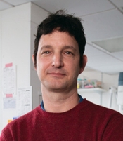 Matthieu Piel合作论文数Institut Pierre Gilles de Gennes, Centre National de la Recherche Scientifique;Institut Curie, Centre National de la Recherche Scientifique5
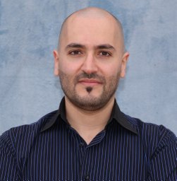 Vito Conte合作论文数Materials Research Group, King’s College London5K. Plak合作论文数5
 Aurélien Roux合作论文数UMR 168 CNRS/Institut Curie, FR-75248 Paris, France5Nargess Khalilgharibi合作论文数4
 Oscar M Lancaster合作论文数4Sushila Ganguli合作论文数4Dimitra Papatziamou合作论文数4Delyan R Mutavchiev合作论文数MRC Lab Mol Cell Biol, UCL4
 Ana Lisica合作论文数London Centre for Nanotechnology, University College London4
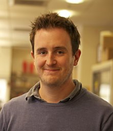 Andrew R Harris合作论文数Department of Mechanical and Aerospace Engineering, Carleton University4
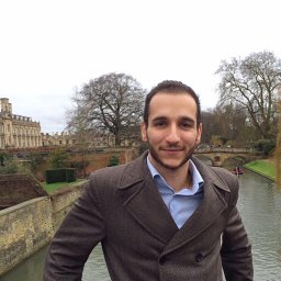 Andrea Dimitracopoulos合作论文数Center for Mathematics, Physics, and Engineering in the Life Sciences and Experimental Biology,;Laboratory for Molecular Cell Biology, Medical Research Council's;Center for Mathematics, Physics, and Engineering in the Life Sciences and Experimental Biology, Medical Research Council's4Almada Pedro合作论文数4
 Rachel McKendry合作论文数London Centre for Nanotechnology, Faculty of Mathematical & Physical Sciences, University College London4
 Agata Nyga合作论文数Centre for Nanotechnology, Biomaterials and Tissue Engineering, University College London4
 Jan Löwe合作论文数Medical Research Council Laboratory of Molecular Biology4
 Mohan Balasubramanian合作论文数University of Warwick4Edward Giniger合作论文数4




