Purpose: To characterize structural and biomechanical changes following ex vivo treatment of rabbit sclera with the bacteriochlorophyll derivative WST11 and near-infrared (NIR) light. Methods: Enucleated left scleras of 10 rabbits were cross-linked by a 20-min topical application of WST11 solution, followed by 30-min NIR irradiation (10 mW/cm(2)). Paired right eyes served as untreated controls. Scleral tangent modulus and ultimate tensile strength were obtained from tensile strip testing. Collagen molecular-level nanostructure was measured using wide-angle X-ray scattering (WAXS). Scleral fibrillar-level microstructure was assessed using second harmonic generation (SHG) and two-photon fluorescence (TPF) imaging. Results: WST11/NIR treatment increased scleral tangent modulus by an average of 79% for strains between 1.5% and 3% (p < 0.05), while ultimate tensile strength increased by 140% (control: 1.719 MPa, treated: 4.134 MPa, p < 0.01). WAXS indicated that collagen molecular spacing (control: 1.485 nm/treated: 1.480 nm, p = 0.65), spatial disorder (47.6 a.u./47.6 a.u., p > 0.99), and anisotropy (0.935 a.u./1.157 a.u., p = 0.09) were not significantly affected by treatment. No marked treatment effects on collagen fibril bundle organization were observable by SHG imaging. TPF imaging revealed additional crimped fibrous structures in the outer half of the tissue depth in treated specimens, indicative of enhanced collagen cross-linking. Conclusions: WST11/NIR treatment significantly increased the stiffness of the ex vivo rabbit sclera. The origin of the stiffening effect is consistent with the formation of collagen cross-links that are predominantly extrafibrillar in nature. Further research is needed to establish the full mechanism, efficacy, and safety of the method as a potential myopia treatment.
High myopia is the leading cause of visual impairment worldwide, driven by excessive axial elongation resulting in biomechanical weakening of the sclera, and increasing the risk of complications such as posterior staphyloma, myopic macular degeneration, retinal detachment, cataract, and glaucoma. Conventional myopia management strategies aim to slow axial elongation in childhood, leaving patients with high myopia at risk of vision-threatening complications as they age. For these individuals, targeted therapies that strengthen the weakened sclera represent a promising, yet still experimental, approach. This review provides an overview of the role of the sclera in myopia progression, emphasizing changes in extracellular matrix composition, collagen organization, biomechanical integrity, and signaling pathways. Investigational scleral therapies are discussed, including posterior scleral reinforcement techniques, crosslinking modalities, pharmacological strategies, and tissue engineering approaches, with discussion of their mechanisms, preclinical and clinical evidence, and translational challenges. Particular attention is given to translational barriers, such as safe delivery to the posterior pole, retinal safety, and the absence of standardized outcome measures that link biomechanical reinforcement to functional benefit. Advances in imaging and in vivo biomechanical assessment, such as polarization-sensitive optical coherence tomography, Brillouin microscopy, and ultrasound-based elastography, hold promise for earlier risk stratification and treatment monitoring. By integrating insights from biomechanics, molecular biology, and therapeutic innovation, this review highlights the sclera as both a key driver of myopia pathology and a promising therapeutic target. Continued interdisciplinary collaboration will be essential to translate these experimental approaches into clinically viable treatments capable of reducing the growing burden of high myopia worldwide.
PURPOSE:To evaluate the effects of extracorporeal ultra-high-fluence corneal cross-linking (ECO-CAIRS) on the dehydration and rehydration behavior of porcine corneal stromal ring segments in an experimental model simulating the preparation of corneal allogenic intrastromal ring segments. SETTING:ELZA Institute, Zurich, Switzerland. METHODS:Corneal ring segments (two per cornea) were prepared from freshly enucleated porcine corneas (n=48) and randomly divided into three groups: controls (no CXL, Jerky technique), extracorporeal ultra-high-fluence CXL at 30 J/cm2, and 60 J/cm2 (n=32 each). After epithelial removal and ring preparation, segments in the control group were soaked in balanced salt solution (BSS), while those in the experimental groups were soaked in riboflavin solution (Ribo-Ker, EMAGine AG, Zug, Switzerland) for 10 minutes. Each group subsequently underwent a total dehydration period of 45 minutes, which included both UV-A irradiation and waiting phases for the CXL-treated groups, followed by a 10-minute rehydration phase using BSS. Segment thickness was measured by calibrated imaging at baseline, after dehydration, and after rehydration. RESULTS:Baseline structural thickness values did not differ significantly between groups. Both CXL-treated groups demonstrated significantly greater dehydration (p<0.01) and reduced rehydration (p<0.01) when compared to controls. No significant differences were found between the two CXL-treated groups. Ultra-high-fluence cross-linking enhanced segment dehydration and slowed rehydration. CONCLUSIONS:ECO-CAIRS effectively modulates the dehydration and rehydration of corneal ring segments, which may improve handling and implantation characteristics during surgery. These findings support the potential clinical benefit of CXL in CAIRS procedures. Further in vivo studies are required to confirm long-term behavior and biomechanical impact.
Keratoconus involves progressive corneal thinning and stromal collagen disorganization that may precede conventional morphological detection. Polarimetric interferometry captures corneal birefringence related to collagen anisotropy and could serve as a non-invasive structural biomarker. We conducted a retrospective observational study of 95 keratoconus patients and 21 healthy controls who underwent corneal topography (Pentacam) and polarimetric interferometry (Lumaxis). Isogyre cross-pattern parameters, widest arm width, narrowest arm width, arm-width standard deviation, and centration indices (M1, M2), were quantified with GeoGebra and correlated with keratometric values, thinnest corneal thickness, Amsler-Krumeich staging, and Belin-Ambrosio ABCD classification. Widest arm width correlated strongly with Pentacam-derived AK stage (Spearman’s ρ = 0.798, p < 0.0001) and maximum keratometry (ρ = 0.730, p < 0.001), and moderately with thinnest corneal thickness (ρ = −0.488, p < 0.001). Arm-width asymmetry correlated inversely with thinnest corneal thickness (ρ = −0.381, p < 0.001). Receiver operating characteristic analysis showed promising discriminative performance: arm-width standard deviation (AUC 0.95) and widest arm (AUC 0.87) differentiated keratoconus from controls. Multivariable analyses confirmed independence from corneal curvature and thickness. Lumaxis-derived isogyre parameters correlate significantly with established keratoconus severity indices, supporting polarimetric interferometry as a complementary modality for assessing corneal stromal disorganization. Prospective longitudinal studies with automated image analysis are warranted.
PURPOSE:To compare the biomechanical stiffening effects of a novel ultra-high fluence corneal cross-linking (CXL) protocol with conventional high-fluence CXL for extracorporeal corneal allogenic intrastromal ring segment optimization (ECO-CAIRS). SETTING:ELZA Institute, Zurich, Switzerland. DESIGN:Experimental, randomized, controlled ex vivo laboratory study using porcine corneas. No masking was applied. METHODS:Eighty-four porcine corneas were divided into four groups: Group A (control), Group B (10 J/cm2), Group C (30 J/cm2), and Group D (60 J/cm2). Following epithelial debridement, Groups B, C, and D were treated with 0.1% hypo-osmolar riboflavin. UV-A irradiation was delivered at fluences of 10 J/cm2 (18 mW/cm2, 9:15 min) for Group B, 30 J/cm2 (30 mW/cm2, 16:40 min) for Group C, and 60 J/cm2 (30 mW/cm2, 33:18 min) for Group D. Corneal biomechanical properties were assessed via stress-strain extensiometry. RESULTS:Mean elastic modulus (5% strain) values were: Group A (1.39 ± 0.49 N/mm), Group B (1.83 ± 0.48 N/mm), Group C (2.25 ± 0.83 N/mm), and Group D (2.31 ± 0.88 N/mm). Ultra-high fluence groups (C and D) showed significantly greater stiffening than Group B (P = 0.041 and P = 0.015), with no significant difference between Groups C and D (P = 1.00). Group B exhibited significantly greater stiffening than Group A (P = 0.023). CONCLUSIONS:Ultra-high fluence CXL enhances biomechanical stiffening compared to conventional high-fluence CXL, supporting its use in ECO-CAIRS. In this extracorporeal context, endothelial safety and haze are irrelevant, while increased stiffness facilitates intrastromal ring insertion and reduces corneal swelling.
Kidney disease, the ninth leading cause of death in the United States, suffers from poor diagnostic efficiency (10%). Traditional biopsies use molecular reagents to enhance diagnostic power but are limited by overlapping spatial and chromatic signals, product quality variability, and additional processing. To address these challenges without disrupting routine diagnostics, we implement label-free imaging modalities—stimulated Raman scattering (SRS), second harmonic generation (SHG), and two-photon fluorescence (TPF)—within a single setup. We identify morphological, lipidomic, and metabolic biomarkers in control and diabetic kidney samples at subcellular resolution. Label-free Stimulated Raman Histology (SRH) reveals distinct collagen morphology, mesangial-glomerular volumes, lipid saturation, redox status, and lipid-protein concentrations previously unrecognized in kidney diseases. Using the same tissue section enhances diagnostic value without compromising limited tissue. These multimodal biomarkers broadly deepen the understanding of kidney disease progression by integrating lipidomic, fibrotic, and metabolic data.
PURPOSE. Donor tissue shortfalls and postsurgical complications are driving novel corneal tissue regeneration approaches. Corneal stromal keratocytes (CSKs) have shown promise in promoting corneal repair and restoring transparency. We investigated the impact of intrastromal CSK injection on corneal ultrastructure and proteoglycan (PG) distribution in a rat injury model. METHODS. Rats were divided into four groups: normal (n = 12), injured (irregular phototherapeutic keratectomy centrally; n = 6), CSK (injured + human CSK intrastromal injection; n = 6), and PBS (injured + PBS injection; n = 6). Three weeks after treatment, corneas were examined by slit-lamp and optical coherence tomography. Corneal ultrastructure was analysed via small-angle x-ray scattering (collagen fibril diameter, interfibrillar spacing and matrix order), transmission electron microscopy with cuprolinic blue before and after chondroitinase digestion (CS/DS and KS PGs), and immunofluorescence staining (lumican and decorin). RESULTS. Irregular phototherapeutic keratectomy caused corneal opacity and significantly disrupted stromal ultrastructure, characterized by increased haze density (P < 0.0001), change in central corneal thickness (P = 0.0005), and interfibrillar spacing (P < 0.0001), along with decreased fibril diameter (P < 0.0001), matrix order (P < 0.0001), CS/DS (P < 0.0001) and KS (P < 0.0001) PGs, lumican, and decorin. CSK injection recovered corneal clarity and native stromal ultrastructure, with haze density (P = 0.8086), change in central corneal thickness (P = 0.9503), fibril diameter (P = 0.1139), interfibrillar spacing (P = 0.5879), matrix order (P = 0.9999), CS/DS (P = 0.9969) and KS (P = 0.2877) PGs, lumican, and decorin returning to normal. In contrast, the PBS group exhibited similar corneal injury responses to the injured group. CONCLUSIONS. CSK injection resolved early stage corneal scarring by restoring stromal collagen arrangement and PG distribution, further endorsing its potential for treating corneal opacities.
This study aimed to investigate the correlation between myopia severity and the stress–strain index (SSI), measured with the Corneal Visualization Scheimpflug Technology (Corvis ST) device. The subjects were divided into two groups, based on both the axial length (AL) and spherical equivalent refraction (SER): 22–26.00 AL group (22 mm < AL < 26.00 mm) associated with SER of less than − 6.00D, and ≥ 26.00 AL group (AL ≥ 26.00 mm) associated with SER over − 6.00D. The differences in the Corvis ST-derived dynamic corneal response parameters and stiffness parameters between the two groups were investigated. The correlation between SSI and AL, SER, age, ratio of AL to radius of corneal curvature (CR) (AL/CR), and axial length minus anterior chamber depth (ACD) (AL-ACD) were analyzed. The SSI (0.95 ± 0.13 in the 22–26.00 AL group and 0.86 ± 0.15 in the ≥ 26.00 AL group) were significantly different between the two groups (P < 0.01). In the ≥ 26.00 AL group, there was evidence of a weak negative correlation between SSI and AL (r = − 0.265, P < 0.01), AL/CR (r = − 0.376, P < 0.01), and AL-ACD (r = − 0.224, P < 0.01); and a weak positive correlation between SSI and SER (r = 0.251, P < 0.01). However, in the 22–26.00 AL group, there was no correlation between SSI and AL, AL-ACD, AL/CR or SER (P > 0.05). SSI was significantly correlated with AL, which is the major determinant of SER, in the ≥ 26.00 AL group. This correlation was not affected with CR and ACD, as both AL/CR and AL-ACD also correlated with SSI at the same degree.
Intrastromal cell therapy utilizing quiescent corneal stromal keratocytes (qCSKs) from human donor corneas emerges as a promising treatment for corneal opacities, aiming to overcome limitations of traditional surgeries by reducing procedural complexity and donor dependency. This investigation demonstrates the therapeutic efficacy of qCSKs in a male rat model of corneal stromal opacity, underscoring the significance of cell-delivery quality and keratocyte differentiation in mediating corneal opacity resolution and visual function recovery. Quiescent CSKs-treated rats display improvements in escape latency and efficiency compared to wounded, non-treated rats in a Morris water maze, demonstrating improved visual acuity, while stromal fibroblasts-treated rats do not. Advanced imaging, including multiphoton microscopy, small-angle X-ray scattering, and transmission electron microscopy, revealed that qCSK therapy replicates the native cornea's collagen fibril morphometry, matrix order, and ultrastructural architecture. These findings, supported by the expression of keratan sulfate proteoglycans, validate qCSKs as a potential therapeutic solution for corneal opacities. This study demonstrates the opacity resolution and visual function improvement following intrastromal injection of quiescent corneal stromal keratocytes but not stromal fibroblasts. The keratocyte therapy recapitulates native cornea's collagen fibril organization and proteoglycans.
Purpose: The study presents an averaged anterior eye geometry model combined with a localised material model that is straightforward, appropriate and amenable for implementation in finite element (FE) modelling.Methods: Both right and left eye profile data of 118 subjects (63 females and 55 males) aged 22-67 years (38.5 +/- 7.6) were used to build an averaged geometry model. Parametric representation of the averaged geometry model was achieved through two polynomials dividing the eye into three smoothly connected volumes. This study utilised the collagen microstructure x-ray data of 6 ex -vivo healthy human eyes, 3 right eyes and 3 left eyes in pairs from 3 donors, 1 male and 2 females aged between 60 and 80 years, to build a localised element-specific material model for the eye.Results: Fitting the cornea and the posterior sclera sections to a 5th-order Zernike polynomial resulted in 21 coefficients. The averaged anterior eye geometry model recorded a limbus tangent angle of 37 degrees at a radius of 6.6 mm from the corneal apex. In terms of material models, the dif-ference between the stresses generated in the inflation simulation up to 15 mmHg in the ring -segmented material model and localised element-specific material model were significantly different (p < 0.001) with the ring-segmented material model recording average Von-Mises stress 0.0168 +/- 0.0046 MPa and the localised element-specific material model recording average Von-Mises stress 0.0144 +/- 0.0025 MPa.Conclusions: The study illustrates an averaged geometry model of the anterior human eye that is easy to generate through two parametric equations. This model is combined with a localised material model that can be used either parametrically through a Zernike fitted polynomial or non -parametrically as a function of the azimuth angle and the elevation angle of the eye globe. Both averaged geometry and localised material models were built in a way that makes them easy to implement in FE analysis without additional computation cost compared to the limbal disconti-nuity so-called idealised eye geometry model or ring-segmented material model.
Mimecan, or osteoglycin, belongs to the family of small leucine-rich proteoglycans. In connective tissues mimecan is implicated in the development and maintenance of normal collagen fibrillar organization. Since collagen fibrils are responsible for tissue reinforcement, the absence of mimecan could lead to abnormal tissue mechanical properties. Here, we carried out a preliminary investigation of possible changes in the mechanical properties of tendons in mice lacking a functional mimecan gene, as a function of age. Tail tendons were dissected from mimecan gene knockout (KO) and wild type (WT) mice at ages 1, 4 and 8 months and mechanical properties evaluated using a microtensile testing equipment. Mimecan gene knockout resulted in changes in tendon elasticity- and fracture-related properties. While tendons of WT mice exhibited enhanced mechanical properties with increasing age, this trend was notably attenuated in mimecan KO tendons, with the exception of fracture strain. When genotype and age were considered as cross factors, the diminution in the mechanical properties of mimecan KO tendons was significant for yield strength, modulus and fracture strength. This effect appeared to affect the mice at 4 month old. These preliminary results suggest that mimecan may have a role in regulating age-dependent mechanical function in mouse tail tendon.
Mechanical loading regulates the functional capabilities of the ocular system, particularly in the sclera ('white of the eye') - the principal load-bearing tissue of the ocular globe. Resident fibroblasts of the scleral eye wall are continuously subjected to fluctuating mechanical strains arising from eye movements, cerebrospinal fluid pressure and, most influentially, intra-ocular pressure (IOP). Whilst fibroblasts are hypothesised to actively participate in scleral biomechanics, to date limited information has been reported on how the macroscopic stresses and strains are transmitted via their cytoskeletal networks. In this study, the effect of applying either a 'physiological load' (simulating healthy IOP) or a 'pathological load' (simulating an elevated glaucomatous IOP) to bovine scleral fibroblasts, as a model of human glaucoma, was conducted to characterise cytoskeletal organi-sation, chromatin condensation and cell dimensions using immunofluorescence confocal microscopy. Quantifi-cation of cell parameters and cytoskeletal element anisotropy were subsequently performed using FibrilTool, and chromatin condensation parameter assessment through a bespoke MATLAB script. The novel findings suggest that physiological load-induced F-actin rearrangement is transient, whereas pathological load, recapitulating in vivo glaucomatous IOP levels, had a reversible and inhibitory influence on remodelling of the cytoskeletal ar-chitecture and, further, induction of chromatin condensation. Ultimately, this could compromise cell behaviour. These findings could provide valuable insight into the mechanism(s) used by scleral fibroblasts to mechanically adapt to support biomechanical tissue integrity, and how it could be potentially modified for therapeutic avenues targeting mechanically mediated ocular pathologies such as glaucoma.
Speckle noise and retinal shadows within OCT B-scans occlude important edges, fine textures and deep tissues, preventing accurate and robust diagnosis by algorithms and clinicians. We developed a single process that successfully removed both noise and retinal shadows from unseen single-frame B-scans within 10.4ms. Mean average gradient magnitude (AGM) for the proposed algorithm was 57.2% higher than current state-of-the-art, while mean peak signal to noise ratio (PSNR), contrast to noise ratio (CNR), and structural similarity index metric (SSIM) increased by 11.1%, 154% and 187% respectively compared to single-frame B-scans. Mean intralayer contrast (ILC) improvement for the retinal nerve fiber layer (RNFL), photoreceptor layer (PR) and retinal pigment epithelium (RPE) layers decreased from 0.362 ± 0.133 to 0.142 ± 0.102, 0.449 ± 0.116 to 0.0904 ± 0.0769, 0.381 ± 0.100 to 0.0590 ± 0.0451 respectively. The proposed algorithm reduces the necessity for long image acquisition times, minimizes expensive hardware requirements and reduces motion artifacts in OCT images.
This study aims to estimate the reduction in collagen fibril density within the central 6 mm radius of keratoconic corneas through the processing of microstructure and videokeratography data. Collagen fibril distribution maps and topography maps were obtained for seven keratoconic and six healthy corneas, and topographic features were assessed to detect and calculate the area of the cone in each keratoconic eye. The reduction in collagen fibril density within the cone area was estimated with reference to the same region in the characteristic collagen fibril maps of healthy corneas. Together with minimum thickness and mean central corneal refractive power, the cone area was correlated with the reduction in the cone collagen fibrils. For the corneas considered, the mean area of keratoconic cones was 3.30 ± 1.90 mm2. Compared with healthy corneas, fibril density in the cones of keratoconic corneas was lower by as much as 35%, and the mean reduction was 17 ± 10%. A linear approximation was developed to relate the magnitude of reduction to the refractive power, minimum corneal thickness and cone area (R2 = 0.95, p < 0.001). Outside the cone area, there was no significant difference between fibril arrangement in healthy and keratoconic corneas. The presented method can predict the mean fibril density in the keratoconic eye's cone area. The technique can be applied in microstructure-based finite-element models of the eye to regulate its stiffness level and the stiffness distribution within the areas affected by keratoconus.
As the eye's main load-bearing connective tissue, the sclera is centrally important to vision. In addition to cooperatively maintaining refractive status with the cornea, the sclera must also provide stable mechanical support to vulnerable internal ocular structures such as the retina and optic nerve head. Moreover, it must achieve this under complex, dynamic loading conditions imposed by eye movements and fluid pressures. Recent years have seen significant advances in our knowledge of scleral biomechanics, its modulation with ageing and disease, and their relationship to the hierarchical structure of the collagen-rich scleral extracellular matrix (ECM) and its resident cells. This review focuses on notable recent structural and biomechanical studies, setting their findings in the context of the wider scleral literature. It reviews recent progress in the development of scattering and bioimaging methods to resolve scleral ECM structure at multiple scales. In vivo and ex vivo experimental methods to characterise scleral biomechanics are explored, along with computational techniques that combine structural and biomechanical data to simulate ocular behaviour and extract tissue material properties. Studies into alterations of scleral structure and biomechanics in myopia and glaucoma are presented, and their results reconciled with associated findings on changes in the ageing eye. Finally, new developments in scleral surgery and emerging minimally invasive therapies are highlighted that could offer new hope in the fight against escalating scleral-related vision disorder worldwide.
Purpose To develop and use a custom virtual fields method (VFM) to assess the biomechanical properties of human prelamina and lamina cribrosa (LC) in vivo. Methods Clinical data of 20 healthy, 20 ocular hypertensive (OHT), 20 primary open-angle glaucoma, and 16 primary angle-closure glaucoma eyes were analyzed. For each eye, the intraocular pressure (IOP) and optical coherence tomography (OCT) images of the optic nerve head (ONH) were acquired at the normal state and after acute IOP elevation. The IOP-induced deformation of the ONH was obtained from the OCT volumes using a three-dimensional tracking algorithm and fed into the VFM to extract the biomechanical properties of the prelamina and the LC in vivo. Statistical measurements and P values from the Mann-Whitney-Wilcoxon tests were reported. Results The average shear moduli of the prelamina and the LC were 64.2 ± 36.1 kPa and 73.1 ± 46.9 kPa, respectively. The shear moduli of the prelamina of healthy subjects were significantly lower than those of the OHT subjects. Comparisons between healthy and glaucoma subjects could not be made robustly due to a small sample size. Conclusions We have developed a methodology to assess the biomechanical properties of human ONH tissues in vivo and provide preliminary comparisons in healthy and OHT subjects. Our proposed methodology may be of interest for glaucoma management.
Computational models of cellular structures generally rely on simplifying approximations and assumptions that limit biological accuracy. This study presents a comprehensive image processing pipeline for creating unified three-dimensional (3D) reconstructions of the cell cytoskeletal networks and nuclei. Confocal image stacks of these cellular structures were reconstructed to 3D isosurfaces (Imaris), then tessellations were simplified to reduce the number of elements in initial meshes by applying quadric edge collapse decimation with preserved topology boundaries (MeshLab). Geometries were remeshed to ensure uniformity (Instant Meshes) and the resulting 3D meshes exported (ABAQUS) for downstream application. The protocol has been applied successfully to fibroblast cytoskeletal reorganisation in the scleral connective tissue of the eye, under mechanical load that mimics internal eye pressure. While the method herein is specifically employed to reconstruct immunofluorescent confocal imaging data, it is also more widely applicable to other biological imaging modalities where accurate 3D cell structures are required.
Purpose We developed a combined biomechanical and hemodynamic model of the human eye to estimate blood flow and oxygen concentration within the lamina cribrosa (LC) and rank the factors that influence LC oxygen concentration. Methods We generated 5000 finite-element eye models with detailed microcapillary networks of the LC and computed the oxygen concentration of the lamina retinal ganglion cell axons. For each model, we varied the intraocular pressure (IOP) from 10 mm Hg to 55 mm Hg in 5-mm Hg increments, the cerebrospinal fluid pressure (13 ± 2 mm Hg), cup depth (0.2 ± 0.1 mm), scleral stiffness (±20% of the mean values), LC stiffness (0.41 ± 0.2 MPa), LC radius (1.2 ± 0.12 mm), average LC pore size (5400 ± 2400 µm2), the microcapillary arrangement (radial, isotropic, or circumferential), and perfusion pressure (50 ± 9 mm Hg). Blood flow was assumed to originate from the LC periphery and drain via the central retinal vein. Finally, we performed linear regressions to rank the influence of each factor on the LC tissue oxygen concentration. Results LC radius and perfusion pressure were the most important factors in influencing the oxygen concentration of the LC. IOP was another important parameter, and eyes with higher IOP had higher compressive strain and slightly lower oxygen concentration. In general, superior–inferior regions of the LC had significantly lower oxygen concentration than the nasal–temporal regions, resulting in an hourglass pattern of oxygen deficiency. Conclusions To the best of our knowledge, this study is the first to implement a comprehensive hemodynamical model of the eye that accounts for the biomechanical forces and morphological parameters of the LC. The results provide further insight into the possible relationship of biomechanical and vascular pathways leading to ischemia-induced optic neuropathy.
Purpose To study the effect of changing heart rate on the ocular pulse and the dynamic biomechanical behaviour of the optic nerve head (ONH) using a comprehensive mathematical model. Methods In a finite element model of a healthy eye, a biphasic choroid consisted of a solid phase with connective tissues and a fluid phase with blood, and the lamina cribrosa (LC) was viscoelastic as characterized by a stress-relaxation test. We applied arterial pressures at 18 ocular entry sites (posterior ciliary arteries) and venous pressures at four exit sites (vortex veins). In the model, the heart rate was varied from 60 bpm to 120 bpm (increment: 20 bpm). We assessed the ocular pulse amplitude (OPA), pulse volume, ONH deformations and the dynamic modulus of the LC at different heart rates. Results With an increasing heart rate, the OPA decreased by 0.04 mmHg for every 10 bpm increase in heart rate. The ocular pulse volume decreased linearly by 0.13 µL for every 10 bpm increase in heart rate. The storage modulus and the loss modulus of the LC increased by 0.014 MPa and 0.04 MPa, respectively, for every 10 bpm increase in heart rate. conclusions In our model, the OPA, pulse volume, and ONH deformations decreased with an increasing heart rate, while the LC became stiffer. The effects of blood pressure / heart rate changes on ONH stiffening may be of interest for glaucoma pathology. Support Singapore Ministry of Education, Academic Research Fund, Tier 2 (R-397-000-280-112). Commercial relationship None
 Keith Meek合作论文数School of Optometry and Vision Sciences, Cardiff University81
 Sally Hayes合作论文数School of Optometry and Vision Sciences, Cardiff University44
 Andrew J. Quantock合作论文数Department of Ophthalmology, Kyoto Prefectural University of Medicine34
 Christina Kamma-Lorger合作论文数19
 Ahmed Elsheikh合作论文数School of Engineering, University of Liverpool16
 Thomas Lykke-Møller Sørensen合作论文数16
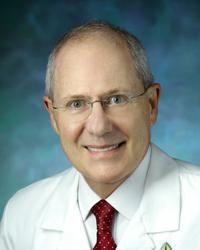 Harry A. Quigley合作论文数Glaucoma Division, Wilmer Eye Institute, School of Medicine, Johns Hopkins University14
 Nicholas J. Terrill合作论文数Diamond Light Source Ltd., Diamond House14
 Robert D Young合作论文数12Jacek K. Pijanka合作论文数12
 Michael Girard合作论文数Department of Ophthalmology, Emory University School of Medicine;Ophthalmic Engineering & Innovation Laboratory, Singapore Eye Research Institute;Emory Eye Center11
 Baptiste Coudrillier合作论文数Department of Mechanical Engineering, Johns Hopkins University11
 Shigeru Kinoshita (木下茂)合作论文数Kyoto Prefectural University of Medicine10
 Chris F Inglehearn合作论文数School of Medicine, Faculty of Medicine and Health, University of Leeds;Division of Molecular Medicine, University of Leeds8Erin P Dooley合作论文数7
 Petar Markov合作论文数7
 Abass Ahmed合作论文数7Sally Dennis合作论文数7Jennifer C. Hiller合作论文数7Jonathan Harris合作论文数6
 Thao D. (Vicky) Nguyen合作论文数Department of Mechanical Engineering, Whiting School of Engineering, Johns Hopkins University6
 Xiaofei Wang (王晓飞)合作论文数School of Biological Science and Medical Engineering, Beihang University6
 Charles Whitford合作论文数School of Engineering, University of Liverpool6
 Sian Morgan合作论文数School of Optometry and Vision Sciences, Cardiff University6
 Paul M Hocking合作论文数The Roslin Institute, The University of Edinburgh/The Royal (Dick) School of Veterinary Studies, The University of Edinburgh6
 Robert D. Young合作论文数OLI Systems Inc6
 James L. Funderburgh合作论文数Center for Vision Restoration, School of Medicine, University of Pittsburgh5
 Perera Shamira A合作论文数Singapore Natl Eye Ctr5
 Aung Tin合作论文数Centre for Vision Research, Duke-NUS Medical School;Singapore National Eye Centre;Singapore Eye Research Institute;Department of Ophthalmology, Yong Loo Lin School of Medicine, National University of Singapore5
 Nicholas White合作论文数Faculty of Tropical Medicine, Mahidol University5
 Emma Blain合作论文数School of Biosciences, Cardiff University5Naoto Yagi合作论文数5Manir Ali合作论文数5
 Sripad Krishna Devalla合作论文数Dept Artificial Intelligence, Univ Malaya4
 Ashkan Eliasy合作论文数Sch Engn, Univ Liverpool4
 Leopold Schmetterer合作论文数School of Chemical and Biomedical Engineering, College of Engineering, Nanyang Technological University4
 Manfred Burghammer合作论文数4Jefferys Joan L合作论文数4
 Hung Tan Pham合作论文数4Julie Albon合作论文数4Hidetoshi Tanioka合作论文数4
 Joram Piatigorsky合作论文数National Eye Institute, National Institutes of Health4Veronique Siegler合作论文数4Wakako Adachi合作论文数3Jennifer Lewis合作论文数3
 Dong Zhou合作论文数3
 Strouthidis Nicholas G合作论文数Singapore Natl Eye Ctr, Singapore Eye Res Inst3
 Paterson Neil G合作论文数3Palka Barbara P合作论文数3




