Rad23 proteins function in both DNA repair and protein stability regulation. As ubiquitinated forms of p53 are stabilized after DNA damage in concert with p53 functional activation, and human Rad23 proteins (hHR23A and B) regulate p53 stability in unstressed cells, the role of hHR23B in post-genotoxin regulation of p53 was investigated. Depletion of hHR23B by specific short interfering RNA before genotoxic exposure attenuated p53, p21 and bax induction, abrogated the accumulation of ubiquitinated p53 and suppressed apoptosis. Expression of ubiquitin derivatives with all lysines mutated except K48 or K63 demonstrated that K48-linked p53-ubiquitin conjugates were specifically induced after DNA damage. hHR23B, along with native and ubiquitinated p53, accumulated in chromatin after genotoxic exposure, and the accumulation of ubiquitinated p53 in chromatin was prevented by hHR23B depletion. Chromatin immunoprecipitation analysis demonstrated that hHR23B and p53 both localized to the p21 promoter shortly after DNA damage. hHR23B thus plays a critical role in the activation and function of p53 after specific genotoxic exposures.
Abrogation of ubiquitin/proteasome-dependent turnover of p53 is critical for its activation. UbL-UBA proteins, including human homolog of Rad23 (hHR23) proteins, may regulate proteasomal degradation of substrates such as p53, due to their ability to interact with both ubiquitinated substrates and the proteasome. siRNA-mediated depletion of hHR23A or hHR23B in human cell lines accelerated p53 degradation. In contrast, overexpression of hHR23 proteins led to the accumulation of ubiquitinated p53, and purified hHR23 proteins also blocked p53 proteasome degradation in vitro. An hHR23-MDM2 complex was identified, suggesting that MDM2 and hHR23 cooperate in the regulation of p53 proteasome degradation. Consistent with this hypothesis, an MDM2 mutant that demonstrated increased binding in vivo to hHR23A was able to ubiquitinate, but not degrade p53. Moreover, the defective phenotype of this MDM2 mutant was rescued by siRNA knockdown of hHR23A. Our data indicate that MDM2 acts at a step in the p53 degradation pathway after ubiquitination, to counteract hHR23 inhibition of p53 turnover. Moreover, our data suggest the possibility that ubiquitin ligase/UbL-UBA protein complexes, as exemplified by the MDM2/hHR23 complex, may serve a general role in regulating substrate degradation by the proteasome.
Yin Yang 1 (YY1) is a transcription factor that plays an essential role in development. However, the full spectrum of YY1's functions and mechanism of action remains unclear. We find that YY1 ablation results in p53 accumulation due to a reduction of p53 ubiquitination in vivo. Conversely, YY1 overexpression stimulates p53 ubiquitination and degradation. Significantly, recombinant YY1 is sufficient to induce Hdm2-mediated p53 polyubiquitination in vitro, suggesting that this function of YY1 is independent of its transcriptional activity. We identify direct physical interactions of YY1 with Hdm2 and p53 and show that the basis for YY1-regulating p53 ubiquitination is its ability to facilitate Hdm2-p53 interaction. Importantly, the tumor suppressor p14ARF compromises the Hdm2-YY1 interaction, which is important for YY1 regulation of p53. Taken together, these findings identify YY1 as a potential cofactor for Hdm2 in the regulation of p53 homeostasis and suggest a possible role for YY1 in tumorigenesis.
Rapid turnover of the tumor suppressor protein p53 requires the MDM2 ubiquitin ligase, and both interact with p300–CREB-binding protein transcriptional coactivator proteins. p53 is stabilized by the binding of p300 to the oncoprotein E1A, suggesting that p300 regulates p53 degradation. Purified p300 exhibited intrinsic ubiquitin ligase activity that was inhibited by E1A. In vitro, p300 with MDM2 catalyzed p53 polyubiquitination, whereas MDM2 catalyzed p53 monoubiquitination. E1A expression caused a decrease in polyubiquitinated but not monoubiquitinated p53 in cells. Thus, generation of the polyubiquitinated forms of p53 that are targeted for proteasome degradation requires the intrinsic ubiquitin ligase activities of MDM2 and p300.
Multiple failures of antiretroviral treatments, as a result of multidrug-resistant virus, have led to a proposal for structured therapeutic interruptions (STI). However, a significant decrease in CD4+ T cells may occur. The aim of our study was to determine the kinetics of T cell subpopulation changes, T cell apoptosis and peripheral blood mononuclear cell proliferation after STI. The impact of resistance mutation disappearance on T cell apoptosis was also studied. Ten patients were enrolled prospectively, and blood sampling was performed at weeks 0, 2, 4, 6, 8 and 12. The mean increase in viral load was 1.3 log(10) copies/ml, ranging from 0.1 to 3.2. CD4+ T cell count decreased to a mean of 80 cells/mm(3) from baseline to week 12. In the same period, CD8+ T cells decreased to a mean of 139 cells/mm(3). A significant increase in both T cell apoptosis and proliferation of mononuclear cells was observed. However, proliferation was an early and brief event. The increase in CD4+ T cell apoptosis was obvious in patients exhibiting complete reversion of resistance mutations to antiviral drugs. Our results suggest that during STI, apoptosis is an overwhelming phenomenon compared with proliferation, and may explain the limited immunological impact of this therapeutic option.
This prospective study investigated the contributions of apoptosis and proliferation of CD4(+) T cells obtained by the introduction of a new antiretroviral treatment for human immunodeficiency virus infection. Virus load; T cell counts; apoptosis of T cell subsets, including naive cells; and proliferation were determined from treatment initiation to the third month in a cohort of patients. An increase in CD4(+) T cell count > or = 100 cells/microL over baseline was considered to be a satisfactory immune reconstitution. Sixty-nine patients completed the protocol, 22 of whom met our definition of a satisfactory immune reconstitution, showing a significantly more pronounced reduction in spontaneous CD4(+) T cell apoptosis at month 1 as well as month 3, compared with the other patients. In contrast, neither Fas-induced apoptosis down-regulation nor Fas-induced increased proliferation capacity was associated with a satisfactory immune reconstitution. Down-regulation of CD4(+) T cell apoptosis by antiretroviral treatment is the main mechanism associated with early CD4(+) T cell increase.
Polymorphonuclear neutrophils (PMNs) are the major source of proteolytic activities involved mainly in tissue injuries observed in chronic inflammatory disorders. High levels of soluble forms of CD23 (the low-affinity receptor for IgE) were found in biological fluids from these patients, and recent reports focused on a CD23-mediated regulation of inflammatory response. In this context, we show here that co-culture of activated PMN with CD23+ B cells resulted in a drastic release of soluble CD23 fragments from the cell surface. This cleavage was inhibited by serine proteases inhibitors, including a1-antitrypsin. We next demonstrated that purified human leukocyte elastase or cathepsin G efficiently cleaved membrane CD23 on B cells with a high specificity. Soluble fragments released by serine proteases-mediated CD23 proteolysis stimulated resting monocytes to produce oxidative burst and proinflammatory cytokine without any co-stimulatory signal. This work strongly supports the idea that the capacity of PMN-derived proteases to release soluble forms of CD23 participates in the inflammatory process mediated by these cells.
In the present study we have compared the binding of two monoclonal antibodies to CD23, EBVCS1 and mAb25, which recognize the stalk and the lectin domain, respectively, on the CD23 molecule. At 4 degreesC, EBVCS1 binds to about 10% of the receptors recognized by mAb25 on the B cell surface. At 37 degreesC, whereas mAb25 reaches its maximal binding within a few seconds, EBVCS1 requires 60 min to bind to the same extent. Stabilization of the oligomeric structure of CD23 with IgE strongly affects in a dose-dependent fashion the number of binding sites seen by EBVCS1 but not the t1/2 to reach them, suggesting that EBVCS1 binds to the coiled coil region through an allosteric mechanism. EBVCS1 rapidly down-modulates the membrane CD23 expression with a coincident increase of CD23-soluble fragments in the culture medium, an effect that is inhibited by IgE. In contrast, mAb25, as well as IgE, protects CD23 from proteolytic cleavage and stimulates its endocytosis. These results suggest that EBVCS1 unravels the coiled coil structure of CD23, rendering it more susceptible to proteolytic attack. This supports the oligomeric model proposed previously (Gould, H., Sutton, B., Edmeades, R., and Beavil, A. (1991) Monogr. Allergy 29, 28-49). The biological significance of these observations is discussed.
 Steven R. Grossman合作论文数Division of Hematology, Oncology and Palliative Care, School of Medicine, Virginia Commonwealth University;Departments of Cancer Biology and Medicine, School of Medicine, Virginia Commonwealth University;Massey Cancer Center, Virginia Commonwealth University4
 J Durant合作论文数3
 Patrizia Dellamonica合作论文数3P. M. Roger合作论文数3J L Cousin合作论文数2A Bernard合作论文数2J.G. Fuzibet合作论文数2Alain Bernard合作论文数2Olivier Munoz合作论文数2Jean-Philippe Breittmayer合作论文数2D. J. Lo合作论文数1philippe halfon合作论文数1C. Ceppi合作论文数1M Batoz合作论文数1N Grenier-Brossette合作论文数1I. Foucault合作论文数1Michel Ticchioni合作论文数1Alain Pesce合作论文数1Jy Bonnefoy合作论文数1e strobert合作论文数1A. Foussat合作论文数1Jp Breittmayer合作论文数1M Song合作论文数1
 Allan Douglas Kirk合作论文数Department of Surgery, Duke University School of Mediane;Duke Cancer Institute, Duke University School of Mediane1Rouquette-Jazdanian Alexandre K合作论文数Institut National de la Santé et de la Recherche Médicale (INSERM) Unit 576, Hôpital de l'Archet I1
 Alton Brad Farris合作论文数Dept Pathol & Lab Med, Emory Univ1
 Peng Yin合作论文数Department of Systems Biology, Harvard Medical School, Harvard University;Molecular Systems Lab, Wyss Institute for Biologically Inspired Engineering, Harvard University;Ultivue, Inc.;NuProbe Global1Donohoe Mary E合作论文数1
 Guangchao Sui (隋广超)合作论文数东北林业大学生命科学学院1
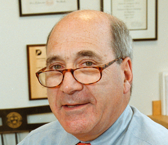 David M. Livingston合作论文数Dana-Farber Cancer Institute, Harvard Medical School;Department of Medicine, Harvard Medical School1Jenkins Joe B合作论文数1
 Christian P. Larsen合作论文数Department of Surgery, School of Medicine, Emory University;Emory Transplant Center, Emory University1
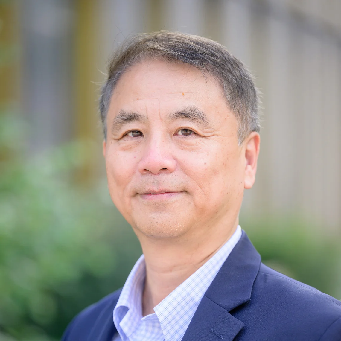 Yang Shi (施扬)合作论文数Ludwig Institute for Cancer Research, Nuffield Department of Medicine, University of Oxford1
 Sylvie Chaillou合作论文数1
 Andrew Kung合作论文数Department of Pediatrics, Memorial Sloan Kettering Cancer Center1Frédéric Sanderson合作论文数1
 Homan Chan合作论文数Division of Biochemistry and Molecular Biology;University of Glasgow1
 Philippe Clevenbergh合作论文数1
 Brigitte Dunais合作论文数1Dominica Calvo合作论文数1Frank V. Leopardi合作论文数1
 Marcel Deckert合作论文数1Eric Cua合作论文数1Alexei F Kisselev合作论文数Norris Cotton Cancer Center, Dartmouth Medical School1
 Nathan R. Wall合作论文数School of Medicine, Loma Linda University;Department of Radiation Medicine, Loma Linda University;Center for Health Disparities & Molecular Medicine, Loma Linda University1
 El Bachir Affar合作论文数1




